Abstract
Cholera toxin (CT) is the prototype for the Vibrio cholerae-Escherichia coli family of heat-labile enterotoxins having an AB5 structure. By substituting amino acids in the enzymatic A subunit that are highly conserved in all members of this family, we constructed 23 variants of CT that exhibited decreased or undetectable toxicity and we characterized their biological and biochemical properties. Many variants exhibited previously undescribed temperature-sensitive assembly of holotoxin and/or increased sensitivity to proteolysis, which in all cases correlated with exposure of epitopes of CT-A that are normally hidden in native CT holotoxin. Substitutions within and deletion of the entire active-site-occluding loop demonstrated a prominent role for His-44 and this loop in the structure and activity of CT. Several novel variants with wild-type assembly and stability showed significantly decreased toxicity and enzymatic activity (e.g., variants at positions R11, I16, R25, E29, and S68+V72). In most variants the reduction in toxicity was proportional to the decrease in enzymatic activity. For substitutions or insertions at E29 and Y30 the decrease in toxicity was 10- and 5-fold more than the reduction in enzymatic activity, but for variants with R25G, E110D, or E112D substitutions the decrease in enzymatic activity was 12- to 50-fold more than the reduction in toxicity. These variants may be useful as tools for additional studies on the cell biology of toxin action and/or as attenuated toxins for adjuvant or vaccine use.
The massive diarrhea characteristic of the disease cholera is in large part due to the action of cholera toxin (CT), produced by Vibrio cholerae of serogroup O1. A better understanding of the structure and function of CT will provide new insights into the pathogenesis of cholera and may aid in the design of safe and effective vaccines against cholera and related diarrheas.
CT is a heterohexameric complex consisting of one A polypeptide and five identical B polypeptides (11). The B pentamer is required for binding to the cell surface receptor ganglioside GM1 (11). The A subunit can be proteolytically cleaved within the single disulfide-linked loop between C187 and C199 to produce the enzymatically active A1 polypeptide (23) and the smaller A2 polypeptide that links fragment A1 to the B pentamer (32). Upon entry into enterocytes by endocytosis and following reduction and translocation, CT-A1 ADP-ribosylates a regulatory G-protein (Gsα), which leads to constitutive activation of adenylate cyclase, increased intracellular concentration of cyclic AMP, and secretion of fluid and electrolytes into the lumen of the small intestine (12). ADP-ribosyl transferase activity of CT is stimulated in vitro by the presence of accessory proteins called ARFs (49), small GTP-binding proteins known to be involved in vesicle trafficking within the eukaryotic cell, but the role of ARFs in the activity of CT in vivo has not yet been determined.
CT is the prototype for the V. cholerae-Escherichia coli family of heat-labile enterotoxins. E. coli heat-labile enterotoxins (LT) are classified into two distinct serogroups (LT-I and LT-II) (reviewed in references 16 and 17). CT is closely related to LT-I. Type I and type II LT have highly homologous A1 polypeptides and moderately homologous A2 polypeptides, but the B polypeptides of LT-II exhibit very low homology to CT or LT-I.
The A1 polypeptide of CT also has limited regions of homology with other ADP-ribosylating toxins (7), including pertussis toxin (PT), diphtheria toxin (DT), and exotoxin A (ET-A). The three-dimensional structures of these toxins have been determined (1, 3, 40–42, 50). All of these ADP-ribosylating toxins have NAD-binding domains with conserved features, but the overall structures of these toxins are not conserved.
Biochemical and genetic analyses of these ADP-ribosylating toxins, and CT and LT in particular, identified several positions where amino acid changes caused inactivation of toxicity (for a review see reference 7). In this study we systematically analyzed the functional importance of selected residues in CT-A that are fully conserved in all members of the V. cholerae-E. coli heat-labile enterotoxin family. Following site-directed mutagenesis of the cloned ctxA gene in E. coli we produced and characterized variant holotoxins with defined amino acid substitutions in CT-A. We identified variants of CT-A that retained the ability to assemble with CT-B to form holotoxins, but they exhibited decreased toxicity or no toxicity. Preliminary data were presented at several meetings, most recently at the 9th European Workshop on Bacterial Protein Toxins (Ste. Maxime, France, 27 June to 2 July 1999). This report emphasizes our findings with novel substitutions in CT that have not been reported previously in any variant of LT-I or LT-II. It also summarizes our comparative studies of substitutions in CT that significantly extend reported findings from previous studies on variants of LT-I or LT-II.
MATERIALS AND METHODS
Bacterial strains, plasmids, and growth conditions.
E. coli TG1 (Amersham Corporation, Arlington Heights, Ill.); TX1, a naladixic acid-resistant derivative of TG1 carrying F′ Tcr lacIq from XL1blue (Stratagene, La Jolla, Calif.) (20); TE1 (TG1 ΔendA F′ Tcr lacIq); and CJ236(F′ Tcr lacIq) (Bio-Rad) were used as hosts for cloning of recombinant plasmids and expression of variant proteins. Plasmid-containing strains were maintained on Luria-Bertani agar plates with antibiotics as required (ampicillin, 50 μg per ml; kanamycin, 25 μg per ml; tetracycline, 10 μg per ml).
Mutagenesis of ctxA gene.
Site-directed mutagenesis using single-stranded uracil-containing templates (25) was used to select for oligonucleotide-derived mutants created in plasmid pMGJ67, an isopropyl-β-d-thiogalactopyranoside (IPTG)-inducible clone of the native CT operon in pBluescript SKII(−) (Stratagene). Mutations were confirmed by DNA sequencing. Some mutations were introduced directly into pARCT4 using the QuickChange mutagenesis method (Stratagene). pARCT4 is an arabinose-inducible clone derived from pAR3 (36) expressing an operon containing the ctxA and ctxB genes with signal sequences derived from the LT-IIb B gene (22) and with each gene independently using the translation initiation sequences derived from T7 gene 10 from vector plasmid pT7-7, a derivative of pT7-1 (44).
One- and two-codon insertion mutations.
Single codon insertions were generated at DdeI restriction sites by partial digestion of pMGJ64 (a derivative of pMGJ67), followed by filling in of the 3-base sticky ends and self-ligation. Two-codon TAB linker insertion mutations were made by adding 6-bp ApaI linkers (GGGCCC) to the ends of RsaI partial digests of pMGJ64 as described in the TAB manual (Pharmacia). Transformants were screened for loss of a single DdeI or RsaI site (and presence of a new ApaI site) and confirmed by DNA sequencing.
Expression of mutant ctxA alleles and purification of variant holotoxins.
Production of each variant holotoxin was tested in 5-ml cultures of Terrific Broth medium (45) in 125-ml Erlenmeyer flasks at 37°C with shaking (200 rpm). Logarithmic phase cells (A600 = 0.8 to 1.0) were induced by the addition of IPTG to 0.4 mM followed by growth overnight. Polymyxin B was added to 1 mg/ml, followed by incubation for 10 min at 37°C. Cells were removed by centrifugation, and the supernatants (periplasmic extracts) were assayed to determine the concentrations of holotoxin and B pentamer as described below. Large-scale cultures of pARCT4 derivatives (1 liter) were grown at 30°C in Terrific Broth to an A600 of 3.0 and induced with 0.5% l-arabinose. After 3 h, cells were harvested and concentrated (25×) extracts were made in Tris-buffered saline (TBS) or TEN (50 mM Tris, 1 mM EDTA, 0.1 M NaCl), pH 7.5, both with 1 mg of polymyxin B/ml. These extracts were passed over a 0.5-ml column of either d-galactose resin (Pierce) or Talon metal affinity resin (Clontech Inc., Palo Alto, Calif.) to bind CT-B subunits, washed with 3 to 5 column volumes of TEN or TBS, and eluted with 1 M d-galactose in TEN or 50 mM imidazole in TBS, respectively. Toxin-containing fractions were pooled, dialyzed against TEN, and stored at 4°C.
Assay for holotoxin antigenicity and toxicity.
CT antigen was determined by ganglioside GM1-dependent solid phase radioimmunoassay (GM1-SPRIA) (19). Briefly, wells of a microtiter plate were incubated with 25-μl samples of 0.15 μM ganglioside GM1 in phosphate-buffered saline prior to blocking nonspecific binding sites with 10% horse serum in phosphate-buffered saline. Periplasmic extracts were serially diluted in these wells. After 60 min at 37°C, unbound antigen was washed away and bound antigen was detected with rabbit antisera (60 min, 37°C) followed by 125I-labeled goat anti-rabbit immunoglobulin G (90 min, 37°C, 150,000 cpm/well, specific activity of approximately 8 mCi/mg). To detect holotoxin, we used monospecific polyclonal rabbit anti-CT-A serum B9. To detect pentameric CT-B (including CT-B in holotoxin), we used rabbit anti-CT-B serum B10. Binding of mouse monoclonal antibody (MAb) was detected with rabbit anti-mouse immunoglobulin G (heavy plus light chains), followed by 125I-labeled goat anti-rabbit immunoglobulin G. GM1 enzyme-linked immunosorbent assays (ELISAs) were performed in a similar manner, except wells of low-binding easy-wash modified flat-bottom ELISA plates (Corning Glassworks, Corning, N.Y.) were coated with 50 μl of 0.15 μM GM1, and bound rabbit antibody was reacted with 1/5,000 goat anti-rabbit-horseradish peroxidase conjugate (Bio-Rad) and detected with OPD substrate (Sigma). Toxicity of culture supernatants was assayed using the mouse Y1 adrenal cell assay (30). One toxic unit is defined as the smallest amount of toxin or supernatant that caused rounding of 75 to 100% of the cells in a well after overnight incubation in RPMI 1640 medium with 10% fetal calf serum.
Western blotting.
Toxin samples were mixed with an equal volume of 2× Laemmli sample buffer, boiled for 5 min in the presence of 3% β-mercaptoethanol, loaded onto sodium dodecyl sulfate (SDS)–12% polyacrylamide gels, and run at a constant 160 V. Proteins were transferred to nitrocellulose by semidry electroblotting as described by the manufacturer (Bio-Rad) and were detected with CT-specific antibodies using an enhanced chemiluminescence kit (Dupont/NEN).
Enzyme assay for ADP-ribosyltransferase activity.
ADP-ribosyltransferase activity was determined using diethylamino(benzylidine-amino)guanidine (DEABAG) as a substrate (34), synthesized in our laboratory. Twenty-five-microliter aliquots of purified toxin variant (activated for 30 min at 30°C with 1/50 [wt/wt] trypsin) was incubated with 200 μl of 2 mM DEABAG in a solution containing 0.1 M K2HPO4 (pH 7.5), 10 mM NAD, and 4 mM dithiothreitol for 2 h. The reaction was stopped by adding 800 μl of a slurry of buffer containing 400 mg of Dowex AG50-X8 resin to bind unreacted substrate. ADP-ribosylated DEABAG in the supernatant was quantitated by fluorescence emission in a DyNAQuant fluorimeter calibrated with DEABAG. For kinetic studies, NAD was titrated over the range 0.25 to 10 mM. To assess activation by ARF, reaction mixtures also included 0.04 mM GTP and 2 μl of a crude preparation of recombinant ARF6 (rARF6). This was made as a whole-cell lysate of E. coli overexpressing an ARF6 cDNA clone (a gift from J. Moss, National Institutes of Health), stored at −20°C in 50% glycerol.
RESULTS
Rationale for site-directed mutagenesis of CT-A1 subunit gene.
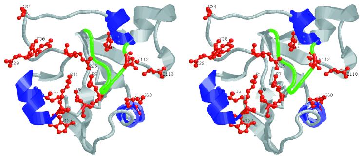
Figure 1 shows a cartoon representation of the predicted active-site domain (A11) of the CT-A1 subunit. Residues that were changed are shown in red in ball-and-stick representation and are numbered. Each residue altered was either completely conserved in CT, LT-I, or LT-II or was a homolog of a residue that was identified as important for NAD binding based on structural alignments of the Cα backbones of CT-A1 or LT-A1 with DT, ET-A, and PT (2).
FIG. 1.
Stereo cartoon representation of the predicted active site of CT-A1. For clarity, only domain A11 is shown (residues 1 to 135). The alpha-helices containing substitutions are shown in blue, and the wt residues substituted are shown in ball-and-stick format in red. Each residue substituted in this study is labeled with the one-letter amino acid code and residue number. The active-site-occluding loop, residues 47 to 56, is shown in green.
Conservative substitutions for Arg-7 (47), Glu-110, and Glu-112 (4) have previously been studied in LT but not biochemically in CT holotoxin, although R7K and E112K variant CT holotoxins have been tested for mucosal adjuvanticity (15). Tyr substitutions for Ser-68 and Val-72 were designed to substitute residues found at the corresponding position in PT where Tyr residues at the homologous positions are predicted to be involved in NAD binding, possibly contributing to the 1,000-fold lower Km value for NAD of PT (cited in reference 28). In LT, different nonconservative substitutions for Ser-68 or Ala-72 were reported to prevent assembly (S68P) or to have no effect on toxicity (S68K or A72R, -H, or -E) (38), although the A72R variant was later reported to show reduced toxicity (13). Homologs of Asp-9 and Arg-11 are also fully conserved and important in PT (37), but substitutions at these positions have not been studied previously in either CT or LT. The hydrophobic Ile residue conserved at position 16 in CT and LT (Val-14 in LT-IIa and LT-IIb), predicted to interact with the nicotinamide moiety of NAD (48), was changed to Ala (nonconservative) or Val (conservative). The Cα backbone trace homolog for Arg-25 in PT is Trp-26, which is also involved in catalysis (5); therefore we converted Arg-25 to Trp or Gly to study the contribution of this residue in CT. Substitutions for Ile-16 and Arg-25 are entirely novel. Glu-29 is fully conserved in the CT-LT family. Although a deletion of this residue in CT has been shown to have greatly reduced toxicity and enzymatic activity (14) for unknown reasons, substitutions for Glu-29 have not been studied previously. His-44, situated in an alpha helix and forming part of a loop that blocks the predicted active site, is also fully conserved among the heat-labile enterotoxins and is located similarly to His-35 of PT (28). His-44 is important in the activity of LT (24), and several nonconservative substitutions in the loop in LT were reported to be less active than that in the wild type (wt) (9). Here we made novel substitutions of Tyr and Ser for His-44 and also deleted the entire loop from 41 to 56 in CT. Four other insertion mutations were also introduced by addition of two-codon linkers (at Y30 and G34) or filling in of restriction sites at T48 (and L153, not shown in Fig. 1). These studies confirmed and significantly extended previous investigations of the structure and function of CT.
Analysis of CT variants.
Initial characterization of each variant holotoxin was performed on crude periplasmic extracts prepared from cells grown at 37°C. While most mutant strains produced wt levels of holotoxin, several mutant strains produced little or no detectable holotoxin under these conditions (R7K, H44Y, H44S, Δ41-56+G, W127S, and L153LL). However, when the growth and induction temperature was reduced to 25 or 30°C, these mutant strains produced normal amounts of holotoxin (data not shown), and the holotoxin variants produced at 25 or 30°C were as stable as wt CT during subsequent incubations at 37°C, as determined by ELISA and SPRIA for immunoreactive holotoxin and Y1 adrenal cell assays for toxicity. Table 1 summarizes the comparative data on holotoxin assembly, toxicity, and enzymatic activity for selected variant CT holotoxins. All holotoxins except the V72Y variant and the conservative substitution variants D9E, I16L, and E29D (not shown) had reduced toxicity, ranging from 50% (E29N, not shown) to less than 1% of the toxicity of wt toxin (R7K, R11K, H44Y, H44S, and E110D+E112D).
TABLE 1.
Toxicity and enzymatic activity of the CT holotoxin variants analyzed in this studya
| CT variant | Holotoxin assembly phenotypeb | Toxicity to Y1 cells (% of wt)c | Enzymatic activity (% of wt) | Ratio of toxicity to enzyme activity |
|---|---|---|---|---|
| wt | + | 100 | 100 | 1.0 |
| R7K | Ts | 0.3 | NT | — |
| D9E | + | 100 | NT | — |
| R11K | + | 0.7 | ND | — |
| I16A | + | 5 | 4 | 1.2 |
| I16L | + | 100 | 96 | 1.0 |
| R25G | + | 6 | 0.5 | 12d |
| R25W | + | 30 | 10 | 3 |
| E29H | + | 5 | 55 | 0.1d |
| Y30WAH | + | 8 | 43 | 0.2d |
| G34GGP | + | 30 | 54 | 0.6 |
| H44Y | Ts | ND | NT | — |
| H44S | Ts | ND | <0.1 | — |
| T48TH | + | 25 | 18 | 1.4 |
| Δ41−56+G | Ts | ND | NT | — |
| S68Y | + | 33 | 11 | 3 |
| V72Y | + | 100 | 68 | 1.5 |
| S68Y+V72Y | + | 5 | 3 | 1.7 |
| E110D | + | 6 | 0.1 | 60d |
| E112D | + | 3 | 0.06 | 50d |
| E110D+E112D | + | <2 | ND | — |
| W127S | Ts | 10 | NT | — |
| L153LL | Ts | 1.5 | 3 | 0.5 |
ND, none detected; NT, not tested; —, not applicable.
Ts, temperature-sensitive, variant CT-A assembles into holotoxin at 26 but not at 37°C; +, assembles normally into holotoxin at 37°C.
Cholera toxin produced in E. coli was unnicked and had a specific activity of 65 to 130 U per μg (one Y1 cell toxic dose [U] equals 7 to 15 ng), compared to 4,000 U per μg for fully nicked native CT from V. cholerae (a Y1 cell toxic dose of 250 pg). Trypsin treatment of unnicked recombinant toxin converted its specific activity to that of native naturally nicked CT (data not shown).
Fivefold or more difference.
For more detailed characterization of these variant holotoxins, the mutations were moved into pARCT4 by subcloning the 552-bp XbaI-ClaI fragment (encoding 95% of the ctxA1 gene) from each mutant plasmid, and variant holotoxins were purified from 1-liter cultures grown at 30°C. Samples of these variant holotoxin preparations were analyzed by SDS-polyacrylamide gel electrophoresis (PAGE) and stained with Coommassie blue (data not shown). Each recombinant holotoxin produced in E. coli was fully stable during assays at 37°C with the CT-A protein in an unnicked form.
Stability of variant holotoxins upon treatment with trypsin.
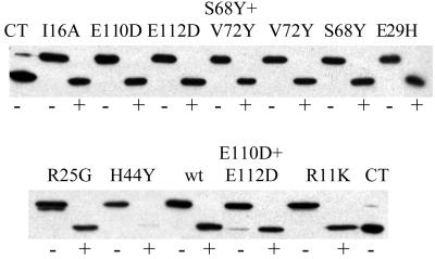
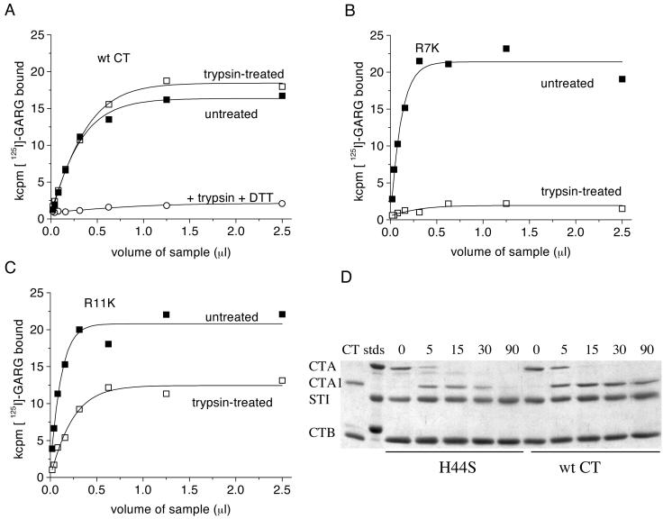
Each variant holotoxin was tested for the ability of its A subunit to be processed by mild trypsin treatment into stable CT-A1 and CT-A2 polypeptides. Untreated or trypsin-treated samples were reduced, denatured, and analyzed by SDS-PAGE and Western blotting (Fig. 2). Most variants were as stable upon trypsin treatment as the wt and generated the expected CT-A1 polypeptide in comparable amounts. Generally, the stability upon trypsinization correlated well with the temperature sensitivity of assembly reported in Table 1. The H44Y variant was highly sensitive to trypsin and was almost completely degraded. The R7K, H44S, and R11K variant holotoxins also showed increased sensitivity to trypsin degradation, to decreasing degrees, as shown in Fig. 3. Analyses by GM1-SPRIA showed that recombinant wt CT treated with trypsin retained its reactivity with anti-CT-A antibody, and the toxin concentrations in the trypsin-treated and untreated samples, as determined by the slope of the curves, were identical (Fig. 3A). The 10% increase in signal at saturation seen after trypsinization probably reflects slightly greater reactivity of the polyclonal antibody with native nicked toxin, the antigen used for immunization.
FIG. 2.
Generation of CT-A1 from CT variant holotoxins by limited treatment with trypsin. Samples of toxin variants were treated with or without trypsin as described in Materials and Methods, and aliquots were analyzed by reducing SDS-PAGE, transferred to nitrocellulose and detected with rabbit anti-CT-A polyclonal antisera by chemiluminescence. Treatment with (+) or without (−) trypsin is denoted by the symbol underneath each lane, and the samples are identified above the lanes by the corresponding toxin variant names. CT represents native, untreated holotoxin from V. cholerae which is ca. 95% nicked.
FIG. 3.
Variability in trypsin sensitivity of selected holotoxin variants. (A through C) Each toxin antigen, treated as shown, was titrated in a GM1-SPRIA and detected with rabbit polyclonal anti-CT-A serum. (A) Recombinant wt CT. (B) R7K variant. (C) R11K variant. (D) Time course of trypsin digestion of H44S variant (left half) compared to that of wt toxin (right half). From left to right, lane CT, native nicked toxin; lane stds, molecular size markers (31, 21.5, and 14.4 kDa); lanes 0, 5, 15, 30, and 90 represent 0, 5, 15, 30, and 90 min of trypsin treatment for each sample. Samples were analyzed by reducing SDS-PAGE and were stained with Coommassie blue. The intense band in all time point samples at 22 kDa is soybean trypsin inhibitor, added to stop the digestion. [125I]-GARG, 125I-labeled goat anti-rabbit immunoglobulin G; DTT, dithiothreitol.
Reduction of a duplicate sample of the trypsin-treated wt toxin with dithiothreitol prior to analysis by GM1-SPRIA led to almost complete loss of the signal (Fig. 3A), presumably reflecting dissociation of the A1 polypeptide from the immobilized A2B5 complex and showing that the trypsin-treated wt sample was in a fully nicked yet stable state. In contrast, the recombinant R7K variant holotoxin lost more than 90% of the signal after brief treatment with trypsin alone (Fig. 3B) without reduction, suggesting that the R7K substitution caused an altered conformation of the A subunit, exposing one or more trypsin-sensitive sites.
The R11K variant holotoxin exhibited an intermediate phenotype, where the immunogenicity of a portion of the sample was stable upon trypsin treatment, but approximately 40% of the molecules were degraded (Fig. 3C), as determined from the plateau signal (free CT-B competes for GM1 binding and reduces the maximum signal) and the slope of the curve (a direct measurement of the concentration of holotoxin). This suggests a slight folding defect in the R11K variant, causing some of the molecules to adopt a wt conformation and the rest to adopt an alternative conformation with exposed trypsin-sensitive sites. The H44S variant, when assayed by reducing SDS-PAGE in a time-limited trypsin treatment format, was nicked by trypsin at the same initial rate as wt CT, but further incubation led to almost complete degradation (Fig. 3D).
Differences in immunoreactivity of representative variant holotoxins.
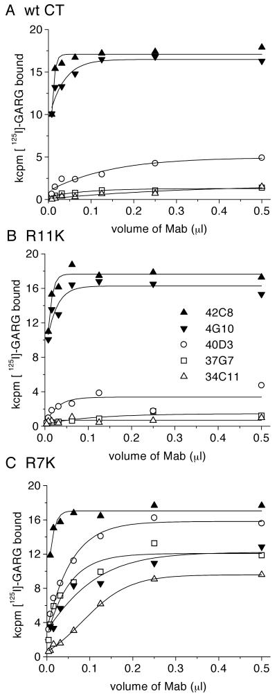
Altered reactivity with MAbs, like differences in susceptibility to proteolytic cleavage, can often reveal subtle differences in conformation of proteins. Among our collection of MAbs that react with conformational epitopes of CT are several that react only with holotoxin by SPRIA, some that react well with holotoxin (by GM1-SPRIA) but poorly with free denatured CT-A (by Western blot), and, conversely, some that react poorly with holotoxin but well with free CT-A (18). When we analyzed the temperature- or trypsin-sensitive variant holotoxins, we consistently found that they had altered patterns of reactivity towards these different classes of MAbs (Fig. 4 and Table 2). Whereas the R11K variant had a slight assembly defect as determined by trypsin sensitivity, it behaved like wt CT in its reactivity with these MAbs, reacting well with 42C8 and 4G10, weakly with 40D3, and poorly with 37G7 and 34C11 (Fig. 4A versus B). In contrast, the R7K variant had a completely different profile, retaining reactivity with 42C8 while losing reactivity to 4G10 and reacting much more strongly with the MAbs that normally give a poor signal with native CT (Fig. 4C). Data for other variants are presented in Table 2, representing the maximum signal obtained for each MAb (0.5 μl of hybridoma supernatant per well). Deletion of the entire loop filling the active site (Δ41-56+G variant) caused almost complete loss of reactivity with group 1 and 2 MAbs that react with holotoxin, suggesting that this loop determines or is essential for the epitope(s) detected by these MAbs. Each variant holotoxin that showed decreased reactivity with group 1 and 2 MAbs also gained reactivity with group 7B MAbs that normally react only with denatured CT-A (bound to plastic or nitrocellulose), suggesting a conformational change resulting in exposure of an epitope of CT-A that is normally hidden in wt holotoxin.
FIG. 4.
Differential MAb reactivity of R7K, R11K, and wt holotoxin variants. Panels A (recombinant CT), B (R11K variant), and C (R7K variant) show the reactivity of saturating amounts of each toxin antigen detected by titration with a battery of anti-CT MAbs in a GM1-SPRIA. Individual MAbs are identified according to the symbols in panel B. [125I]-GARG, 125I-labeled goat anti-rabbit immunoglobulin G.
TABLE 2.
Altered conformations of selected holotoxin variants as assessed by MAb reactivitya
| CT variant or property assayed | MAb reactivity
|
Rabbit polyclonal serum | |||
|---|---|---|---|---|---|
| Group 1 (17F7) | Group 2 (4G10 and 42C8)d | Group 7A (14E7) | Group 7B (34C11 and 37G7)e | ||
| Propertiesb | |||||
| CT neutralization | − | − | + | − | ++ |
| CTRIA | ++ | ++ | ++ | + | ++ |
| CT-ARIA | − | − | + | ++ | ++ |
| CT-AWestern | − | − | − | + | ++ |
| CT variantsc | |||||
| wt | ++++ | ++++ | ++++ | − | ++++ |
| R7K | ++++ | ++/++++ | ++++ | ++/+++ | ++++ |
| R11K | ++++ | ++++ | ++++ | − | ++++ |
| H44Y | ++ | ++ | ++++ | +++ | ++++ |
| H44S | ++++ | ++++/++ | ++++ | +++ | ++++ |
| Δ41−56+G | + | −/+ | + | +++ | +++ |
| W127S | ++++ | +++ | +++ | − | ++++ |
| L153LL | ++++ | ++++ | ++++ | ++++ | ++++ |
Saturating amounts of antigen bound in GM1-SPRIA were assayed for antibody reactivity.
From reference 18. CTRIA and CT-ARIA refer to direct SPRIA assays using plates coated with native CT holotoxin or purified CT-A subunit, respectively. CT-AWestern refers to reactivity with CTA1 polypeptide in a Western blot. −, negative; +, positive; ++, strongly positive. Group 1 and 2 MAbs react with native holotoxin only; group 7A MAbs react strongly with holotoxin and weakly with denatured CT-A and are the only group with toxin-neutralizing activity; group 7B MAbs react weakly or not at all with holotoxin but strongly with denatured CT-A.
Maximum signal expressed semiquantitatively in units of 1,000 cpm of 125I-labeled goat anti-rabbit Immunoglobulin G bound. −, <1; +, 1 to 3; ++, 3 to 8; +++, 8 to 15; ++++, >15.
For results with two sets of data, the first set refers to reactivity to 4G10 and the second set refers to reactivity to 42C8.
For results with two sets of data, the first set refers to reactivity to 34C11 and the second set refers to reactivity to 37G7.
Enzymatic activity of variant holotoxins.
Holotoxin variants that were able to be nicked by trypsin were tested for ADP-ribosyltransferase activity. CT ADP-ribosylates DEABAG in an NAD-dependent manner that is absolutely dependent upon reduction and is greatly increased by both nicking and addition of ARF and GTP. Basal levels of activity for trypsin-activated variants without addition of ARF6 or GTP are shown in Table 1 as percentages of wt activity. We were unable to detect any enzymatic activity for the R11K, H44S, or E110D+E112D variant holotoxins. Generally, the level of enzyme activity correlated well with Y1 toxicity data. Exceptions included substitutions for R25, E110, and E112 that had notably less enzymatic activity than toxicity, and conversely, the E29H and Y30WAH substitutions that had significantly more enzymatic activity than toxicity.
Kinetic studies of enzyme activity of holotoxin variants.
DEABAG assays were done with various NAD concentrations to obtain apparent Km and Vmax values for selected holotoxin variants from Lineweaver-Burk plots of 1/V0 against 1/[NAD]. In independent experiments with wt CT, we obtained apparent Kms (5 to 8 mM; Table 3) that were similar to those from other reports in the literature (1.1 to 5.6 mM), depending on the acceptor substrate (27, 29, 32, 36). The E29H variant showed a slight increase in Km for NAD in the absence of ARF6. Other variants had too little activity to give reliable data under these conditions. With the addition of rARF6 and GTP, the Km for NAD with wt CT decreased significantly and the Vmax increased (Table 3), as has been reported previously (34). Significant increases in Km for NAD compared to wt CT were observed for the R11K and I16A variants, while all other variants tested had Kms for NAD that remained close to that of the wt yet had reduced Vmax values, as expected for variants with reduced enzymatic activity.
TABLE 3.
Summary of kinetic data for selected CT holotoxin variants
| CT variant | Amount of toxin assayed (μg) | Presence of rARF6 + GTP | Apparent Km (mM NAD)a | Apparent Vmax (nmol of ADPR- DEABAG/min/μg)a |
|---|---|---|---|---|
| Native CT | 0.5 | − | 5–8 | 0.9–1.4 |
| E29H | 0.75–1.0 | − | 11–13b | 0.8 |
| Native CT | 0.075 | + | 0.33–2.0 | 2.8–5.6 |
| Recombinant CT | 0.075 | + | 0.74 | 3.6 |
| R11K | 20 | + | 19b | 0.04 |
| 116A | 1–2 | + | 13–16b | 0.25–1.0 |
| E29H | 0.075 | + | 0.7 | 1.2–1.9 |
| Y30WAH | 0.2 | + | 1.6 | 1.4 |
| G34GGP | 0.2 | + | 1.0 | 1.6 |
| S68Y | 1.0 | + | 1.5–3.0 | 0.3–0.6 |
Values show ranges obtained in independent experiments.
Estimates only; maximum NAD concentration tested was 10 mM.
DISCUSSION
By concentrating on residues that are highly conserved in the A subunits of the CT-LT family of enterotoxins, we identified several novel residues of CT-A that are critical to the structure and function of CT holotoxin. Novel substitution or insertion mutants for E29, Y30, E110, or E112 differentially affected toxicity and enzymatic activity in vitro without detectably affecting holotoxin structure, while the effects of other substitution mutants could be accounted for, at least in part, by previously undocumented alterations in holotoxin structure.
Many of these substitutions are located in or adjacent to the proposed active-site NAD-binding cleft (7) and can reasonably be assumed to exert their effects directly on NAD binding (R7K, R11K, I16A) or catalytic activity (H44S, E110D, E112D). Analysis of the three-dimensional structures of members of the CT-LT family predicts that the residues equivalent to Glu-112 in CT correspond to the single active-site glutamates of PT (Glu-129), DT (Glu-148), and ET-A (Glu-153). In studies with an isolated LT-A subunit, however, both Glu-110 and Glu-112 were required for full enzymatic activity (4, 27), since individual aspartate substitutions at these positions reduced activity 20- and 100-fold, respectively. We saw very similar results on toxicity with the individual aspartate-substituted mutant CT holotoxins but saw much greater effects on enzymatic activity. Only when both glutamates were replaced with aspartate did we see complete loss of toxicity and enzymatic activity. Clearly the members of the CT-LT family of ADP-ribosylating toxins differ significantly from the other ADP-ribosylating toxins in the details of their catalytic activity.
In crystallographic studies, the R7K variant of LT differed significantly in structure from wt LT (47) and was more sensitive to proteolysis (29). Our biochemical and immunological data showed similar changes in the R7K mutant of CT, supporting a role for R7K both in maintaining holotoxin structure and in NAD binding. The novel R11K variant was significantly more stable than the R7K variant, yet it showed a similar reduction in toxicity as well as an increased apparent Km for NAD. The conservative R11K substitution had a much greater effect on toxin activity (<1% of wt) than did a nonconservative substitution of the corresponding residue in PT (R13L) which showed 25% of wt toxicity (37), emphasizing the differences in functional importance of conserved residues between these toxins. At the equivalent of position 16 in CT, PT and the LT-IIb toxin have Val, whereas CT, LT-I, and LT-IIa have Ile. Replacing Ile-16 with Ala in CT reduced activity 20- to 25-fold compared to that of the wt and significantly increased the Km for NAD, while the conservative Leu substitution had no effect, providing the first direct evidence for the importance of a highly hydrophobic residue at this position for NAD binding. These data provide novel, direct experimental support for the structure-based hypothesis of van den Akker et al. (47, 48) that Ile-16 in LT (and, by inference, in CT) is involved in a hydrophobic interaction with the adenine ring of NAD.
We also changed the amino acids at positions 25, 68, and 72 in CT to residues found at homologous positions in PT that were proposed to participate in NAD binding (2) to test the hypothesis that the poor affinity of CT for NAD (Km of several millimolars [10]) could be improved by modifying its predicted NAD-binding site to resemble more closely that of PT, which has a several-micromolar Km for NAD (28). In both CT and LT-I, Arg-25 has hydrophobic interactions with residue 55 that are proposed to influence the movement of the active-site-occluding loop 47 to 56 (47). The R25W variant, which may maintain some of these hydrophobic interactions with His-55, retained significantly greater enzymatic activity and toxicity than the R25G variant, although both were less active than the wt. The single substitutions S68Y and V72Y decreased the toxicity and enzymatic activity of the variant holotoxins slightly to moderately, but together they produced 20- and 30-fold reductions, respectively. The S68Y variant, in the presence of recombinant ARF6, showed a slight increase in the apparent Km for NAD and a 10-fold reduction in the apparent Vmax compared to those of the wt. These substitutions at positions 25, 68, and 72 in CT therefore generally had adverse effects on enzymatic activity and/or toxicity but not on the structure of the CT variants, and their phenotypes did not suggest that they had significantly increased affinity for NAD. Other investigators (6) showed that CT-A1 variants with substitutions in the conserved β3 strand (YVSTS, residues 59 to 63) of the putative NAD-binding site (2) also affected the structure of the variant CT-A1 polypeptides and markedly decreased their enzymatic activity, but they did not study the effects of these substitutions in variant holotoxins.
Data from our laboratory and others support the critical role played by the active-site-occluding loop in the activity of the heat-labile enterotoxins (8, 9, 47). Substitutions for His-44 in CT decreased the stability of variant holotoxins and abolished enzyme activity in a manner similar to that reported for the H44A variant of LT (24). The proposal of Kato et al. (24) that His-44 interacts directly with the catalytic residue Glu-112 is unlikely given the distance between His-44 and Glu-112 in the crystal structures of both LT and CT. Instead, His-44 may act indirectly to activate a water nucleophile that interacts with Glu-112, as proposed by Rising and Schramm (39). In LT, substitutions within the active-site-occluding loop of residues that are found in the related LT-II toxins also affect structure and function (8). The fact that normally cryptic epitopes recognized by our group 7B MAbs were exposed in the His-44 holotoxin variants, in the Δ41-56 deletion variant, and in all temperature-sensitive holotoxin variants with significantly reduced activity (R7K, L153LL) suggests a common structural or folding defect in these variants. We see a slight effect on enzymatic activity and toxicity with a single residue insertion on the external surface of this loop in CT (T48TH). In a recent study, we independently identified an H44Y variant that affected the interaction of CT-A1 with ARF6 in a bacterial two-hybrid system (21). All other substitutions in the present study with detectable enzymatic activity were stimulated by ARF6, suggesting that ARF6 does not interact directly with the catalytically important residues.
Surprisingly, substitutions for Glu-29 and Tyr-30 and the replacement of L153 by LL, which are all well removed from the proposed active site, significantly affected toxicity and enzymatic activity. The Y30WAH and E29H variant holotoxins retained high levels of enzymatic activity yet had 5- to 10-fold lower toxicity, respectively, suggesting an additional role for these residues in vivo, possibly for toxin entry, trafficking, or substrate interaction. An explanation for the phenotypes of the two other variants produced in this study, W127S and L153LL, is more difficult. Both showed temperature-sensitive assembly defects. Although the W127S holotoxin formed at the permissive temperature and appeared near-wt in its reactivity with anti-CT MAbs, it showed only 10% toxicity. The L153LL variant holotoxin behaved like the other structurally altered variants, displaying nonnative MAb epitopes and greatly reduced toxicity. This residue is well removed from the active site on the “backside” of the A1 subunit. The L153LL insertion variant may exert its effects on toxin assembly by affecting interaction of the A1 polypeptide with the A2 subunit, but it is not clear how it dramatically affects the enzymatic and toxic activities of CT.
In summary, we have identified a diverse group of novel holotoxins with variant CT-A subunits that have altered biological properties, which in some cases include greatly reduced toxicity. The present study significantly extends the available information on the effects of structural changes in CT-A on the biological and enzymatic activities of CT. Several of the CT variants that exhibited differential effects on toxicity and enzyme activity should be useful for future studies on the structural basis for interaction of CT with target cells. One or more of these variants may prove useful as an immunological adjuvant. In this regard, our collaborators recently showed that the E29H variant can act as an effective mucosal adjuvant for immunization against respiratory syncytial virus (46).
ACKNOWLEDGMENTS
This work was supported in part by Public Health Service grant AI31940 from the National Institutes of Health.
The experiments reported here were initiated in the Department of Microbiology and Immunology at the Uniformed Services University of the Health Sciences, Bethesda, Md., and were completed at the University of Colorado.
REFERENCES
- 1.Allured V S, Collier R J, Carroll S F, McKay D B. Structure of exotoxin A of Pseudomonas aeruginosa at 3.0 angstrom resolution. Proc Natl Acad Sci USA. 1986;83:1320–1324. doi: 10.1073/pnas.83.5.1320. [DOI] [PMC free article] [PubMed] [Google Scholar]
- 2.Bell C E, Eisenberg D. Crystal structure of diphtheria toxin bound to nicotinamide adenine dinucleotide. Biochemistry. 1996;35:1137–1149. doi: 10.1021/bi9520848. [DOI] [PubMed] [Google Scholar]
- 3.Choe S, Bennett M J, Fujii G, Curmi P M G, Kantardjieff K A, Collier R J, Eisenberg D. The crystal structure of diphtheria toxin. Nature. 1992;357:216–222. doi: 10.1038/357216a0. [DOI] [PubMed] [Google Scholar]
- 4.Cieplak W, Jr, Mead D J, Messer R J, Grant C C R. Site-directed mutagenic alteration of potential active-site residues of the A subunit of Escherichia coli heat-labile enterotoxin—evidence for a catalytic role for glutamic acid 112. J Biol Chem. 1995;270:30545–30550. doi: 10.1074/jbc.270.51.30545. [DOI] [PubMed] [Google Scholar]
- 5.Cortina G, Barbieri J T. Role of tryptophan 26 in the NAD glycohydrolase reaction of the S-1 subunit of pertussis toxin. J Biol Chem. 1989;264:17322–17328. [PubMed] [Google Scholar]
- 6.Dolan K M, Lindenmayer G, Olson J C. Functional comparison of the NAD binding cleft of ADP-ribosylating toxins. Biochemistry. 2000;39:8266–8275. doi: 10.1021/bi992856q. [DOI] [PubMed] [Google Scholar]
- 7.Domenighini M, Rappuoli R. Three conserved consensus sequences identify the NAD-binding site of ADP-ribosylating enzymes, expressed by eukaryotes, bacteria and T-even bacteriophages. Mol Microbiol. 1996;21:667–674. doi: 10.1046/j.1365-2958.1996.321396.x. [DOI] [PubMed] [Google Scholar]
- 8.Feil I K, Platas A A, van den Akker F, Reddy R, Merritt E A, Storm D R, Hol W G. Stepwise transplantation of an active site loop between heat-labile enterotoxins LT-II and LT-I and characterization of the obtained hybrid toxins. Protein Eng. 1998;11:1103–1109. doi: 10.1093/protein/11.11.1103. [DOI] [PubMed] [Google Scholar]
- 9.Feil I K, Reddy R, De Haan L, Merritt E A, van den Akker F, Storm D R, Hol W G J. Protein engineering studies of A-chain loop 47–56 of Escherichia coli heat-labile enterotoxin point to a prominent role of this loop for cytotoxicity. Mol Microbiol. 1996;20:823–832. doi: 10.1111/j.1365-2958.1996.tb02520.x. [DOI] [PubMed] [Google Scholar]
- 10.Galloway T S, van Heyningen S. Binding of NAD+ by cholera toxin. Biochem J. 1987;244:225–230. doi: 10.1042/bj2440225. [DOI] [PMC free article] [PubMed] [Google Scholar]
- 11.Gill D M. The arrangement of subunits in cholera toxin. Biochemistry. 1976;15:1242–1248. doi: 10.1021/bi00651a011. [DOI] [PubMed] [Google Scholar]
- 12.Gill D M, Meren R. ADP-ribosylation of membrane proteins catalyzed by cholera toxin: basis of the activation of adenylate cyclase. Proc Natl Acad Sci USA. 1978;75:3050–3054. doi: 10.1073/pnas.75.7.3050. [DOI] [PMC free article] [PubMed] [Google Scholar]
- 13.Giuliani M M, Del G G, Giannelli V, Dougan G, Douce G, Rappuoli R, Pizza M. Mucosal adjuvanticity and immunogenicity of LTR72, a novel mutant of Escherichia coli heat-labile enterotoxin with partial knockout of ADP-ribosyltransferase activity. J Exp Med. 1998;187:1123–1132. doi: 10.1084/jem.187.7.1123. [DOI] [PMC free article] [PubMed] [Google Scholar]
- 14.Glineur C, Locht C. Importance of ADP-ribosylation in the morphological changes of PC12 cells induced by cholera toxin. Infect Immun. 1994;62:4176–4185. doi: 10.1128/iai.62.10.4176-4185.1994. [DOI] [PMC free article] [PubMed] [Google Scholar]
- 15.Hagiwara Y, Komase K, Chen Z, Matsuo K, Suzuki Y, Aizawa C, Kurata T, Tamura S. Mutants of cholera toxin as an effective and safe adjuvant for nasal influenza vaccine. Vaccine. 1999;17:2918–2926. doi: 10.1016/s0264-410x(99)00135-8. [DOI] [PubMed] [Google Scholar]
- 16.Holmes R K. Heat-labile enterotoxins (Escherichia coli) In: Montecucco C, Rappuoli R, editors. Guidebook to protein toxins and their use in cell biology. Oxford, England: Oxford University Press; 1997. [Google Scholar]
- 17.Holmes R K, Jobling M G, Connell T D. Cholera toxin and related enterotoxins of Gram-negative bacteria. In: Moss J, Iglewski B, Vaughan M, Tu A, editors. Handbook of natural toxins: bacterial toxins and virulence factors in disease. Vol. 8. New York, N.Y: Marcel Dekker, Inc; 1995. pp. 225–256. [Google Scholar]
- 18.Holmes R K, Twiddy E M. Characterization of monoclonal antibodies that react with unique and cross-reacting determinants of cholera enterotoxin and its subunits. Infect Immun. 1983;42:914–923. doi: 10.1128/iai.42.3.914-923.1983. [DOI] [PMC free article] [PubMed] [Google Scholar]
- 19.Jobling M G, Holmes R K. Analysis of structure and function of the B subunit of cholera toxin by the use of site directed mutagenesis. Mol Microbiol. 1991;5:1755–1767. doi: 10.1111/j.1365-2958.1991.tb01925.x. [DOI] [PubMed] [Google Scholar]
- 20.Jobling M G, Holmes R K. Fusion proteins containing the A2 domain of cholera toxin assemble with B polypeptides of cholera toxin to form immunoreactive and functional holotoxin-like chimeras. Infect Immun. 1992;60:4915–4924. doi: 10.1128/iai.60.11.4915-4924.1992. [DOI] [PMC free article] [PubMed] [Google Scholar]
- 21.Jobling M G, Holmes R K. Identification of motifs in cholera toxin A1 polypeptide that are required for its interaction with human ADP-ribosylation factor 6 in a bacterial two-hybrid system. Proc Natl Acad Sci USA. 2000;97:14662–14667. doi: 10.1073/pnas.011442598. [DOI] [PMC free article] [PubMed] [Google Scholar]
- 22.Jobling M G, Palmer L M, Erbe J L, Holmes R K. Construction and characterization of versatile cloning vectors for efficient delivery of native foreign proteins to the periplasm of Escherichia coli. Plasmid. 1997;38:158–173. doi: 10.1006/plas.1997.1309. [DOI] [PubMed] [Google Scholar]
- 23.Kassis S, Hagmann J, Fishman P H, Chang P P, Moss J. Mechanism of action of cholera toxin on intact cells. Generation of A1 peptide and activation of adenylate cyclase. J Biol Chem. 1982;257:12148–12152. [PubMed] [Google Scholar]
- 24.Kato M, Imamura S, Kawase H, Miyama A, Tsuji T. Histidine-44 of the A subunit of Escherichia coli enterotoxin is involved in its enzymatic and biological activities. FEMS Microbiol Lett. 1997;152:219–225. doi: 10.1111/j.1574-6968.1997.tb10431.x. [DOI] [PubMed] [Google Scholar]
- 25.Kunkel T A. Rapid and efficient site specific mutagenesis without phenotypic selection. Proc Natl Acad Sci USA. 1985;82:488–492. doi: 10.1073/pnas.82.2.488. [DOI] [PMC free article] [PubMed] [Google Scholar]
- 26.Larew J S, Peterson J E, Graves D J. Determination of the kinetic mechanism of arginine-specific ADP-ribosyltransferases using a high performance liquid chromatographic assay. J Biol Chem. 1991;266:52–57. [PubMed] [Google Scholar]
- 27.Lobet Y, Cluff C W, Cieplak W. Effect of site-directed mutagenic alterations on ADP-ribosyltransferase activity of the A subunit of Escherichia coli heat-labile enterotoxin. Infect Immun. 1991;59:2870–2879. doi: 10.1128/iai.59.9.2870-2879.1991. [DOI] [PMC free article] [PubMed] [Google Scholar]
- 28.Locht C, Antoine R. A proposed mechanism of ADP-ribosylation catalyzed by the pertussis toxin S1 subunit. Biochimie. 1995;77:333–340. doi: 10.1016/0300-9084(96)88143-0. [DOI] [PubMed] [Google Scholar]
- 29.Magagnoli C, Manetti R, Fontana M R, Giannelli V, Giuliani M M, Rappuoli R, Pizza M. Mutations in the A subunit affect yield, stability, and protease sensitivity of nontoxic derivatives of heat-labile enterotoxin. Infect Immun. 1996;64:5434–5438. doi: 10.1128/iai.64.12.5434-5438.1996. [DOI] [PMC free article] [PubMed] [Google Scholar]
- 30.Maneval D R, Colwell R R, Grays S W J, Donta S T. A tissue culture method for the detection of bacterial enterotoxins. J Tissue Cult Methods. 1981;6:85–90. [Google Scholar]
- 31.Mekalanos J J, Collier R J, Romig W R. Enzymatic activity of cholera toxin. I. New method of assay and the mechanism of ADP-ribosyl transfer. J Biol Chem. 1979;254:5849–5854. [PubMed] [Google Scholar]
- 32.Mekalanos J J, Collier R J, Romig W R. Enzymatic activity of cholera toxin. II. Relationships to proteolytic processing, disulfide bond reduction and subunit composition. J Biol Chem. 1979;254:5855–5861. [PubMed] [Google Scholar]
- 33.Murayama T, Tsai S-C, Adamik R, Moss J, Vaughan M. Effects of temperature on ADP-ribosylation factor stimulation of cholera toxin activity. Biochemistry. 1993;32:561–566. doi: 10.1021/bi00053a022. [DOI] [PubMed] [Google Scholar]
- 34.Narayanan J, Hartman P A, Graves D J. Assay of heat-labile enterotoxins by their ADP-ribosyltransferase activities. J Clin Microbiol. 1989;27:2414–2419. doi: 10.1128/jcm.27.11.2414-2419.1989. [DOI] [PMC free article] [PubMed] [Google Scholar]
- 35.Osborne J C, Jr, Stanley S J, Moss J. Kinetic mechanism of two NAD:arginine ADP-ribosyltransferases: the soluble, salt-stimulated transferase from turkey erythrocytes and choleragen, a toxin from Vibrio cholerae. Biochemistry. 1985;24:5235–5240. doi: 10.1021/bi00340a042. [DOI] [PubMed] [Google Scholar]
- 36.Pérez-Pérez J, Gutiérrez J. An arabinose-inducible expression vector, pAR3, compatible with ColE1-derived plasmids. Gene. 1995;158:141–142. doi: 10.1016/0378-1119(95)00127-r. [DOI] [PubMed] [Google Scholar]
- 37.Pizza M, Covacci A, Bartolini A, Perugini M, Nencioni L, de Magistris M T, Barbieri J T, Rappuoli R. Mutants of pertussis toxin suitable for vaccine development. Science. 1989;246:497–500. doi: 10.1126/science.2683073. [DOI] [PubMed] [Google Scholar]
- 38.Pizza M, Domenighini M, Hol W, Gianelli V, Fontana M R, Giuliani M M, Magagnoli C, Peppoloni S, Manetti R, Rappuoli R. Probing the structure-activity relationship of Escherichia coli LT-A by site-directed mutagenesis. Mol Microbiol. 1994;14:51–60. doi: 10.1111/j.1365-2958.1994.tb01266.x. [DOI] [PubMed] [Google Scholar]
- 39.Rising K A, Schramm V L. Transition state analysis of NAD+ hydrolysis by the cholera toxin catalytic subunit. J Am Chem Soc. 1997;119:27–37. [Google Scholar]
- 40.Sixma T K, Kalk K H, van Zanten B A M, Dauter Z, Kingma J, Witholt B, Hol W G. Refined structure of Escherichia coli heat-labile enterotoxin, a close relative of cholera toxin. J Mol Biol. 1993;230:890–918. doi: 10.1006/jmbi.1993.1209. [DOI] [PubMed] [Google Scholar]
- 41.Sixma T K, Pronk S E, Kalk K H, Wartna E S, van Zanten B A, Witholt B, Hol W G. Crystal structure of a cholera toxin-related heat-labile enterotoxin of Escherichia coli. Nature. 1991;351:371–377. doi: 10.1038/351371a0. [DOI] [PubMed] [Google Scholar]
- 42.Stein P E, Boodhoo A, Armstrong G D, Cockle S A, Klein M H, Read R J. The crystal structure of pertussis toxin. Structure. 1994;2:45–57. doi: 10.1016/s0969-2126(00)00007-1. [DOI] [PubMed] [Google Scholar]
- 43.Stock J B, Ninfa A J, Stock A M. Protein phosphorylation and regulation of adaptive responses in bacteria. Microbiol Rev. 1989;53:450–490. doi: 10.1128/mr.53.4.450-490.1989. [DOI] [PMC free article] [PubMed] [Google Scholar]
- 44.Tabor S, Richardson C C. A bacteriophage T7 RNA polymerase/promoter system for the controlled exclusive expression of specific genes. Proc Natl Acad Sci USA. 1985;82:1074–1078. doi: 10.1073/pnas.82.4.1074. [DOI] [PMC free article] [PubMed] [Google Scholar]
- 45.Tartof K D, Hobbs C A. Improved media for growing plasmid and cosmid clones. Focus. 1987;9:12. [Google Scholar]
- 46.Tebbey P W, Scheuer C A, Peek J A, Zhu D, LaPierre N A, Green B A, Phillips E D, Ibraghimov A R, Eldridge J H, Hancock G E. Effective mucosal immunization against respiratory syncytial virus using purified F protein and a genetically detoxified cholera holotoxin, CT-E29H. Vaccine. 2000;18:2723–2734. doi: 10.1016/s0264-410x(00)00058-x. [DOI] [PubMed] [Google Scholar]
- 47.van den Akker F, Merritt E A, Pizza M G, Domenighini M, Rappuoli R, Hol W G J. The Arg7 Lys mutant of heat-labile enterotoxin exhibits great flexibility of active site loop 47–56 of the A subunit. Biochemistry. 1995;34:10996–11004. doi: 10.1021/bi00035a005. [DOI] [PubMed] [Google Scholar]
- 48.van den Akker F, Sarfaty S, Twiddy E M, Connell T D, Holmes R K, Hol W G J. Crystal structure of a new heat-labile enterotoxin, LT-IIb. Structure. 1996;4:665–678. doi: 10.1016/s0969-2126(96)00073-1. [DOI] [PubMed] [Google Scholar]
- 49.Welsh C F, Moss J, Vaughan M. ADP-ribosylation factors: a family of guanine nucleotide-binding proteins that activate cholera toxin and regulate vesicular transport. In: Moss J, Iglewski B, Vaughan M, Tu A, editors. Handbook of natural toxins: bacterial toxins and virulence factors in disease. Vol. 8. New York, N.Y: Marcel Dekker, Inc; 1995. pp. 257–280. [Google Scholar]
- 50.Zhang R G, Scott D L, Westbrook M L, Nance S, Spangler B D, Shipley G G, Westbrook E M. The three-dimensional crystal structure of cholera toxin. J Mol Biol. 1995;251:563–573. doi: 10.1006/jmbi.1995.0456. [DOI] [PubMed] [Google Scholar]


