Abstract
Macrophage migration inhibitory factor (MIF) is a proinflammatory cytokine with a pleiotropic spectrum of biological functions implicated in the pathogenesis of cancer and inflammatory diseases. MIF is constitutively present in several cell types and non-lymphoid tissues and is secreted after acute stress or inflammation. MIF triggers the release of proinflammatory cytokines, overrides the anti-inflammatory effects of glucocorticoids, and exerts chemokine function, resulting in increased migration and recruitment of leukocytes into inflamed tissue. Despite this, MIF is a challenging target for therapeutic intervention because of its ubiquitous nature and presence in the circulation and tissue of healthy individuals. Oxidized MIF (oxMIF) is an immunologically distinct disease-related structural isoform found in the plasma and tissues of patients with inflammatory diseases and in solid tumor tissues. MIF converts to oxMIF in an oxidizing, inflammatory environment. This review discusses the biology and activity of MIF and the potential for autoimmune disease and cancer modification by targeting oxMIF. Anti-oxMIF antibodies reduce cancer cell invasion/migration, angiogenesis, proinflammatory cytokine production, and ERK and AKT activation. Anti-oxMIF antibodies also elicit apoptosis and alter immune cell function and/or migration. When co-administered with a glucocorticoid, anti-oxMIF antibodies produced a synergistic response in inflammatory models. Anti-oxMIF antibodies therefore counterregulate biological activities attributed to MIF. oxMIF expression has been observed in inflammatory diseases (eg, sepsis, psoriasis, asthma, inflammatory bowel disease, and systemic lupus erythematosus) and oxMIF has been detected in ovarian, colorectal, lung, and pancreatic cancers. In contrast to MIF, oxMIF is specifically detected in plasma and/or tissues of diseased patients, but not in healthy individuals. Therefore, as a druggable isoform of MIF, oxMIF represents a potential new therapeutic target in inflammatory diseases and cancer. Fully human, monoclonal anti-oxMIF antibodies have been shown to selectively bind oxMIF in preclinical and phase I studies; however, additional clinical assessments are necessary to validate their use as either a monotherapy or in combination with standard-of-care regimens (ie, immunomodulatory agents/checkpoint inhibitors, anti-angiogenic drugs, chemotherapeutics, and glucocorticoids).
Keywords: tumor microenvironment; review; therapies, investigational; inflammation; inflammation mediators
Introduction
Oxidized macrophage migration inhibitory factor (oxMIF) is a disease-related conformational isoform of macrophage migration inhibitory factor (MIF) that can be selectively targeted in cancer and inflammatory diseases. 1–3 MIF—first described as a soluble immune cell-derived factor in 19664—is a key mediator in the pathogenesis of cancers and inflammatory diseases.5–9 MIF is a pro-inflammatory cytokine with a pleiotropic spectrum of associated biological functions.10 Extracellular MIF binding to CD74 in heterocomplex with CD44, chemokine receptors CXCR2, CXCR4, and/or CXCR7,10–14 among other activities, initiates activation of the mitogen-activated protein kinase (MAPK)/extracellular signal-regulated kinase (ERK) and/or phosphoinositide 3-kinase (PI3K)-protein kinase B (AKT) intracellular signaling pathways,5 10 12–16 which prompts downstream pro-inflammatory and pro-tumorigenic effects. The extracellular MIF/CD74 interaction increases c-Jun phosphorylation and pro-inflammatory activator protein 1 (AP-1) transcription.10 17 18 In contrast, intracellular MIF independently binds c-Jun activation domain binding protein-1 (JAB1) and negatively regulates AP-1 transcription and c-Jun amino terminal kinase (JNK) activities, indicating an immunosuppressive role.10 19 20 The balance between extracellular versus intracellular MIF levels/activities may determine the pro-inflammatory versus anti-inflammatory phenotype, respectively.10
Under normal circumstances, preformed MIF is constitutively present in several cell types (eg, monocytes, macrophages, lymphocytes, eosinophils, epithelial cells, and endothelial cells)19 21–23 and non-lymphoid tissues (eg, pituitary gland, lung, liver, kidney, spleen, adrenal gland, and skin)19 21 22 (online supplemental figure 1). Preformed MIF is rapidly secreted from intracellular pools after acute stress or inflammatory stimulation (eg, bacterial lipopolysaccharide (LPS), tumor necrosis factor (TNF), or interferon-γ (IFN-γ)).19 21–24 Once released, MIF triggers the release of pro-inflammatory cytokines,19 21 23–26 overrides anti-inflammatory effects of glucocorticoids,19 24 26–28 and enhances chemokine function, resulting in increased migration and recruitment of leukocytes into inflamed tissue.11 29 Acting in parallel with endogenous glucocorticoids, MIF controls the threshold and magnitude of immune and inflammatory responses.19 24 26–28 These distinctive features and functions of MIF distinguish it from other cytokines or hormonal mediators.
jitc-2022-005475supp001.pdf (310KB, pdf)
MIF is a critical upstream regulator of innate immunity and inflammation, and excess MIF expression causes exaggerated inflammation and immunopathology, including cancer.1 2 19 23 24 30–40 For example, MIF is implicated in inflammatory diseases such as sepsis,19 24 30 41 psoriasis,1,1 42 asthma,1 inflammatory bowel disease,1 arthritis,23 31 43–45 and systemic lupus erythematosus (SLE).23 46 Additionally, high expression of MIF is found in certain solid tumors (ie, colorectal cancer, gastric cancer, non-small cell lung cancer (NSCLC), ovarian cancer, esophageal squamous cell carcinoma, pancreatic cancer, melanoma, neuroblastoma, and osteosarcoma) and is associated with high tumor burden and grade, increased metastasis risk, and poor prognosis.2 33–40 47–54 Evidence indicates that MIF alleles are associated with incidence and/or severity of these conditions.23 24 33 44 55–62 Functional MIF gene-promoter polymorphisms (eg, CATT repetition represented 5–8 times and a single nucleotide polymorphism, -173G/C) make individuals susceptible to elevated MIF expression and the consequent exaggerated immune and inflammatory responses, or—in a few instances—result in protection from (or less aggressive) disease.63 64 For example, individuals with the MIF-173*C allele have increased susceptibility to systemic-onset juvenile idiopathic arthritis, psoriasis, SLE, and prostate cancer.44 55 57 59 62 In contrast, significantly fewer patients with Crohn’s disease who were heterozygous for the MIF-173G→C substitution than patients who were homozygous had upper gastrointestinal tract involvement, and the 5-CATT repeat allele (vs the 6, 7, or 8 repeat alleles) correlated with low disease severity in patients with rheumatoid arthritis.56 65
Despite its role in inflammatory disease and cancer pathways, MIF is a challenging target for therapeutic intervention because of its ubiquitous nature and presence in the circulation and tissue of healthy individuals.2 However, evidence indicates that MIF occurs in two immunologically distinct, conformational isoforms: oxMIF, which is a disease-related structural isoform found predominantly in plasma and tissues of patients with inflammatory diseases and in tumor tissues and corresponding metastases, and reduced MIF (redMIF), which is present in plasma, cells, and tissues of healthy and diseased individuals.1
MIF is a 115-amino acid polypeptide, with a homotrimer de facto structure that is toroidal in shape with a central, solvent-filled pore.66 Unlike other cytokines, MIF has two evolutionarily conserved catalytic activities—a tautomerase activity and a thiol-protein oxidoreductase (TPOR) activity—that are carried out by two distinct catalytic centers.66 67 The TPOR activity is mediated through a conserved cysteine (Cys)-56-alanine-leucine-Cys-59 (CALC) motif located in the central pore of MIF.67 The tautomerase activity, which has no known function, is facilitated by the conserved N-terminal proline.67 68 Some of the pro-inflammatory activities of MIF are inhibited by targeting the N-terminal proline, which is involved in receptor binding.69–74 MIF is also a poly ADP ribose polymerase-1-dependent, apoptosis-inducing factor-associated nuclease.75 76 MIF nuclease activity promotes tumor growth in cancer cells77 and neurodegeneration and neuronal cell death in post-traumatic brain injury.76
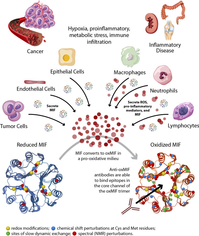
MIF is modified covalently and structurally, which changes MIF bioactivity. For example, one modification of MIF—mediated by myeloperoxidase (MPO)-derived hypochlorous acid—is oxidation of the N-terminal proline, which eliminates tautomerase activity but spares pro-inflammatory activities.68 Furthermore, MIF can interconvert between redMIF and oxMIF, and antibodies have been shown to selectively bind oxMIF but not redMIF (figure 1). 1 66 78 One of these antibodies (imalumab) is a recombinant, fully human, immunoglobulin G1 monoclonal antibody. Imalumab binds to an epitope covering the CALC motif and has been tested in clinical studies.3 66 79 Distal to the CALC motif is a third Cys (Cys-80), which operates as a switch, converting redMIF to oxMIF through post-translational modification.66 80 81 Redox-sensitive amino acids within MIF (eg, Cys-80, lysine (Lys)-66) were identified as latent sites of functional control.81 Mutational analyses showed loss of these redox-responsive amino acids attenuated activation of CD74 and altered MIF enzymatic activity.81
Figure 1.
Redox-dependent macrophage migration inhibitory factor (MIF) structure. Hypoxic pro-inflammatory conditions are common in cancer and inflammatory disease microenvironments. Macrophages, neutrophils, and other immune cells contribute to the generation of ROS and pro-inflammatory mediators. Under these oxidizing conditions, MIF, which is secreted by tumor cells, as well as endothelial cells, epithelial cells, and other immune cells, can convert to oxMIF. oxMIF has increased redox modifications (yellow dots) and chemical shift perturbations at Cys and Met residues (blue dots), more sites of slow dynamic exchange (green dots), and more spectral (NMR) perturbations (red dots) compared with redMIF. Once in the oxMIF isoform, anti-oxMIF antibodies can access and bind to epitopes in the core channel of the trimer. Cys, cysteine; Met, methionine; MIF, macrophage migration inhibitory factor; NMR, nuclear magnetic resonance; oxMIF, oxidized macrophage migration inhibitory factor; ROS, reactive oxygen species.
Using MIF-specific and oxMIF-specific antibodies, total MIF—but not oxMIF—was detected in plasma (by quantitative ELISA), and in the cytosol (by immunoprecipitation and western blot analysis), and on the surface of immune cells (by flow cytometry), from healthy controls.1 In contrast, oxMIF was detected in plasma and on the surface of granulocytes, macrophages, and NK cells (but not T or B cells) only under inflammatory disease conditions.1 Furthermore, using immunohistochemistry methods which prevented an artificial conversion of MIF to oxMIF (eg, by fixatives or oxidative agents), oxMIF-specific antibodies detected oxMIF in primary tumors (eg, pancreatic, colorectal, ovarian, and lung) but not in adjacent non-tumoural tissue; and in corresponding metastatic tissue.2 oxMIF was also detected via immunohistochemistry in metastases from biopsied patients in the imalumab phase I trial; results were interpreted using digital pathology algorithms.3 Therefore, oxMIF represents a potential new therapeutic target in solid tumors and inflammatory diseases. Here, we discuss the biology and activity of MIF, as well as the potential for autoimmune disease and cancer modification by targeting oxMIF.
MIF biology and disease modification by targeting oxMIF
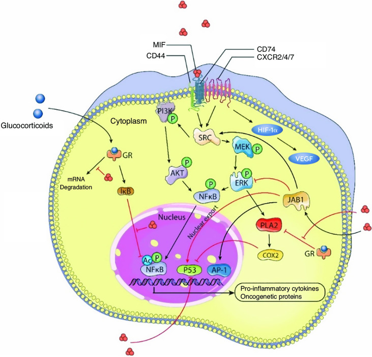
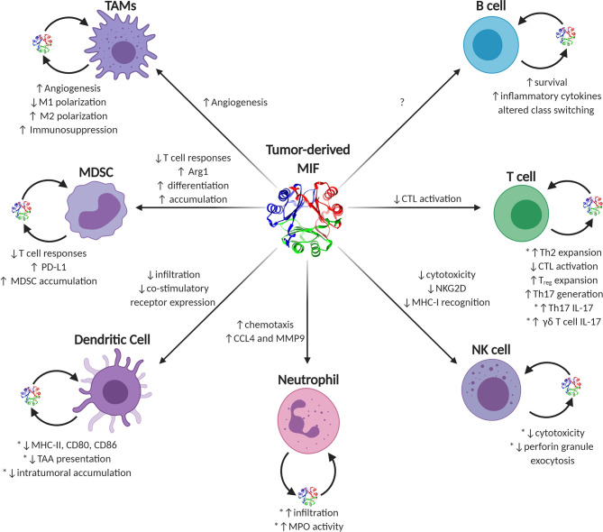
Owing to its pleiotropic action, MIF promotes inflammation, cell proliferation, and inhibition of cell death by apoptosis (figure 2).19 82 During the early phases of tumor development, MIF/oxMIF elicits pro-inflammatory immune responses in the tumor microenvironment.2 10 In contrast, during later-stage disease the effects of MIF more closely mimic wound-resolution activity, increasing neovascularization and immune evasion.10 In addition, MIF regulates migration, activation, differentiation, and reprogramming of immune and non-immune cells (figures 2 and 3).10 19 82 For example, inhibition of MIF in solid tumors efficiently repolarizes tumor-associated macrophages (TAMs) within tumors from an immunosuppressive and pro-angiogenic/pro-tumorigenic phenotype to an immunostimulatory and non-angiogenic/anti-tumor phenotype.10
Figure 2.
Role of MIF in activation of anti-apoptotic, pro-angiogenic, and pro-proliferative pathways. MIF binds the ligand-binding protein CD74 and the signal transducer CD44. The MIF-CD74-CD44 complex activates transcription factors that regulate the Src proto-oncogene, non-receptor tyrosine kinase (SRC) and ERK-MAPK pathways, which control gene expression and cellular proliferation. The MIF-CD74 interaction activates the AKT pathway via the mediation of kinases SRC and PI3K. MIF also interacts with JAB1 to activate the JNK and works as a co-activator of the transcription factor AP-1. MIF also limits the immunosuppressive effects of glucocorticoid and revokes the glucocorticoid-mediated inhibition of PLA2 and arachidonic acid production. MIF can inactivate p53-mediated apoptosis and growth arrest, and many pieces of evidence connect MIF with inflammation and tumorigenesis. AP-1, activator protein 1; ERK, extracellular signal-regulated kinase; GR, glucocorticoid receptor; IκB, inhibitor of NF-κB; JNK, JUN N-terminal kinase; MAPK, mitogen-activated protein kinase; MIF, macrophage migration inhibitory factor; NF-κB, nuclear factor kappa B. Figure has been modified from Osipyan A, Chen D, Dekker FJ. Epigenetic regulation in macrophage migration inhibitory factor (MIF)-mediated signaling in cancer and inflammation. Drug Discov Today 2021;26(7):1728–1734.
Figure 3.
Known and putative effects of MIF on tumor-infiltrating immune cells. Graphical depiction of immune effector cells known to be influenced by MIF. Sources of intratumoral MIF include both paracrine acting tumor-secreted and autocrine acting immune cell-secreted MIF. Phenotypes ascribed to tumor-derived MIF on individual cell types are listed next to each corresponding arrow to each cell type, while autocrine-associated activities are noted next to each cell with the caveat that those activities validated using recombinant MIF sources are noted with an asterisk. CCL4, inflammatory chemokine (C-C motif) ligand 4; CTL, cytotoxic T lymphocyte; MDSC, myeloid-derived suppressor cells; MHC, major histocompatibility complex; MIF, macrophage migration inhibitory factor; MMP9, matrix metalloproteinase 9; MPO, myeloperoxidase; NK, natural killer; PD-L1, programmed death-ligand 1; TAA, tumor-associated antigens; TAMs, tumor-associated macrophages; Th, T helper. Figure has been reprinted from Noe JT, Mitchell RA. MIF-dependent control of tumor immunity. Front Immunol 2020;11:609948.10
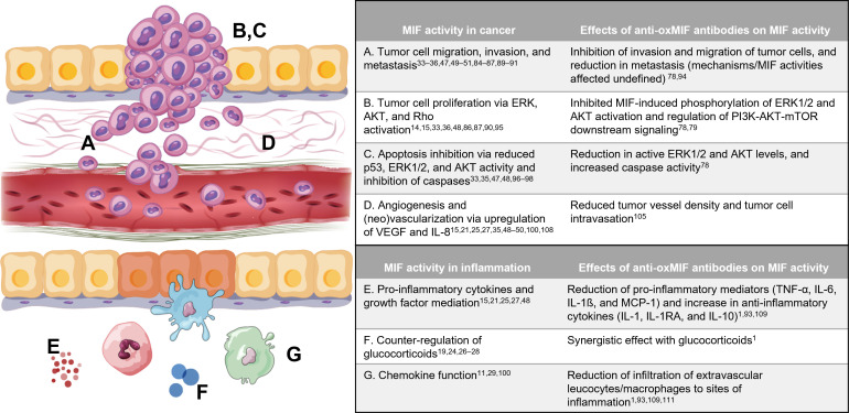
The role of MIF in tumor invasion and metastasis, tumor cell proliferation, apoptosis, angiogenesis, pro-inflammatory mediator production, counterregulation of glucocorticoids, and chemokine function, and the effects of anti-oxMIF antibodies on these activities are summarized in figure 4 and below.
Figure 4.
In vitro and in vivo studies demonstrate that MIF activity can be regulated by targeting oxMIF. AKT, protein kinase B; ERK, extracellular signal-regulated kinases; IL, interleukin; IL-1RA, IL-1 receptor agonist; MIF, macrophage migration inhibitory factor; MCP-1, monocyte chemoattractant protein-1; mTOR, mammalian target of rapamycin; TNF-α, tumor necrosis factor-alpha; VEGF, vascular endothelial growth factor. Tumor image has been modified from Novikov NM, Zolotaryova SY, Gautreau AM et al. Mutational drivers of cancer cell migration and invasion. Br J Cancer 2021;124:102–114 (http://creativecommons.org/licenses/by/4.0/).
Tumor cell migration, invasion, and metastasis
Several biological activities of MIF favor cancer development, growth, and metastasis, including sustained ERK activation, activation of the AKT pathway, cyclooxygenase-2/prostaglandin E-2 (PGE2) induction, p53 inhibition, and endothelial cell proliferation and differentiation.5 10 14 19 82 83 MIF plays a critical role in tumor cell migration, invasion, and metastasis10 19 82 83 as demonstrated in in vitro and in vivo studies, including studies of pancreatic,36 47 84–86 prostate,33 lung,34 52 colorectal,35 87 88 ovarian,48 89 esophageal squamous cell carcinoma,49 53 osteosarcoma,50 melanoma,51 and nasopharyngeal cancers.90 91 MIF overexpression was associated with cancer cell invasion and particularly aggressive disease.33 35 36 47 53 86 91 Potential mechanisms for the role of MIF in tumor cell migration, invasion, and metastasis include: the dependence of liver premetastatic niche formation on tumor exosome-derived MIF during pancreatic ductal adenocarcinoma (PDAC) metastasis84; activation of AKT serine/threonine kinase and ERK36; activation of cyclin D1 expression36; activation of matrix metalloproteinase expression36 88; regulation of the tumor microenvironment (eg, cytokines, angiogenic factors, and chemokines)48; induced epithelial-to-mesenchymal transition (evidenced by decreased E-cadherin and increased mesenchymal markers)47 53; loss of cell adhesion and anchorage-dependent growth34; promotion of RAC1 activity and subsequent tumor cell motility through lipid raft stabilization34; increased vascular endothelial growth factor (VEGF) expression and formation of new tumor vasculature87; decreased CD74 and Tiam1 expression87; JNKII-dependent and nuclear factor-κB (NF-κB)-dependent induction of release by macrophages89; and activation of the Rho pathway.88
Blocking MIF activity using antibodies, small interfering ribonucleic acids (siRNAs), or microRNAs inhibited invasion and reduced metastasis in multiple studies.33 34 36 78 83 86 87 90 92 Anti-oxMIF antibodies, which have binding regions that include the MIF oxidoreductase motif, were effective at inhibiting the capacity of prostate cancer cells to invade and migrate through extracellular matrix components.78 93 In the same study, anti-oxMIF antibodies reduced active ERK1/2 and AKT levels. Finally, in a study of bevacizumab-resistant glioblastomas, xenografts expressed high levels of MIF, and exposure to imalumab reduced tumor cell invasion in Matrigel and bioengineered three-dimensional hyaluronic acid-based assays.94
Tumor cell proliferation
The role of MIF in tumor cell proliferation has been demonstrated in various in vitro and in vivo studies involving multiple tumor types (eg, pancreatic, colorectal, prostate, ovarian, nasopharyngeal, gastric, and melanoma).15 33 36 47 48 51 86 87 90 95 96 MIF triggers cell proliferation, at least in part, by activating MAPK/ERK and AKT intracellular signaling.15 16 95 Both endogenous and exogenous MIF stimulate cellular proliferation in association with MAPK/ERK activation; resulting sustained cytoplasmic phospholipase A2 activation leads to the production of arachidonic acid, which is a precursor for the synthesis of prostaglandins and leukotrienes.15 MIF knockdown suppressed proliferation of DU-145 (human prostate cancer) cells, and MIF-specific inhibition suppressed proliferation and tumor growth (in part, by inhibiting angiogenesis) in a prostate cancer xenograft model.33 Furthermore, MIF knockdown, which suppressed proliferation of PDAC cells, also significantly inhibited activation of ERK and AKT signaling.36
In an in vitro study in PC3 (human prostate cancer) cells, anti-oxMIF antibodies inhibited MIF-induced phosphorylation of ERK1/2 and AKT activation.78 Additionally, satisfactory tissue penetration of imalumab was observed in pretherapy and on-therapy advanced solid tumor biopsies, with regulation of PI3K/AKT/mammalian target of rapamycin (mTOR) downstream signaling, in all biopsy-evaluable patients.3 79
Apoptosis
Initially, MIF was shown to inhibit apoptosis by overcoming p53 activity in mouse embryonic fibroblasts, macrophages from LPS-stimulated mice, and MIF-transfected murine macrophages.97 98 Further research revealed several MIF-involved mechanisms associated with apoptosis. Tumors arising from MIF knockdown in PDAC cells and ID8 (murine ovarian cancer) syngeneic tumors had significantly increased apoptosis, which was associated with reduced AKT phosphorylation and increased p53 phosphorylation.48 MIF, through interaction with Jun activation domain-binding protein-1/constitutive photomorphogenic-9 signalosome, modulated activator protein-1 activity and cell proliferation, and inactivated p53.19 82 Murine CT26 and LoVo (colon carcinoma) cells incubated with a MIF-specific inhibitor reduced cell proliferation, suggesting that increased MIF expression in cancer may inhibit p53-mediated apoptosis.35 Blocking the MIF-CD74 interaction decreased ERK1/2 activation and increased apoptosis in DU-145 (prostate cancer) cells.33 MIF overexpression in Capan 2 and Panc 1 (pancreatic cancer) cell lines decreased apoptosis, as measured by lower caspase-3 and caspase-7 activity relative to controls.47 Similarly, MIF activation of the PI3K/AKT pathway led to inactivation of pro-apoptotic proteins in mouse embryonic fibroblasts and NIH/3T3 (immortalized fibroblasts), HeLa (cervical carcinoma), and various breast cancer cell lines14 and expression of anti-apoptotic proteins in chronic lymphocytic leukemia B cells.99 MIF knockdown in EC9706 and NE6-T (esophageal squamous cell carcinoma) cells increased apoptosis with elevated levels of cleaved caspase-3.53
Reduced levels of active ERK1/2 and AKT favor caspase activation, thereby promoting apoptosis.78 When PC3 cells were incubated with anti-oxMIF antibodies, which reduced active ERK1/2 and AKT levels, increased caspase activity was detected in lysates.78 In addition, satisfactory tissue penetration of imalumab was observed along with apoptosis in biopsy-evaluable patients in a phase I study.79
Angiogenesis and neovascularization
MIF is an angiogenesis-promoting factor during neoplastic transformation and neovascularization, the latter of which is controlled by angiogenic factors (eg, VEGF) that are secreted mainly by tumor cells and is essential for tumor growth.100–102 In addition, MIF activates hypoxia-induced factor-1a (HIF-1α), directly and indirectly, which leads to interleukin (IL)-8 and VEGF expression.54 The role of MIF in angiogenesis was evaluated in various in vitro and in vivo studies of multiple tumor types, including colon/colorectal cancer, prostate cancer, lymphoma, osteosarcoma, melanoma, and esophageal squamous cell carcinoma.33 35 49–51 88 92 100 103 104 In these studies, MIF stimulated angiogenesis in a MAPK/ERK-dependent and PI3K/AKT-dependent manner92 100 and induced angiogenic factors (eg, VEGF, IL-8).35 49 50 100 Additionally, MIF serum levels correlated with serum VEGF levels and microvessel density correlated with MIF expression.49 50 Furthermore, angiogenesis and tumor (out)growth were suppressed when MIF activity was blocked (eg, MIF siRNA, MIF-specific inhibitor, anti-MIF monoclonal antibodies, and MIF-deficient tumors).33 35 50 88 100 103 104 Together, these findings suggest MIF participates in tumor angiogenesis in an autocrine fashion.49 100 Reduced vessel density and tumor cell intravasation were observed in tumors from human prostate cancer cell-line xenografted mice that were treated with a novel anti-oxMIF antibody105; however, additional studies are needed to evaluate whether the HIF-1α/VEGF axis contributed to these effects and whether MIF-dependent effects on stromal macrophages contribute to these phenotypes.49 106 107
Pro-inflammatory cytokine and growth factor mediation
As a pro-inflammatory cytokine, MIF promotes production of pro-inflammatory and pro-tumorigenic mediators (eg, TNF-α, nitric oxide, and PGE2, and the cytokines IL-1β, IL-6, and IFN-γ).26 35 49 50 100 108 oxMIF-specific antibodies reduced TNF-α and IL-6 levels in an endotoxic shock model.93 Reductions in TNF-α, IL-6, and monocyte chemoattractant protein-1 were observed after treatment with oxMIF antibodies in in vivo animal models of chronic inflammation. 1 109 Additionally, imalumab downregulated TNF-α and IL-6 in murine models of inflammatory disease.1 93 109 In a phase I study of multiple solid tumors, satisfactory tissue penetration of imalumab was observed, along with regulation of TNF-α signaling and anti-inflammatory cytokines (IL-1 and IL-10), in biopsy-evaluable patients.79
Counterregulation of glucocorticoids
On stimulation by glucocorticoids, MIF is secreted from monocytes and macrophages.26–28 A hallmark of MIF is its ability to override the immunosuppressive effects of glucocorticoids on activation of immune cells and production of pro-inflammatory cytokines, which, in turn, promotes and aggravates local and systemic inflammatory responses mediated by macrophages and monocytes.19 24 26–28 Proposed mechanisms for the physiological antagonistic effect of MIF on glucocorticoids include: interference with glucocorticoids at a transcriptional and post-transcriptional level; counteracting the steroid-mediated induction of cytosolic inhibitor of NF-κB (IκB), thus antagonizing the effect of glucocorticoids on the NF-κB signal-transduction pathway; inhibiting the regulation of cytokine production at the transcriptional and post-transcriptional level; and activating the ERK1/ERK2 pathway, which, in turn, induces cytoplasmic phospholipase A2—a key target of the anti-inflammatory effects of glucocorticoids.19
Combination treatment with a MIF inhibitor and glucocorticoid (dexamethasone) acted synergistically in an in vitro inflammatory model to attenuate LPS-induced TNF-α release, as well as in vivo in an experimental autoimmune encephalitis model, decreasing disease scores.110 In mouse models of acute and chronic enterocolitis, anti-oxMIF antibodies reduced disease severity and, when administered with a glucocorticosteroid (dexamethasone) in a rat model of crescentic glomerulonephritis, had a synergistic effect, resulting in greater renal improvement than with dexamethasone monotherapy.1
Chemokine function
Chemokines coordinate the activation and recruitment of leukocytes during inflammation.11 As mentioned, MIF is a cognate ligand for CD74 and a non-cognate ligand for the functional chemokine receptors CXCR2 and CXCR4, as well as CXCR7.11 13 82 However, not all target cells (eg, neutrophils) express CD74.11 MIF also binds to CXCR2 or CXCR4, or to CXCR2/CD74 and CXCR4/CD74 receptor complexes.10 11 82 By interaction with CXCR2, MIF promotes the recruitment of monocytes and neutrophils; via CXCR4, chemotactic activity extends to T cells.11 In intravital microscopy studies, C-C motif chemokine ligand 2-induced leukocyte adhesion and transmigration were reduced in MIF and CD74 knockdown mice.29 Reduced migration was associated with attenuated MAPK phosphorylation, RhoA GTPase activity, and actin polymerization in MIF-deficient and CD74-deficient macrophages, which also exhibited reduced chemotaxis. Furthermore, elevated MAPK phosphatase-1 levels were present in MIF-deficient macrophages, potentially explaining the reduced MAPK phosphorylation in MIF-deficient cells. These findings indicate MIF and CD74 promote macrophage migration; however, they do so through distinct mechanisms. In studies of acute colitis-associated colorectal cancer (mouse model), CD68-positive macrophage/monocyte infiltration was decreased in MIF knockdown tumors, supporting MIF as a chemokine that facilitates macrophage recruitment.100 In the adjacent epithelium, macrophages specifically infiltrated tumors, suggesting it regulated chemotaxis of tumor-associated macrophages to promote tumorigenesis.
In an experimental evaluation of anti-oxMIF antibodies in T-cell-mediated immune responses, ear swelling in challenged mice was significantly reduced following treatment.93 In the aforementioned rat model of crescentic glomerulonephritis, anti-oxMIF antibody treatment resulted in fewer infiltrating macrophages, directly linking to the inhibition of MIF chemokine functions.1 In vitro, immobilized anti-oxMIF antibody BaxB01 bound to rat oxMIF with high affinity and reduced rat macrophage migration.109 In addition, treatment with BaxB01 in an experimentally induced rat glomerulonephritis model significantly reduced histopathological glomerular crescent formation, suggesting the anti-oxMIF antibodies altered immune cell function and/or migration.109 Finally, oxMIF—by binding to CXCR2 on neutrophils and peripheral blood mononuclear cells—prolonged neutrophil survival by exerting anti-apoptotic effects.111 When evaluated in murine lungs, single-point mutations in redox-modifiable (Cys-80) and redox-sensitive (Lys-66) MIF residues reduced total immune cell recruitment compared with wild-type MIF.81
oxMIF as a targeted therapy and diagnostic tool
Due to constitutive expression and presence in the circulation and/or tissues of healthy individuals, a targeted MIF therapy is challenging.2 However, specific expression of oxMIF in various inflammatory diseases and cancers allows for targeted inhibition of the disease-related biological activities associated with MIF. 1 2
oxMIF in inflammatory disease
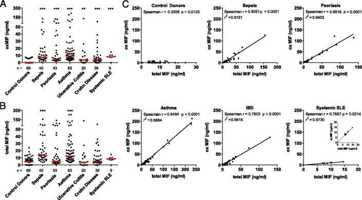
Compared with healthy controls, overexpression of MIF and high oxMIF levels were observed in sepsis, psoriasis, asthma, inflammatory bowel disease, and SLE (figure 5). 1 19 23 24 30–32 41 44–46 For example, in patients with severe sepsis, median MIF plasma levels were approximately 4–5 times higher than in healthy blood donors.30 Similarly, oxMIF plasma levels were significantly elevated in patients with septicemia compared with healthy controls.1 In patients with ulcerative colitis or Crohn’s disease, significantly higher levels of MIF and oxMIF were detected in biopsies from acute inflammation sites compared with non-lesion biopsy sites or healthy controls.1 MIF also was overexpressed in patients with SLE, and elevated plasma levels of oxMIF were observed in a subpopulation of patients with SLE (with systemic exacerbations of SLE without renal involvement) but not in patients with SLE in remission or lupus nephritis.1 23 46 Finally, in the aforementioned rat glomerulonephritis model, one dose of BaxB01 created an exposure-related, anti-inflammatory environment by inhibiting glomerular cellular inflammation.109 Treatment significantly reduced proteinuria and diminished histopathological glomerular crescent formation, indicating BaxB01 altered immune cell function and migration.109
Figure 5.
Presence of oxMIF in the circulation of patients with inflammatory disease and healthy controls. (A) Plasma levels of oxMIF in samples from healthy controls and patients with acute or chronic inflammatory diseases. (B) Plasma levels of total MIF in the same samples. Individual values and medians (red lines) are shown. We used the Kruskal-Wallis test followed by Dunn’s multiple correction test for statistical analyses. **P≤0.01; ***p≤0.001. (C) oxMIF levels plotted against total MIF levels for each individual plasma sample. Spearman’s correlation analysis showed a significant correlation between MIF and oxMIF levels for each disease, but not the healthy control group. IBD, inflammatory bowel disease; MIF, macrophage migration inhibitory factor; oxMIF, oxidized MIF; SLE, systemic lupus erythematosus. Figure has been reprinted from Thiele M, Kerschbaumer RJ, Tam FWK, et al. Selective targeting of a disease-related conformational isoform of macrophage migration inhibitory factor ameliorates inflammatory conditions. J Immunol 2015;195:2343–52.
MIF has been implicated in conditions with a neuroinflammatory component such as Parkinson’s disease and amyotrophic lateral sclerosis, where MIF has a protective effect, and Alzheimer’s disease and mild cognitive impairment, where conflicting data exist regarding protective versus harmful effects.112 113 The presence of herpes simplex virus type 1 in the brain increases the risk of Alzheimer’s disease.114 Interestingly, in a study of patients with herpes and associated Alzheimer’s disease, MIF had antiherpetic activity and oxMIF, which was present in postmortem brains, was postulated to be an overlapping factor, linking herpes simplex virus type 1-induced cellular changes to Alzheimer’s disease-like cellular pathology.115
oxMIF in cancer
High levels of MIF are present in certain solid tumors (eg, colorectal cancer, gastric cancers, NSCLC, ovarian cancer, esophageal squamous cell carcinoma, pancreatic cancer, melanoma, neuroblastoma, and osteosarcoma) and are associated with high tumor burden and grade, increased metastasis risk, and poor prognosis.2 33–40 47–51 For example, MIF was significantly overexpressed in primary ovarian cancer tissue, where its expression increased with grade of malignancy and correlated with the high MIF levels present in metastatic ascites.37 Similarly, oxMIF was detected in primary tumor tissue and in the circulation of women with ovarian cancer, with weak-to-strong cytoplasmic and membranous oxMIF staining in the apical papillary tumor cells, papillary projection cells, and the tumor stroma.2 oxMIF was most prominent in adenocarcinomas, serous adenocarcinomas, and mucinous cystadenocarcinomas, and no oxMIF was detected in normal ovarian tissue (online supplemental figure 2A).2 In addition, high levels of oxMIF were detected in ascites (online supplemental figure 2B).
jitc-2022-005475supp002.pdf (296.9KB, pdf)
High MIF levels also are present in the serum, colorectal tissue, and metastases of patients with colorectal cancer, where increased serum MIF concentrations correlated with severity of colorectal cancer and with the extent of metastasis.35 Likewise, oxMIF was detected in primary colorectal tumors and liver metastases, with a pronounced presence in vessel-like structures (online supplemental figure 2C).2 Moreover, in a phase I study of imalumab in patients with metastatic colorectal adenocarcinoma, plasma levels of total MIF correlated with oxMIF in oxMIF-positive patients, and significantly lower levels of oxMIF versus total MIF were detected in healthy tissue.3
MIF is highly expressed in lung tissue of patients with lung cancer, where high MIF levels correlated with poor prognosis.39 52 MIF-knockdown and MIF-mutant (lacking enzymatic activity) murine models resulted in attenuated lung tumor growth.116 MIF knockdown also reduced cell migration in human squamous cell carcinoma and human lung adenocarcinoma cell lines.34 52 oxMIF also was detected in lung cancer tissue and was most prominent in adenocarcinomas and squamous cell carcinomas (online supplemental figure 2D).2
In PDAC, MIF is overexpressed, and higher MIF levels correlate with lower survival—independent of tumor grade—and enhanced tumor aggression through its involvement in the epithelial-to-mesenchymal transition.38 47 Additionally, MIF was an independent predictor of prognosis in univariate and multivariate analyses.47 The potential for MIF as a prognostic marker is supported by the finding that MIF is highly expressed in PDAC-derived exosomes and plasma from patients with stage I PDAC before liver metastasis, and blocking MIF prevented liver premetastatic niche formation and exosome-induced PDAC metastasis.84 oxMIF was detected in pancreatic intraepithelial neoplasia, including during early tumor stages, with a more pronounced presence at later stages of disease (online supplemental figure 2E and 2F).2
Finally, in a study of bevacizumab-resistant glioblastomas, high levels of oxMIF were expressed in resistant xenografts, and exposure to imalumab reduced invasion of bevacizumab-naïve and bevacizumab-resistant glioblastomas in Matrigel and bioengineered three-dimensional hyaluronic acid-based assays.94 These findings indicate MIF oxidation was increased in the tumor microenvironment, facilitating invasion in a means targetable by imalumab.
Anti-oxMIF therapy
Fully human, monoclonal anti-oxMIF antibodies were developed, and those that did not bind redMIF and recognized distinct MIF epitopes (amino acids 50–68, including the oxidoreductase motif, and C-terminus, amino acids 86–102) were shown to neutralize the pro-inflammatory activities of MIF in vivo.2 93 In addition, inhibition of oxMIF promoted cancer cell apoptosis and inhibited cancer cell proliferation in vitro and in vivo.78 The effects of inhibiting oxMIF with an anti-oxMIF antibody were confirmed in a phase I study.3 After treatment with imalumab, one-quarter (13/50; 26%) of heavily pretreated patients with late-stage, solid tumor cancers had stable disease (SD) based on RECIST V.1.1 criteria.3 Furthermore, among the patients with SD, almost two-thirds (8/13; 62%) had SD lasting >4 months (NSCLC, n=2; ovarian cancer, n=2; colorectal cancer, n=1; esophageal cancer, n=1; and cancer of the parotid gland, n=1). Since many cancer treatment strategies are limited because of narrow therapeutic indices, toxicities, and development of tumor resistance to chemotherapeutic agents, regimens comprising a targeted therapy and ≥1 chemotherapeutic agent often are used. In in vitro and in vivo studies, anti-oxMIF antibodies sensitized cancer cells to cytotoxic agents and improved antitumorigenic effects (eg, decreased cell proliferation and increased apoptosis).2 These anti-oxMIF antibodies exerted their effects by inhibiting cell proliferation and survival signaling pathways, reducing active ERK1/2 and AKT levels, and promoting activation of caspase-3.78 Additional clinical studies are needed to further investigate the role of oxMIF as a therapeutic target.
Discussion
oxMIF exhibits pathological properties of MIF (consistent with the published literature) that can be modified with oxMIF-specific antibodies (figure 4). oxMIF is specifically detected in plasma and/or tissues of diseased patients, but not in healthy individuals. 1 2 Therefore, oxMIF is a promising drug target and diagnostic biomarker in certain inflammatory diseases and solid tumors.1 2
Targeting MIF with biologics and small molecules is challenging because such entities are intercepted by the excess MIF protein in the circulation and in normal tissues and, therefore, are impaired in exerting beneficial effects. Furthermore, non-pathological functions of MIF, which contribute to redox-homeostasis and wound-healing processes, could be impaired by entities that do not distinguish between MIF and oxMIF.117 Nevertheless, MIF is the precursor to oxMIF, justifying the concept that elevated MIF levels contribute to the development and maintenance of inflammatory and immunopathological processes. The identification of effective anti-oxMIF antibodies was obviously not predictable. Of the 145 anti-MIF antibodies originally analyzed from a phage display system, fewer than 10 demonstrated efficacy in in vitro and in vivo models of inflammation.93 Subsequently, it was found that these effective antibodies only showed binding specificity for MIF under pro-oxidative conditions, which is where the name ‘oxMIF’ originated.1 Unfolding of the protein under oxidizing conditions involves conformational changes that apparently expose epitopes in the MIF trimer that are not accessible to anti-oxMIF antibodies under normal (non-oxidizing) conditions.81 Recent studies confirmed that an oxidizing environment destabilizes MIF to unfolding relative to redMIF and modulates an equilibrium between the two MIF isoforms.81
Mutational analyses of latent redox-dependent, functionally important MIF residues demonstrated the propensity for MIF to toggle its confirmation and binding partners depending on the redox environment.80 81 Similar redox-dependent conformational changes have not been demonstrated for the MIF homolog D-dopachrome tautomerase (MIF-2), which has low sequence identity (35%).118 However, mutations in allosteric residues Pro-1, Ser-62, and Phe-100 that occupy the same positions as Pro-1, His-62, and Tyr-99 in MIF also affected structure, dynamics, and biological function of MIF-2, demonstrating a conserved control of structure-dependent functions for MIF superfamily members.118 119 Elsewhere in nature, redox-dependent sensors exist with isoform-specific functions,120–123 the evolutionary implications of which are yet to be understood. Redox sensitivity likely affects both intracellular and extracellular interactions of MIF and could provide additional insight into its pro-inflammatory mechanisms and pleiotropic spectrum of functions.19 81 Many in vitro studies on signaling, receptor binding, migration, etc, were performed using recombinant MIF, which raises the question: to what extent can these effects be attributed to oxMIF? Working with redox-sensitive proteins is complex and prone to interference. Several parameters may have unknowingly contributed to results from historical in vitro studies. The MIF protein preparation itself may have contained MIF molecules that have adopted the oxMIF structure during certain purification/refolding procedures. In cell-based assays, which may be especially complex in co-culture settings, secreted reactive oxygen species (ROS)/reactive nitrogen species or different enzyme activities may have contributed to an oxidative environment, eliciting oxMIF formation. The impact of culture media compositions should also be considered.
In acute inflammatory responses, neutrophils are activated and ROS, including MPO-derived hypochlorous acid and hypothiocyanous acid, are produced.68 The mechanism by which MIF is converted to oxMIF in the tumor microenvironment is unknown but may involve tumor-associated neutrophils and MPO activity at inflammatory sites where both MIF and neutrophil-derived MPO are present.67 68 Several concepts for post-translational modifications of MIF have been proposed; however, additional studies are needed to fully define the process(es).67
While MIF is detected at various levels in the circulation, on cell surfaces, and in the cytoplasm of cells under normal, healthy conditions, oxMIF is predominantly expressed under inflammatory conditions, such as acute and chronic inflammatory diseases or solid tumors.1 2 Specific inhibition of oxMIF is a potent method of interfering with the biological activities of MIF (figure 4). In inflammatory diseases, the prevalence of oxMIF and evidence supporting the effects of anti-oxMIF antibodies is especially promising given the synergistic effect of oxMIF/MIF inhibition with glucocorticoids. It would be interesting to evaluate whether murine monoclonal anti-MIF antibodies that have shown beneficial effects in animal models of inflammation and cancer (eg, clone III.D.9) have a higher affinity or specificity for oxMIF relative to MIF.32 103 124 125 Clinical evaluation of anti-oxMIF antibodies as a late-stage cancer therapeutic showed modest anti-tumor activity,3 which could be enhanced by optimizing biophysiochemical properties and immunomodulatory functions of such antibodies and by rational combinations with standard-of-care regimens.126 Emerging nanomedicine-based approaches127 may also enhance therapeutic impacts of anti-oxMIF antibodies. Nanomaterials exerting ROS-scavenging and anti-inflammation effects as observed in an in vivo atherosclerosis mouse model,128 could act synergistically with anti-oxMIF-based therapeutic approaches. However, further studies are warranted to validate these hypotheses.
As discussed, MIF exerts its effects by binding to CD74, which triggers recruitment of CD44, resulting in activation of the MAPK/ERK and PI3K/AKT pathways.10 12–14 82 MIF also binds directly to CXCR2 or CXCR4, or CXCR2/CD74 and CXCR4/CD74 receptor complexes, resulting in activation of the MAPK/ERK and PI3K/AKT pathways.10 11 82 Effects of oxMIF could also be mediated by these same receptors. However, acknowledging that the term ‘oxMIF’ was originally defined in terms of antibody specificity and not in terms of an elucidated biological conversion mechanism is important.1 93 oxMIF is detected at sites of inflammation and in the circulation of patients with chronic inflammatory diseases, suggesting the post-translationally modified form of MIF can leave inflamed tissues, depending on the disease and stage of inflammation. In contrast, oxMIF is more likely to be retained in primary tumors and corresponding metastases in most tumor types. 2 3 The soluble oxMIF form generated by neutrophil-MPO-mediated oxidation of MIF with hypochlorous acid was reported to bind to CXCR2, thereby triggering the release of IL-6 and CXCL8 by peripheral blood mononuclear cells and contributing to neutrophil survival.111 Binding and structural analyses are needed to determine whether oxMIF also triggers cell signaling via CD74/CD44 and whether inhibitory effects of anti-oxMIF antibodies on pro-proliferative downstream signaling pathways (eg, ERK and/or AKT) are mediated by this receptor complex. The impact of oxidation of Cys-80 and/or Lys-66 on CD74 binding and cell signaling is yet to be determined.
Imalumab (first-generation anti-oxMIF antibody) showed promise in preclinical studies (in vitro and in vivo) and in a phase I study.2 3 78 93 Imalumab neutralized the biological activity of oxMIF in relevant pathways (eg, PI3K/AKT/mTOR signaling),3 penetrated and accumulated in tumors and metastases during treatment,1 and colocalized with oxMIF in tumor cells and stroma, with efficient target binding by end of first cycle of treatment.3 Clinical evaluation of imalumab was prematurely halted; however, targeted development of improved, second-generation, anti-oxMIF antibodies continues, and these antibodies are being evaluated in preclinical and clinical proof-of-concept studies.
The early phase imalumab clinical trial included patients with advanced solid tumors including metastatic lung, colorectal, and ovarian carcinomas.3 These cancer types are newly diagnosed at annual rates of 2,200,000, 1,150,000, and 314,000129 with 5-year mortality rates of 79%, 35%, and 51%,130 respectively, presenting an unmet need for more effective therapies. The benign toxicity of oxMIF therapy3 should allow for combination therapy and integration into standard-of-care regimens without exacerbating patient toxicity. In the case of inflammatory diseases, these indications are often treated with steroids. Although not yet evaluated in clinical trials, concomitant steroid and anti-oxMIF therapy would ideally capitalize on MIF’s counteracting of glucocorticoids in order to provide steroid-sparing benefits.131
Conclusion
Pathological properties of MIF are based on biological functions, which are evidently altered by anti-oxMIF antibodies. Therefore, oxMIF reflects a druggable isoform of MIF in inflammatory diseases and cancer. Considering the versatile mechanisms of action of MIF and its role in inflammation and cancer, anti-oxMIF modalities could be applied as monotherapy and/or in combination with standard-of-care regimens (ie, immunomodulatory agents/checkpoint inhibitors, anti-angiogenic drugs, chemotherapeutics, and glucocorticoids/nonsteroidal anti-inflammatory drugs). Additional clinical assessments are necessary to validate these hypotheses.
Acknowledgments
Medical writing and editorial assistance were provided by Maribeth Bogush, PhD, and Julie O’Grady, BA, of The Medicine Group, LLC (New Hope, PA, USA) in accordance with Good Publication Practice guidelines. Funding for medical writing and manuscript preparation was provided by OncoOne Research & Development GmbH (Vienna, Austria).
Footnotes
Contributors: MT conceived this manuscript. MT, SCD, and RAM made significant contributions to the design, drafting, and critical revision of the manuscript. MT, SCD, and RAM approved the final version for submission.
Competing interests: MT is an employee of OncoOne Research & Development GmbH. SCD has nothing to declare. RAM is an inventor on patents pertaining to MIF inhibition.
Provenance and peer review: Commissioned; externally peer reviewed.
Supplemental material: This content has been supplied by the author(s). It has not been vetted by BMJ Publishing Group Limited (BMJ) and may not have been peer-reviewed. Any opinions or recommendations discussed are solely those of the author(s) and are not endorsed by BMJ. BMJ disclaims all liability and responsibility arising from any reliance placed on the content. Where the content includes any translated material, BMJ does not warrant the accuracy and reliability of the translations (including but not limited to local regulations, clinical guidelines, terminology, drug names and drug dosages), and is not responsible for any error and/or omissions arising from translation and adaptation or otherwise.
Ethics statements
Patient consent for publication
Not applicable.
Ethics approval
Not applicable.
References
- 1.Thiele M, Kerschbaumer RJ, Tam FWK, et al. Selective targeting of a disease-related conformational isoform of macrophage migration inhibitory factor ameliorates inflammatory conditions. J Immunol 2015;195:2343–52. 10.4049/jimmunol.1500572 [DOI] [PMC free article] [PubMed] [Google Scholar]
- 2.Schinagl A, Thiele M, Douillard P, et al. Oxidized macrophage migration inhibitory factor is a potential new tissue marker and drug target in cancer. Oncotarget 2016;7:73486–96. 10.18632/oncotarget.11970 [DOI] [PMC free article] [PubMed] [Google Scholar]
- 3.Mahalingam D, Patel MR, Sachdev JC, et al. Phase I study of imalumab (BAX69), a fully human recombinant antioxidized macrophage migration inhibitory factor antibody in advanced solid tumours. Br J Clin Pharmacol 2020;86:1836–48. 10.1111/bcp.14289 [DOI] [PMC free article] [PubMed] [Google Scholar]
- 4.Bloom BR, Bennett B. Mechanism of a reaction in vitro associated with delayed-type hypersensitivity. Science 1966;153:80–2. 10.1126/science.153.3731.80 [DOI] [PubMed] [Google Scholar]
- 5.Kang I, Bucala R. The immunobiology of MIF: function, genetics and prospects for precision medicine. Nat Rev Rheumatol 2019;15:427–37. 10.1038/s41584-019-0238-2 [DOI] [PubMed] [Google Scholar]
- 6.Nishihira J. Molecular function of macrophage migration inhibitory factor and a novel therapy for inflammatory bowel disease. Ann N Y Acad Sci 2012;1271:53–7. 10.1111/j.1749-6632.2012.06735.x [DOI] [PMC free article] [PubMed] [Google Scholar]
- 7.Adamali H, Armstrong ME, McLaughlin AM, et al. Macrophage migration inhibitory factor enzymatic activity, lung inflammation, and cystic fibrosis. Am J Respir Crit Care Med 2012;186:162–9. 10.1164/rccm.201110-1864OC [DOI] [PMC free article] [PubMed] [Google Scholar]
- 8.Tilstam PV, Qi D, Leng L, et al. MIF family cytokines in cardiovascular diseases and prospects for precision-based therapeutics. Expert Opin Ther Targets 2017;21:671–83. 10.1080/14728222.2017.1336227 [DOI] [PMC free article] [PubMed] [Google Scholar]
- 9.Kevill KA, Bhandari V, Kettunen M, et al. A role for macrophage migration inhibitory factor in the neonatal respiratory distress syndrome. J Immunol 2008;180:601–8. 10.4049/jimmunol.180.1.601 [DOI] [PubMed] [Google Scholar]
- 10.Noe JT, Mitchell RA. MIF-dependent control of tumor immunity. Front Immunol 2020;11:609948. 10.3389/fimmu.2020.609948 [DOI] [PMC free article] [PubMed] [Google Scholar]
- 11.Bernhagen J, Krohn R, Lue H, et al. MIF is a noncognate ligand of CXC chemokine receptors in inflammatory and atherogenic cell recruitment. Nat Med 2007;13:587–96. 10.1038/nm1567 [DOI] [PubMed] [Google Scholar]
- 12.Shi X, Leng L, Wang T, et al. Cd44 is the signaling component of the macrophage migration inhibitory factor-CD74 receptor complex. Immunity 2006;25:595–606. 10.1016/j.immuni.2006.08.020 [DOI] [PMC free article] [PubMed] [Google Scholar]
- 13.Leng L, Bucala R. Insight into the biology of macrophage migration inhibitory factor (MIF) revealed by the cloning of its cell surface receptor. Cell Res 2006;16:162–8. 10.1038/sj.cr.7310022 [DOI] [PubMed] [Google Scholar]
- 14.Lue H, Thiele M, Franz J, et al. Macrophage migration inhibitory factor (MIF) promotes cell survival by activation of the Akt pathway and role for CSN5/JAB1 in the control of autocrine MIF activity. Oncogene 2007;26:5046–59. 10.1038/sj.onc.1210318 [DOI] [PubMed] [Google Scholar]
- 15.Mitchell RA, Metz CN, Peng T, et al. Sustained mitogen-activated protein kinase (MAPK) and cytoplasmic phospholipase A2 activation by macrophage migration inhibitory factor (MIF). regulatory role in cell proliferation and glucocorticoid action. J Biol Chem 1999;274:18100–6. 10.1074/jbc.274.25.18100 [DOI] [PubMed] [Google Scholar]
- 16.Wen Y, Cai W, Yang J, et al. Targeting macrophage migration inhibitory factor in acute pancreatitis and pancreatic cancer. Front Pharmacol 2021;12:638950. 10.3389/fphar.2021.638950 [DOI] [PMC free article] [PubMed] [Google Scholar]
- 17.Lue H, Dewor M, Leng L, et al. Activation of the JNK signalling pathway by macrophage migration inhibitory factor (MIF) and dependence on CXCR4 and CD74. Cell Signal 2011;23:135–44. 10.1016/j.cellsig.2010.08.013 [DOI] [PMC free article] [PubMed] [Google Scholar]
- 18.Coleman AM, Rendon BE, Zhao M, et al. Cooperative regulation of non-small cell lung carcinoma angiogenic potential by macrophage migration inhibitory factor and its homolog, D-dopachrome tautomerase. J Immunol 2008;181:2330–7. 10.4049/jimmunol.181.4.2330 [DOI] [PMC free article] [PubMed] [Google Scholar]
- 19.Calandra T, Roger T. Macrophage migration inhibitory factor: a regulator of innate immunity. Nat Rev Immunol 2003;3:791–800. 10.1038/nri1200 [DOI] [PMC free article] [PubMed] [Google Scholar]
- 20.Kleemann R, Hausser A, Geiger G, et al. Intracellular action of the cytokine MIF to modulate AP-1 activity and the cell cycle through Jab1. Nature 2000;408:211–6. 10.1038/35041591 [DOI] [PubMed] [Google Scholar]
- 21.Calandra T, Bernhagen J, Mitchell RA, et al. The macrophage is an important and previously unrecognized source of macrophage migration inhibitory factor. J Exp Med 1994;179:1895–902. 10.1084/jem.179.6.1895 [DOI] [PMC free article] [PubMed] [Google Scholar]
- 22.Bacher M, Meinhardt A, Lan HY, et al. Migration inhibitory factor expression in experimentally induced endotoxemia. Am J Pathol 1997;150:235–46. [PMC free article] [PubMed] [Google Scholar]
- 23.Bilsborrow JB, Doherty E, Tilstam PV, et al. Macrophage migration inhibitory factor (MIF) as a therapeutic target for rheumatoid arthritis and systemic lupus erythematosus. Expert Opin Ther Targets 2019;23:733–44. 10.1080/14728222.2019.1656718 [DOI] [PMC free article] [PubMed] [Google Scholar]
- 24.Baugh JA, Donnelly SC. Macrophage migration inhibitory factor: a neuroendocrine modulator of chronic inflammation. J Endocrinol 2003;179:15–23. 10.1677/joe.0.1790015 [DOI] [PubMed] [Google Scholar]
- 25.Bernhagen J, Mitchell RA, Calandra T, et al. Purification, bioactivity, and secondary structure analysis of mouse and human macrophage migration inhibitory factor (MIF). Biochemistry 1994;33:14144–55. 10.1021/bi00251a025 [DOI] [PubMed] [Google Scholar]
- 26.Bacher M, Metz CN, Calandra T, et al. An essential regulatory role for macrophage migration inhibitory factor in T-cell activation. Proc Natl Acad Sci U S A 1996;93:7849–54. 10.1073/pnas.93.15.7849 [DOI] [PMC free article] [PubMed] [Google Scholar]
- 27.Calandra T, Bernhagen J, Metz CN, et al. MIF as a glucocorticoid-induced modulator of cytokine production. Nature 1995;377:68–71. 10.1038/377068a0 [DOI] [PubMed] [Google Scholar]
- 28.Bucala R. MIF rediscovered: cytokine, pituitary hormone, and glucocorticoid-induced regulator of the immune response. Faseb J 1996;10:1607–13. 10.1096/fasebj.10.14.9002552 [DOI] [PubMed] [Google Scholar]
- 29.Fan H, Hall P, Santos LL, et al. Macrophage migration inhibitory factor and CD74 regulate macrophage chemotactic responses via MAPK and Rho GTPase. J Immunol 2011;186:4915–24. 10.4049/jimmunol.1003713 [DOI] [PMC free article] [PubMed] [Google Scholar]
- 30.Lehmann LE, Novender U, Schroeder S, et al. Plasma levels of macrophage migration inhibitory factor are elevated in patients with severe sepsis. Intensive Care Med 2001;27:1412–5. 10.1007/s001340101022 [DOI] [PubMed] [Google Scholar]
- 31.Ichiyama H, Onodera S, Nishihira J, et al. Inhibition of joint inflammation and destruction induced by anti-type II collagen antibody/lipopolysaccharide (LPS)-induced arthritis in mice due to deletion of macrophage migration inhibitory factor (MIF). Cytokine 2004;26:187–94. 10.1016/j.cyto.2004.02.007 [DOI] [PubMed] [Google Scholar]
- 32.de Jong YP, Abadia-Molina AC, Satoskar AR, et al. Development of chronic colitis is dependent on the cytokine MIF. Nat Immunol 2001;2:1061–6. 10.1038/ni720 [DOI] [PubMed] [Google Scholar]
- 33.Meyer-Siegler KL, Iczkowski KA, Leng L, et al. Inhibition of macrophage migration inhibitory factor or its receptor (CD74) attenuates growth and invasion of DU-145 prostate cancer cells. J Immunol 2006;177:8730–9. 10.4049/jimmunol.177.12.8730 [DOI] [PubMed] [Google Scholar]
- 34.Rendon BE, Roger T, Teneng I, et al. Regulation of human lung adenocarcinoma cell migration and invasion by macrophage migration inhibitory factor. J Biol Chem 2007;282:29910–8. 10.1074/jbc.M704898200 [DOI] [PubMed] [Google Scholar]
- 35.He X-X, Chen K, Yang J, et al. Macrophage migration inhibitory factor promotes colorectal cancer. Mol Med 2009;15:1–10. 10.2119/molmed.2008.00107 [DOI] [PMC free article] [PubMed] [Google Scholar]
- 36.Wang D, Wang R, Huang A, et al. Upregulation of macrophage migration inhibitory factor promotes tumor metastasis and correlates with poor prognosis of pancreatic ductal adenocarcinoma. Oncol Rep 2018;40:2628–36. 10.3892/or.2018.6703 [DOI] [PMC free article] [PubMed] [Google Scholar]
- 37.Krockenberger M, Dombrowski Y, Weidler C, et al. Macrophage migration inhibitory factor contributes to the immune escape of ovarian cancer by down-regulating NKG2D. J Immunol 2008;180:7338–48. 10.4049/jimmunol.180.11.7338 [DOI] [PMC free article] [PubMed] [Google Scholar]
- 38.Tan L, Ye X, Zhou Y, et al. Macrophage migration inhibitory factor is overexpressed in pancreatic cancer tissues and impairs insulin secretion function of β-cell. J Transl Med 2014;12:92. 10.1186/1479-5876-12-92 [DOI] [PMC free article] [PubMed] [Google Scholar]
- 39.Tomiyasu M, Yoshino I, Suemitsu R. Quantification of macrophage migration inhibitory factor mRNA expression in non-small cell lung cancer tissues and its clinical significance. Clin Cancer Res 2002;8:3755–9. [PubMed] [Google Scholar]
- 40.He X-X, Yang J, Ding Y-W, et al. Increased epithelial and serum expression of macrophage migration inhibitory factor (MIF) in gastric cancer: potential role of MIF in gastric carcinogenesis. Gut 2006;55:797–802. 10.1136/gut.2005.078113 [DOI] [PMC free article] [PubMed] [Google Scholar]
- 41.Bozza M, Satoskar AR, Lin G, et al. Targeted disruption of migration inhibitory factor gene reveals its critical role in sepsis. J Exp Med 1999;189:341–6. 10.1084/jem.189.2.341 [DOI] [PMC free article] [PubMed] [Google Scholar]
- 42.Shimizu T, Nishihira J, Mizue Y, et al. High macrophage migration inhibitory factor (MIF) serum levels associated with extended psoriasis. J Invest Dermatol 2001;116:989–90. 10.1046/j.0022-202x.2001.01366.x [DOI] [PubMed] [Google Scholar]
- 43.Meazza C, Travaglino P, Pignatti P, et al. Macrophage migration inhibitory factor in patients with juvenile idiopathic arthritis. Arthritis Rheum 2002;46:232–7. [DOI] [PubMed] [Google Scholar]
- 44.Donn RP, Shelley E, Ollier WE, et al. A novel 5'-flanking region polymorphism of macrophage migration inhibitory factor is associated with systemic-onset juvenile idiopathic arthritis. Arthritis Rheum 2001;44:1782–5. [DOI] [PubMed] [Google Scholar]
- 45.Sampey AV, Hall PH, Mitchell RA, et al. Regulation of synoviocyte phospholipase A2 and cyclooxygenase 2 by macrophage migration inhibitory factor. Arthritis Rheum 2001;44:1273–80. [DOI] [PubMed] [Google Scholar]
- 46.Foote A, Briganti EM, Kipen Y, et al. Macrophage migration inhibitory factor in systemic lupus erythematosus. J Rheumatol 2004;31:268–73. [PubMed] [Google Scholar]
- 47.Funamizu N, Hu C, Lacy C, et al. Macrophage migration inhibitory factor induces epithelial to mesenchymal transition, enhances tumor aggressiveness and predicts clinical outcome in resected pancreatic ductal adenocarcinoma. Int J Cancer 2013;132:785–94. 10.1002/ijc.27736 [DOI] [PMC free article] [PubMed] [Google Scholar]
- 48.Hagemann T, Robinson SC, Thompson RG, et al. Ovarian cancer cell-derived migration inhibitory factor enhances tumor growth, progression, and angiogenesis. Mol Cancer Ther 2007;6:1993–2002. 10.1158/1535-7163.MCT-07-0118 [DOI] [PubMed] [Google Scholar]
- 49.Ren Y, Law S, Huang X, et al. Macrophage migration inhibitory factor stimulates angiogenic factor expression and correlates with differentiation and lymph node status in patients with esophageal squamous cell carcinoma. Ann Surg 2005;242:55–63. 10.1097/01.sla.0000168555.97710.bb [DOI] [PMC free article] [PubMed] [Google Scholar]
- 50.Han I, Lee MR, Nam KW, et al. Expression of macrophage migration inhibitory factor relates to survival in high-grade osteosarcoma. Clin Orthop Relat Res 2008;466:2107–13. 10.1007/s11999-008-0333-1 [DOI] [PMC free article] [PubMed] [Google Scholar]
- 51.Shimizu T, Abe R, Nakamura H, et al. High expression of macrophage migration inhibitory factor in human melanoma cells and its role in tumor cell growth and angiogenesis. Biochem Biophys Res Commun 1999;264:751–8. 10.1006/bbrc.1999.1584 [DOI] [PubMed] [Google Scholar]
- 52.Koh HM, Kim DC, Kim Y-M, et al. Prognostic role of macrophage migration inhibitory factor expression in patients with squamous cell carcinoma of the lung. Thorac Cancer 2019;10:2209–17. 10.1111/1759-7714.13198 [DOI] [PMC free article] [PubMed] [Google Scholar]
- 53.Liu R-M, Sun D-N, Jiao Y-L, et al. Macrophage migration inhibitory factor promotes tumor aggressiveness of esophageal squamous cell carcinoma via activation of Akt and inactivation of GSK3β. Cancer Lett 2018;412:289–96. 10.1016/j.canlet.2017.10.018 [DOI] [PubMed] [Google Scholar]
- 54.Cavalli E, Ciurleo R, Petralia MC, et al. Emerging role of the macrophage migration inhibitory factor family of cytokines in neuroblastoma. Pathogenic effectors and novel therapeutic targets? Molecules 2020;25:1194. 10.3390/molecules25051194 [DOI] [PMC free article] [PubMed] [Google Scholar]
- 55.Donn RP, Plant D, Jury F, et al. Macrophage migration inhibitory factor gene polymorphism is associated with psoriasis. J Invest Dermatol 2004;123:484–7. 10.1111/j.0022-202X.2004.23314.x [DOI] [PubMed] [Google Scholar]
- 56.Baugh JA, Chitnis S, Donnelly SC, et al. A functional promoter polymorphism in the macrophage migration inhibitory factor (MIF) gene associated with disease severity in rheumatoid arthritis. Genes Immun 2002;3:170–6. 10.1038/sj.gene.6363867 [DOI] [PubMed] [Google Scholar]
- 57.Razzaghi MR, Mazloomfard MM, Malekian S, et al. Association of macrophage inhibitory factor -173 gene polymorphism with biological behavior of prostate cancer. Urol J 2019;16:32–6. 10.22037/uj.v0i0.3968 [DOI] [PubMed] [Google Scholar]
- 58.Illescas O, Gomez-Verjan JC, García-Velázquez L, et al. Macrophage migration inhibitory factor -173 G/C polymorphism: a global meta-analysis across the disease spectrum. Front Genet 2018;9:55. 10.3389/fgene.2018.00055 [DOI] [PMC free article] [PubMed] [Google Scholar]
- 59.Morales-Zambrano R, Bautista-Herrera LA, De la Cruz-Mosso U, et al. Macrophage migration inhibitory factor (MIF) promoter polymorphisms (-794 CATT5-8 and -173 G>C): association with MIF and TNFα in psoriatic arthritis. Int J Clin Exp Med 2014;7:2605–14. [PMC free article] [PubMed] [Google Scholar]
- 60.Sreih A, Ezzeddine R, Leng L, et al. Dual effect of the macrophage migration inhibitory factor gene on the development and severity of human systemic lupus erythematosus. Arthritis Rheum 2011;63:3942–51. 10.1002/art.30624 [DOI] [PMC free article] [PubMed] [Google Scholar]
- 61.Sánchez E, Gómez LM, Lopez-Nevot MA, et al. Evidence of association of macrophage migration inhibitory factor gene polymorphisms with systemic lupus erythematosus. Genes Immun 2006;7:433–6. 10.1038/sj.gene.6364310 [DOI] [PubMed] [Google Scholar]
- 62.De la Cruz-Mosso U, Bucala R, Palafox-Sánchez CA, et al. Macrophage migration inhibitory factor: association of -794 CATT5-8 and -173 G>C polymorphisms with TNF-α in systemic lupus erythematosus. Hum Immunol 2014;75:433–9. 10.1016/j.humimm.2014.02.014 [DOI] [PMC free article] [PubMed] [Google Scholar]
- 63.Sumaiya K, Langford D, Natarajaseenivasan K, et al. Macrophage migration inhibitory factor (MIF): a multifaceted cytokine regulated by genetic and physiological strategies. Pharmacol Ther 2022;233:108024. 10.1016/j.pharmthera.2021.108024 [DOI] [PubMed] [Google Scholar]
- 64.Yang J, Li Y, Zhang X. Meta-Analysis of macrophage migration inhibitory factor (MIF) gene -173G/C polymorphism and inflammatory bowel disease (IBD) risk. Int J Clin Exp Med 2015;8:9570–4. [PMC free article] [PubMed] [Google Scholar]
- 65.Dambacher J, Staudinger T, Seiderer J, et al. Macrophage migration inhibitory factor (MIF) -173G/C promoter polymorphism influences upper gastrointestinal tract involvement and disease activity in patients with Crohn's disease. Inflamm Bowel Dis 2007;13:71–82. 10.1002/ibd.20008 [DOI] [PubMed] [Google Scholar]
- 66.Trivedi-Parmar V, Jorgensen WL. Advances and insights for small molecule inhibition of macrophage migration inhibitory factor. J Med Chem 2018;61:8104–19. 10.1021/acs.jmedchem.8b00589 [DOI] [PMC free article] [PubMed] [Google Scholar]
- 67.Schindler L, Dickerhof N, Hampton MB, et al. Post-Translational regulation of macrophage migration inhibitory factor: basis for functional fine-tuning. Redox Biol 2018;15:135–42. 10.1016/j.redox.2017.11.028 [DOI] [PMC free article] [PubMed] [Google Scholar]
- 68.Dickerhof N, Schindler L, Bernhagen J, et al. Macrophage migration inhibitory factor (MIF) is rendered enzymatically inactive by myeloperoxidase-derived oxidants but retains its immunomodulatory function. Free Radic Biol Med 2015;89:498–511. 10.1016/j.freeradbiomed.2015.09.009 [DOI] [PubMed] [Google Scholar]
- 69.Senter PD, Al-Abed Y, Metz CN, et al. Inhibition of macrophage migration inhibitory factor (MIF) tautomerase and biological activities by acetaminophen metabolites. Proc Natl Acad Sci U S A 2002;99:144–9. 10.1073/pnas.011569399 [DOI] [PMC free article] [PubMed] [Google Scholar]
- 70.Dios A, Mitchell RA, Aljabari B, et al. Inhibition of MIF bioactivity by rational design of pharmacological inhibitors of MIF tautomerase activity. J Med Chem 2002;45:2410–6. 10.1021/jm010534q [DOI] [PubMed] [Google Scholar]
- 71.Rajasekaran D, Zierow S, Syed M, et al. Targeting distinct tautomerase sites of d-dT and MIF with a single molecule for inhibition of neutrophil lung recruitment. Faseb J 2014;28:4961–71. 10.1096/fj.14-256636 [DOI] [PMC free article] [PubMed] [Google Scholar]
- 72.Al-Abed Y, VanPatten S. Mif as a disease target: iso-1 as a proof-of-concept therapeutic. Future Med Chem 2011;3:45–63. 10.4155/fmc.10.281 [DOI] [PubMed] [Google Scholar]
- 73.Ouertatani-Sakouhi H, El-Turk F, Fauvet B, et al. Identification and characterization of novel classes of macrophage migration inhibitory factor (MIF) inhibitors with distinct mechanisms of action. J Biol Chem 2010;285:26581–98. 10.1074/jbc.M110.113951 [DOI] [PMC free article] [PubMed] [Google Scholar]
- 74.Lubetsky JB, Dios A, Han J, et al. The tautomerase active site of macrophage migration inhibitory factor is a potential target for discovery of novel anti-inflammatory agents. J Biol Chem 2002;277:24976–82. 10.1074/jbc.M203220200 [DOI] [PubMed] [Google Scholar]
- 75.Wang Y, An R, Umanah GK, et al. A nuclease that mediates cell death induced by DNA damage and poly(ADP-ribose) polymerase-1. Science 2016;354:6872. 10.1126/science.aad6872 [DOI] [PMC free article] [PubMed] [Google Scholar]
- 76.Ruan Z, Lu Q, Wang JE, et al. Mif promotes neurodegeneration and cell death via its nuclease activity following traumatic brain injury. Cellular and Molecular Life Sciences 2022;79:39. 10.1007/s00018-021-04037-9 [DOI] [PMC free article] [PubMed] [Google Scholar]
- 77.Wang Y, Chen Y, Wang C, et al. Mif is a 3' flap nuclease that facilitates DNA replication and promotes tumor growth. Nat Commun 2021;12:2954. 10.1038/s41467-021-23264-z [DOI] [PMC free article] [PubMed] [Google Scholar]
- 78.Hussain F, Freissmuth M, Völkel D, et al. Human anti-macrophage migration inhibitory factor antibodies inhibit growth of human prostate cancer cells in vitro and in vivo. Mol Cancer Ther 2013;12:1223–34. 10.1158/1535-7163.MCT-12-0988 [DOI] [PubMed] [Google Scholar]
- 79.Mahalingam D, Patel MR, Sachdev JC, et al. First-In-Human, phase I study assessing imalumab (Bax69), a first-in-class anti-oxidized macrophage migration inhibitory factor (oxMIF) antibody in advanced solid tumors. Journal of Clinical Oncology 2015;33:2518. 10.1200/jco.2015.33.15_suppl.2518 [DOI] [Google Scholar]
- 80.Schinagl A, Kerschbaumer RJ, Sabarth N, et al. Role of the cysteine 81 residue of macrophage migration inhibitory factor as a molecular redox switch. Biochemistry 2018;57:1523–32. 10.1021/acs.biochem.7b01156 [DOI] [PubMed] [Google Scholar]
- 81.Skeens E, Gadzuk-Shea M, Shah D, et al. Redox-Dependent structure and dynamics of macrophage migration inhibitory factor reveal sites of latent allostery. Structure 2022;30:840–50. 10.1016/j.str.2022.03.007 [DOI] [PubMed] [Google Scholar]
- 82.Song S, Xiao Z, Dekker FJ, et al. Macrophage migration inhibitory factor family proteins are multitasking cytokines in tissue injury. Cell Mol Life Sci 2022;79:105. 10.1007/s00018-021-04038-8 [DOI] [PMC free article] [PubMed] [Google Scholar]
- 83.Conroy H, Mawhinney L, Donnelly SC. Inflammation and cancer: macrophage migration inhibitory factor (MIF)--the potential missing link. QJM 2010;103:831–6. 10.1093/qjmed/hcq148 [DOI] [PMC free article] [PubMed] [Google Scholar]
- 84.Costa-Silva B, Aiello NM, Ocean AJ, et al. Pancreatic cancer exosomes initiate pre-metastatic niche formation in the liver. Nat Cell Biol 2015;17:816–26. 10.1038/ncb3169 [DOI] [PMC free article] [PubMed] [Google Scholar]
- 85.Yang S, He P, Wang J, et al. A novel MIF signaling pathway drives the malignant character of pancreatic cancer by targeting NR3C2. Cancer Res 2016;76:3838–50. 10.1158/0008-5472.CAN-15-2841 [DOI] [PMC free article] [PubMed] [Google Scholar]
- 86.Cheng B, Wang Q, Song Y, et al. MIF inhibitor, iso-1, attenuates human pancreatic cancer cell proliferation, migration and invasion in vitro, and suppresses xenograft tumour growth in vivo. Sci Rep 2020;10:6741. 10.1038/s41598-020-63778-y [DOI] [PMC free article] [PubMed] [Google Scholar]
- 87.Wu L-H, Xia HH-X, Ma W-Q, et al. Macrophage migration inhibitory factor siRNA inhibits hepatic metastases of colorectal cancer cells. Front Biosci 2017;22:1365–78. 10.2741/4549 [DOI] [PubMed] [Google Scholar]
- 88.Sun B, Nishihira J, Yoshiki T, et al. Macrophage migration inhibitory factor promotes tumor invasion and metastasis via the Rho-dependent pathway. Clin Cancer Res 2005;11:1050–8. 10.1158/1078-0432.1050.11.3 [DOI] [PubMed] [Google Scholar]
- 89.Hagemann T, Wilson J, Kulbe H, et al. Macrophages induce invasiveness of epithelial cancer cells via NF-kappa B and JNK. J Immunol 2005;175:1197–205. 10.4049/jimmunol.175.2.1197 [DOI] [PubMed] [Google Scholar]
- 90.Liu N, Jiang N, Guo R, et al. MiR-451 inhibits cell growth and invasion by targeting MIF and is associated with survival in nasopharyngeal carcinoma. Mol Cancer 2013;12:1–8. 10.1186/1476-4598-12-123 [DOI] [PMC free article] [PubMed] [Google Scholar]
- 91.Pei X-J, Wu T-T, Li B, et al. Increased expression of macrophage migration inhibitory factor and DJ-1 contribute to cell invasion and metastasis of nasopharyngeal carcinoma. Int J Med Sci 2014;11:106–15. 10.7150/ijms.7264 [DOI] [PMC free article] [PubMed] [Google Scholar]
- 92.Amin MA, Volpert OV, Woods JM, et al. Migration inhibitory factor mediates angiogenesis via mitogen-activated protein kinase and phosphatidylinositol kinase. Circ Res 2003;93:321–9. 10.1161/01.RES.0000087641.56024.DA [DOI] [PubMed] [Google Scholar]
- 93.Kerschbaumer RJ, Rieger M, Völkel D, et al. Neutralization of macrophage migration inhibitory factor (MIF) by fully human antibodies correlates with their specificity for the β-sheet structure of MIF. J Biol Chem 2012;287:7446–55. 10.1074/jbc.M111.329664 [DOI] [PMC free article] [PubMed] [Google Scholar]
- 94.Hoffman D, Nguyen A, Jahangiri A, et al. ANGI-07. oxidized MIF drives glioblastoma cell invasion during bevacizumab resistance through unique signaling mechanisms. Neuro Oncol 2017;19:vi23. 10.1093/neuonc/nox168.086 [DOI] [Google Scholar]
- 95.Li G-Q, Xie J, Lei X-Y, et al. Macrophage migration inhibitory factor regulates proliferation of gastric cancer cells via the PI3K/Akt pathway. World J Gastroenterol 2009;15:5541–8. 10.3748/wjg.15.5541 [DOI] [PMC free article] [PubMed] [Google Scholar]
- 96.Denz A, Pilarsky C, Muth D, et al. Inhibition of MIF leads to cell cycle arrest and apoptosis in pancreatic cancer cells. J Surg Res 2010;160:29–34. 10.1016/j.jss.2009.03.048 [DOI] [PubMed] [Google Scholar]
- 97.Hudson JD, Shoaibi MA, Maestro R, et al. A proinflammatory cytokine inhibits p53 tumor suppressor activity. J Exp Med 1999;190:1375–82. 10.1084/jem.190.10.1375 [DOI] [PMC free article] [PubMed] [Google Scholar]
- 98.Mitchell RA, Liao H, Chesney J, et al. Macrophage migration inhibitory factor (MIF) sustains macrophage proinflammatory function by inhibiting p53: regulatory role in the innate immune response. Proc Natl Acad Sci U S A 2002;99:345–50. 10.1073/pnas.012511599 [DOI] [PMC free article] [PubMed] [Google Scholar]
- 99.Binsky I, Haran M, Starlets D, et al. Il-8 secreted in a macrophage migration-inhibitory factor- and CD74-dependent manner regulates B cell chronic lymphocytic leukemia survival. Proc Natl Acad Sci U S A 2007;104:13408–13. 10.1073/pnas.0701553104 [DOI] [PMC free article] [PubMed] [Google Scholar]
- 100.Klemke L, De Oliveira T, Witt D, et al. Hsp90-stabilized MIF supports tumor progression via macrophage recruitment and angiogenesis in colorectal cancer. Cell Death Dis 2021;12:155. 10.1038/s41419-021-03426-z [DOI] [PMC free article] [PubMed] [Google Scholar]
- 101.Hanahan D, Folkman J. Patterns and emerging mechanisms of the angiogenic switch during tumorigenesis. Cell 1996;86:353–64. 10.1016/S0092-8674(00)80108-7 [DOI] [PubMed] [Google Scholar]
- 102.Pradeep CR, Sunila ES, Kuttan G. Expression of vascular endothelial growth factor (VEGF) and VEGF receptors in tumor angiogenesis and malignancies. Integr Cancer Ther 2005;4:315–21. 10.1177/1534735405282557 [DOI] [PubMed] [Google Scholar]
- 103.Chesney J, Metz C, Bacher M, et al. An essential role for macrophage migration inhibitory factor (MIF) in angiogenesis and the growth of a murine lymphoma. Mol Med 1999;5:181–91. 10.1007/BF03402061 [DOI] [PMC free article] [PubMed] [Google Scholar]
- 104.Ogawa H, Nishihira J, Sato Y, et al. An antibody for macrophage migration inhibitory factor suppresses tumour growth and inhibits tumour-associated angiogenesis. Cytokine 2000;12:309–14. 10.1006/cyto.1999.0562 [DOI] [PubMed] [Google Scholar]
- 105.Landlinger C, Mirkina I, Maurer B. ON203: a new antibody targeting the oxidized form of macrophage migration inhibitory factor demonstrates antitumorigenic activity in preclinical models. poster presented at: EACR 2022; June 20-23: Seville, Spain. Available: https://nwm.covr.be/cmEpostersV2/index.html#/PosterDetail/1070
- 106.Girard E, Strathdee C, Trueblood E, et al. Macrophage migration inhibitory factor produced by the tumour stroma but not by tumour cells regulates angiogenesis in the B16-F10 melanoma model. Br J Cancer 2012;107:1498–505. 10.1038/bjc.2012.392 [DOI] [PMC free article] [PubMed] [Google Scholar]
- 107.Yaddanapudi K, Putty K, Rendon BE, et al. Control of tumor-associated macrophage alternative activation by macrophage migration inhibitory factor. J.i. 2013;190:2984–93. 10.4049/jimmunol.1201650 [DOI] [PMC free article] [PubMed] [Google Scholar]
- 108.Zhang J, Zhang G, Yang S, et al. Macrophage migration inhibitory factor regulating the expression of VEGF-C through MAPK signal pathways in breast cancer MCF-7 cell. World J Surg Oncol 2016;14:51. 10.1186/s12957-016-0797-5 [DOI] [PMC free article] [PubMed] [Google Scholar]
- 109.Höllriegl W, Bauer A, Baumgartner B, et al. Pharmacokinetics, disease-modifying activity, and safety of an experimental therapeutic targeting an immunological isoform of macrophage migration inhibitory factor, in rat glomerulonephritis. Eur J Pharmacol 2018;820:206–16. 10.1016/j.ejphar.2017.12.040 [DOI] [PubMed] [Google Scholar]
- 110.Bloom J, Sun S, Al-Abed Y. MIF, a controversial cytokine: a review of structural features, challenges, and opportunities for drug development. Expert Opin Ther Targets 2016;20:1463–75. 10.1080/14728222.2016.1251582 [DOI] [PubMed] [Google Scholar]
- 111.Schindler L, Zwissler L, Krammer C, et al. Macrophage migration inhibitory factor inhibits neutrophil apoptosis by inducing cytokine release from mononuclear cells. J Leukoc Biol 2021;110:893–905. 10.1002/JLB.3A0420-242RRR [DOI] [PubMed] [Google Scholar]
- 112.Petralia MC, Battaglia G, Bruno V, et al. The role of macrophage migration inhibitory factor in Alzheimer's disease: conventionally pathogenetic or unconventionally protective? Molecules 2020;25:291. 10.3390/molecules25020291 [DOI] [PMC free article] [PubMed] [Google Scholar]
- 113.Chen X, Chen Y, Qi D, et al. Multifaceted interconnections between macrophage migration inhibitory factor and psychiatric disorders. Prog Neuropsychopharmacol Biol Psychiatry 2022;112:110422. 10.1016/j.pnpbp.2021.110422 [DOI] [PubMed] [Google Scholar]
- 114.Protto V, Marcocci ME, Miteva MT, et al. Role of HSV-1 in Alzheimer's disease pathogenesis: a challenge for novel preventive/therapeutic strategies. Curr Opin Pharmacol 2022;63:102200. 10.1016/j.coph.2022.102200 [DOI] [PubMed] [Google Scholar]
- 115.Müller-Schiffmann A, Torres F, Kitaygorodskyy A. Macrophage migration inhibitory factor is a valid drug target at the intersection of herpes simplex virus 1 replication and Alzheimer’s disease-relevant cellular pathology. BioRxiv 2021;09459903. [Google Scholar]
- 116.Mawhinney L, Armstrong ME, O’Reilly C, et al. Macrophage migration inhibitory factor (MIF) enzymatic activity and lung cancer. Mol Med 2014;20:729–35. 10.2119/molmed.2014.00136 [DOI] [PMC free article] [PubMed] [Google Scholar]
- 117.Thiele M, Bernhagen J. Link between macrophage migration inhibitory factor and cellular redox regulation. Antioxid Redox Signal 2005;7:1234–48. 10.1089/ars.2005.7.1234 [DOI] [PubMed] [Google Scholar]
- 118.Chen E, Reiss K, Shah D, et al. A structurally preserved allosteric site in the MIF superfamily affects enzymatic activity and CD74 activation in D-dopachrome tautomerase. J Biol Chem 2021;297:101061. 10.1016/j.jbc.2021.101061 [DOI] [PMC free article] [PubMed] [Google Scholar]
- 119.Chen E, Shah D, Bhandari V, et al. A structurally preserved allosteric site in the MIF superfamily affects enzymatic activity and CD74 activation in D‐dopachrome tautomerase. The FASEB Journal 2022;36. 10.1096/fasebj.2022.36.S1.L7711 [DOI] [PMC free article] [PubMed] [Google Scholar]
- 120.Messina S, De Simone G, Ascenzi P. Cysteine-Based regulation of redox-sensitive Ras small GTPases. Redox Biol 2019;26:101282. 10.1016/j.redox.2019.101282 [DOI] [PMC free article] [PubMed] [Google Scholar]
- 121.Klotz L-O, Sánchez-Ramos C, Prieto-Arroyo I, et al. Redox regulation of FoxO transcription factors. Redox Biol 2015;6:51–72. 10.1016/j.redox.2015.06.019 [DOI] [PMC free article] [PubMed] [Google Scholar]
- 122.Tabata F, Wada Y, Kawakami S, et al. Serum albumin redox states: more than oxidative stress biomarker. Antioxidants 2021;10:503. 10.3390/antiox10040503 [DOI] [PMC free article] [PubMed] [Google Scholar]
- 123.Careccia G, Saclier M, Tirone M, et al. Rebalancing expression of HMGB1 redox isoforms to counteract muscular dystrophy. Sci Transl Med 2021;13:eaay8416. 10.1126/scitranslmed.aay8416 [DOI] [PubMed] [Google Scholar]
- 124.Bernhagen J, Bacher M, Calandra T, et al. An essential role for macrophage migration inhibitory factor in the tuberculin delayed-type hypersensitivity reaction. J Exp Med 1996;183:277–82. 10.1084/jem.183.1.277 [DOI] [PMC free article] [PubMed] [Google Scholar]
- 125.Mikulowska A, Metz CN, Bucala R, et al. Macrophage migration inhibitory factor is involved in the pathogenesis of collagen type II-induced arthritis in mice. J Immunol 1997;158:5514–7. [PubMed] [Google Scholar]
- 126.Schinagl A, Thiele M, Mirkina I. 313 / 4 - Novel bioengineered monoclonal antibodies targeting oxidized macrophage migration inhibitory factor as anti-cancer therapeutics and companion diagnostics. Proceedings of the 113th Annual Meeting of the American Association for Cancer Research. New Orleans LA: Philadelphia (PA): AACR, 2022. [Google Scholar]
- 127.Dai H, Fan Q, Wang C. Recent applications of immunomodulatory biomaterials for disease immunotherapy. Exploration 2022;20:20210157. 10.1002/EXP.20210157 [DOI] [PMC free article] [PubMed] [Google Scholar]
- 128.Zhang Y, Yin Y, Zhang W, et al. Reactive oxygen species scavenging and inflammation mitigation enabled by biomimetic Prussian blue analogues boycott atherosclerosis. J Nanobiotechnology 2021;19:1–13. 10.1186/s12951-021-00897-2 [DOI] [PMC free article] [PubMed] [Google Scholar]
- 129.Sung H, Ferlay J, Siegel RL, et al. Global cancer statistics 2020: GLOBOCAN estimates of incidence and mortality worldwide for 36 cancers in 185 countries. CA Cancer J Clin 2021;71:209–49. 10.3322/caac.21660 [DOI] [PubMed] [Google Scholar]
- 130.Siegel RL, Miller KD, Fuchs HE, et al. Cancer statistics, 2021. CA Cancer J Clin 2021;71:7–33. 10.3322/caac.21654 [DOI] [PubMed] [Google Scholar]
- 131.Morand EF, Bucala R, Leech M. Macrophage migration inhibitory factor: an emerging therapeutic target in rheumatoid arthritis. Arthritis Rheum 2003;48:291–9. 10.1002/art.10728 [DOI] [PubMed] [Google Scholar]
Associated Data
This section collects any data citations, data availability statements, or supplementary materials included in this article.
Supplementary Materials
jitc-2022-005475supp001.pdf (310KB, pdf)
jitc-2022-005475supp002.pdf (296.9KB, pdf)


