Abstract
Bacillus subtilis displays a complex adaptive response to the presence of reactive oxygen species. To date, most proteins that protect against reactive oxygen species are members of the peroxide-inducible PerR and ςB regulons. We investigated the function of two B. subtilis homologs of the Xanthomonas campestris organic hydroperoxide resistance (ohr) gene. Mutational analyses indicate that both ohrA and ohrB contribute to organic peroxide resistance in B. subtilis, with the OhrA protein playing the more important role in growing cells. Expression of ohrA, but not ohrB, is strongly and specifically induced by organic peroxides. Regulation of ohrA requires the convergently transcribed gene, ohrR, which encodes a member of the MarR family of transcriptional repressors. In an ohrR mutant, ohrA expression is constitutive, whereas expression of the neighboring ohrB gene is unaffected. Selection for mutant strains that are derepressed for ohrA transcription identifies a perfect inverted repeat sequence that is required for OhrR-mediated regulation and likely defines an OhrR binding site. Thus, B. subtilis contains at least three regulons (ςB, PerR, and OhrR) that contribute to peroxide stress responses.
Elevated levels of reactive oxygen species (ROS) can damage proteins, DNA, and lipids and eventually lead to cell death. These ROS include hydrogen peroxide, superoxide anion, hydroxyl radical, and organic hydroperoxides. Bacteria have numerous enzymes to detoxify ROS (36), including catalases, superoxide dismutases, alkyl hydroperoxide reductase, and related peroxidases of the AhpC/thiol-specific antioxidant (TSA) family.
In Bacillus subtilis, there are several well-characterized systems that defend the cell against oxidants. Oxidatively stressed cells induce the synthesis of KatA, the major vegetative catalase (5, 15). A second catalase, KatB, is induced upon starvation or as part of the ςB-dependent general stress response (17). A third catalase, KatX, is found in endospores (4, 30). B. subtilis also encodes a peroxide-inducible alkyl hydroperoxide reductase, encoded by the ahpCF operon (1, 7). Superoxide dismutase is encoded by the sodA gene (22, 23), which affects resistance to superoxide generating compounds and also participates in the maturation of the spore coat (21).
Alkyl hydroperoxide reductase (AhpCF) is the best-studied enzyme that can detoxify organic hydroperoxides (24) and is the founding member of the large AhpC/TSA family of peroxidases (11). The AhpC subunit reduces peroxides to the corresponding alcohols and it, in turn, is reduced by the AhpF flavoprotein (16, 25, 31, 32). Other members of the AhpC/TSA protein family can be reduced by thioredoxin and are referred to as thioredoxin-dependent peroxidases (TPx) (9, 10, 33). While most members of the AhpC/TSA family have two active site cysteine residues that are oxidized to a disulfide during each catalytic cycle, some related proteins have a single redox active cysteine (1 Cys peroxiredoxin proteins) and are reduced by an unknown electron donor. In addition to ahpC, B. subtilis contains three additional genes (ytgI, ygaF, and ykuU) that encode members of the AhpC/TSA family, but the functions of these genes have not yet been studied. A similar set of paralogs is found in yeast, which expresses five distinct members of the AhpC/TSA protein family which vary in subcellular localization (29).
Recently, a new type of organic hydroperoxide resistance (ohr) gene has been isolated from Xanthomonas campestris (27). The ohr mutant is more sensitive to organic hydroperoxides than is the wild type; however, it does not display sensitivity to hydrogen peroxide and superoxide generators (27). The Ohr protein is a member of a conserved family of proteins of largely uncharacterized function (OsmC/Ohr family [3]). Consistent with a role in organic peroxide detoxification, Ohr proteins have two conserved cysteine residues that are catalytically important, but Ohr proteins are not obviously homologous to the AhpC/TSA family of enzymes (3). There are two homologs of Ohr in B. subtilis; these homologs are encoded by the yklA and ykzA genes, but mutations in these genes have not been reported to have an effect on resistance to ROS (38).
In general, most enzymes that function in resistance to ROS are either inducible by oxidative stress or synthesized as part of a stationary-phase adaptative response. For example, Escherichia coli OxyR is a global peroxide regulator that can activate the expression of hydroperoxidase I (KatG), alkyl hydroperoxide reductase (AhpCF), a DNA-binding protein (Dps), and other resistance proteins (36). In B. subtilis, a similar peroxide stress response is regulated by PerR, a hydrogen peroxide- and metal ion-sensing repressor of the genes encoding KatA, AhpCF, MrgA (a Dps homolog), and heme biosynthesis enzymes (8). Interestingly, in both organisms, resistance to ROS is upregulated upon starvation. This stationary-phase induction of oxidant defenses is regulated by ςS in Escherichia coli and by the general stress response regulator, ςB, in B. subtilis.
We demonstrate here that the two B. subtilis ohr homologs, yklA and ykzA, are both involved in organic hydroperoxide resistance, and we therefore rename these genes ohrA and ohrB, respectively. In addition, we show that the intervening gene, ohrR (formerly ykmA), encodes an organic peroxide-sensing repressor (OhrR) for ohrA. In contrast, expression of ohrB is part of the ςB-dependent general stress regulon (38).
MATERIALS AND METHODS
Bacterial strains and growth conditions.
The bacterial strains used in this study are listed in Table 1. All E. coli and B. subtilis strains were grown in Luria-Bertani (LB) medium with appropriate antibiotics (100 μg of ampicillin, 100 μg of spectinomycin, 10 μg of chloramphenicol, 8 μg of neomycin, and 1 μg of erythromycin per ml and 25 μg of lincomycin per ml for macrolide-lincosamine-streptogramin B [MLS] resistance) at 37°C with vigorous shaking.
TABLE 1.
Strains, plasmids, and primers used in this study
| Strain, plasmid, or primer | Relevant characteristics | Relevant mutation(s) | Reporter | Reference, source, or derivation |
|---|---|---|---|---|
| Strains | ||||
| B. subtilis | ||||
| CU1065 | W168 attSPβ trpC2 | 37 | ||
| ZB307A | W168 SPβ2Δ2::Tn917::pBSK10Δ6 | 40 | ||
| BFS1816 | 168 wild-type ohrA-lacZ | ohrA | ohrA | 38 |
| BFS1818 | 168 wild-type ohrB-lacZ | ohrB | ohrB | 38 |
| HB574 | CU1065 ohrA-lacZ | ohrA | ohrA | This work |
| HB575 | CU1065 ohrB-lacZ | ohrB | ohrB | This work |
| HB1703 | CU1065 ahpC::Tn10 (1603) | ahpC | 7 | |
| HB2000 | CU1065 ohrR::kan | ohrR | This work | |
| HB2001 | HB574 ohrR::kan | ohrR, ohrA | ohrA | This work |
| HB2002 | HB575 ohrR::kan | ohrR, ohrB | ohrB | This work |
| HB2003 | HB574 ohrB::pGEMCAT | ohrA, ohrB | ohrA | This work |
| HB2006 | ZB307A SPβc2Δ2::Tn917::φ(ohrR′-cat-lacZ) | ohrR | This work | |
| HB2007 | ZB307A SPβc2Δ2::Tn917::φ(ohrA′-cat-lacZ) | ohrA | This work | |
| HB2008 | HB574 ahpC::Tn10 | ohrA, ahpC | ohrA | This work |
| HB2009 | HB575 ahpC::Tn10 | ohrB, ahpC | ohrB | This work |
| HB2010 | HB2003 ahpC::Tn10 | ohrA, ohrB, ahpC | ohrA | This work |
| HB2011 | CU1065 SPβc2Δ2::Tn917::φ(ohrR′-cat-lacZ) | ohrR | This work | |
| HB2012 | CU1065 SPβc2Δ2::Tn917::φ(ohrA′-cat-lacZ) | ohrA | This work | |
| HB2013 | HB2000 SPβc2Δ2::Tn917::φ(ohrR′-cat-lacZ) | ohrR | ohrR | This work |
| HB2014 | HB2000 SPβc2Δ2::Tn917::φ(ohrA′-cat-lacZ) | ohrR | ohrA | This work |
| HB2031 | CU1065 SPβc2Δ2::Tn917::φ(ohrA∗-cat-lacZ) | ohrA | This work | |
| HB2044 | HB2000 SPβc2Δ2::Tn917::φ(ohrA∗-cat-lacZ) | ohrR | ohrA | This work |
| HB6506 | HB1000 ahpC::Tn10(1603) | ahpC | 7 | |
| E. coli | ||||
| DH5α | φ80lacZΔM15 recA1 endA1 gyrA96 thi-1 hsdR17 (rK−, mK+) supE44 relA1 deoR Δ(lacZYA-argF)U169 | Lab stock | ||
| GM 2163 | F−ara-14 leuB6 thi-1 fhuA31 lacY1 tsx-78 galK2 galT22 supE44 hisG4 rpsL1 (Strr) xyl-5 mtl-1 dam13::Tn9 (Cmr) dcm-6 mcrB1 hsdR2 (rK−, mK+) mcrA | NEB | ||
| Plasmids | ||||
| pGEM-cat | pGEM-3zf(+)-cat-1 (carrying Cmr gene) | 39 | ||
| pGEM-mA | pGEM-3zf(+) with PstI-SphI containing ohrA′-ohrR-ohrB | This work | ||
| pBC-zA | pBCSK (Stratagene) containing ohrB | This work | ||
| pJPM122 | cat-lacZ operon fusion vector for SPβ | 34 | ||
| pDG792 | pMTL23 containing Kanr cassette | 19 | ||
| pMF1 | pGEM-mA containing the BamHI-BglII Kanr cassette (1.6 kb) from pDG792 at BclI site in ohrR | ohrR | This work | |
| pMF2 | pGEM-cat containing intergenic SphI-EcoRI fragment of ohrB | This work | ||
| pMF3 | pJPM122 with ohrA promoter | This work | ||
| pMF4 | pJPM122 with ohrR promoter | This work | ||
| Primers | ||||
| 366 | 5′-ACTCTCCGTCGCTATTGTAACCAG-3′ | Lab stock | ||
| 495 (forward) | 5′-CGGGATCCTAGCGGGGTAATGTTCAATG-3′ | This work | ||
| 496 (reverse) | 5′-CCGAATTCAAAAGCGGTTGACATTCCAG-3′ | This work | ||
| 497 (forward) | 5′-CGGGATCCTGTATTGCTTTGTCATCTCC-3′ | This work | ||
| 519 (reverse) | 5′-CGGGATCCAAATCAAGAACACCGTCATC-3′ | This work | ||
| 527 (forward) | 5′-GGTGAACACCATGGAAAATAAATT-3′ | This work | ||
| 528 (reverse) | 5′-CCGGATCCGTTGCTGAATAAATAAA-3′ | This work | ||
| 529 (reverse) | 5′-CGGGATCCAATGACCTTTCCTTCTCTTC-3′ | This work | ||
| 530 (reverse) | 5′-CCCAAGCTTAAATCAAGAACACCGTCATC-3′ | This work | ||
| 531 (forward) | 5′-CGGGATCCTATATTGGGGGAATGAAAAA-3′ | This work | ||
| 535 | 5′-GTACATATTGTCGTTAGAAC-3′ | This work | ||
| 536 (reverse) | 5′-AATGTCAACCGCTTTTTCT-3′ | This work | ||
| PE | 5′-AACGCGGTCTGATCAAATGA-3′ | This work |
Construction of ohrA and ohrB mutant strains.
Previously, yklA::pMUTIN and ykzA::pMUTIN strains (BFS1816 and BFS1818) were described that contain insertional disruptions in each gene that result in transcriptional fusions to lacZ (38). Chromosomal DNA from BFS1816 (ohrA-lacZ) or BFS1818 (ohrB-lacZ) was transformed into CU1065 with selection for MLS resistance to generate strains HB574 and HB575, respectively. The presence of lacZ at the desired site was confirmed by PCR.
The ohrB gene was cloned into BamHI and EcoRV-digested pBCSK (Stratagene) as a 593-bp PCR product extending 162 bp upstream and 21 bp downstream of the ohrB reading frame, generating plasmid pBC-zA. To create an ohrA ohrB double mutant, plasmid pMF2 was constructed by subcloning a 189-bp SphI-EcoRI fragment of ohrB from pBC-zA into pGEM-cat at the SphI-EcoRI sites. pMF2 was transformed into HB574 with selection for chloramphenicol resistance to generate HB2003. The presence of the ohrB::pMF2 disruption was confirmed by PCR of chromosomal DNA.
To introduce an ahpC mutation into the ohrA (HB574), ohrB (HB575), and ohrA ohrB (HB2003) mutant backgrounds, chromosomal DNA containing ahpC::Tn10 (ahpC1603) (from strain HB6506 [7]) was transformed into HB574, HB575, and HB2003 to create HB2008, HB2009, and HB2010, respectively.
Construction of an ohrR (ykmA) mutant.
The region of the B. subtilis chromosome containing the ohrA, ohrR, and ohrB genes was amplified by PCR to generate plasmid pYK15. A region extending from the PstI site internal to ohrA to the SphI site internal to ohrB, and therefore containing the entire ohrR gene, into pGEM-3zf to generate pGEM-mA. To construct an ohrR mutant, a kanamycin cassette from pDG792 (19) was subcloned into the BclI site internal to ohrR in pGEM-mA, generating pMF1. An ohrR mutant, HB2000, was constructed by transformation of linearized-pMF1 into CU1065 with selection for kanamycin resistance. HB2001 and HB2002 were generated by transforming ohrR::kan into HB574 and HB575, respectively. All strains were checked by PCR.
Construction of ohrA-cat-lacZ and ohrR-cat-lacZ fusions in SPβ.
To construct an ohrA-cat-lacZ fusion, the ohrA promoter was amplified by PCR with primers 495 and 529. A BamHI site was introduced into primer 529, and this PCR fragment contains internal HindIII sites. After BamHI-HindIII digestion, this fragment was cloned into pJPM122 after digestion with BamHI-HindIII to generate pMF3. To generate pMF4 containing an ohrR-cat-lacZ operon fusion, the ohrR promoter was amplified by PCR with primers 497 and 530 and cloned into pJPM122 as described above. pMF3 and pMF4 were transformed into strain ZB307A to transfer the promoter-cat-lacZ fusions into the SPβc2Δ2::Tn917::pBSK10Δ6 prophage by double cross over recombination. Using phage transduction, the operon fusions were transferred to CU1065 to generate HB2012 (SPβ ohrA-cat-lacZ) and HB2011 (SPβ ohrR-cat-lacZ) and into the ohrR mutant strain to generate HB2014 and HB2013.
RNA isolation and Northern hybridization.
Cells were grown to mid log phase (optical density at 600 nm of [OD600] = 0.4). Oxidants and chemicals used for induction were 100 μM cumene hydroperoxide (CHP), 100 μM tert-butyl hydroperoxide, 100 μM H2O2, 4% ethanol, or 4% NaCl. After 15 min of treatment, the cells were placed immediately on ice and centrifuged at 10,000 rpm at 4°C. Total RNA was isolated using RNAwiz RNA isolation kit (Ambion). Then, 10 μg of total RNA was loaded onto a 1% formaldehyde gel. The separated RNA was then transferred to a nylon membrane and hybridized with radiolabeled probe at 42°C overnight in ULTRAhyb solution (Ambion). The ohrA probe was prepared by HinfI digestion of the PCR product generated from primers 531 and 496. A 314-bp HinfI fragment containing the ohrA coding region was purified from an agarose gel and labeled with [α-32P]dATP and the Klenow fragment of DNA polymerase. The ohrB probe was prepared from an internal 200-bp SphI-to-EcoRI fragment isolated from pBC-zA. The ohrR probe was prepared from HinfI digestion products of the PCR fragment generated from primers 527 and 536. This PCR product contains the coding region of ohrR, which has two internal HinfI restriction sites. HinfI fragments were labeled by the fill-in method with [α-32P]dATP. Membranes were washed twice with 2× SSC (1× SSC is 0.15 M NaCl plus 0.015 sodium citrate) plus 0.1% sodium dodacyl sulfate (SDS) for 5 min at 42°C, followed by two washes with 0.1× SSC–0.1% SDS for 15 min at 42°C.
Primer extension.
RNA was prepared using a hot phenol extraction protocol. A total of 10 μg of RNA was annealed with the 32P-labeled oligonucleotide PE (Table 1). Primer extension reactions were performed using the Ready-To-Go You-Prime First-Strand Beads Kit (Amersham Pharmacia Biotech) according to the manufacturer's instructions.
β-Galactosidase assays.
Cells were grown overnight in LB medium containing appropriate antibiotic(s) and then diluted 1:100 in the same medium. Samples of 1 ml were harvested at an OD600 of ca. 0.4 and assayed for β-galactosidase essentially as described earlier (26).
Disk diffusion assay.
Cell were grown overnight in LB medium containing appropriate antibiotic(s) and then diluted 1:100 in the same medium. Then, 100 μl of cells at an OD600 of ca. 0.4 were mixed with 3 ml of LB containing 0.75% agar and poured onto plates containing 15 ml of LB agar with appropriate antibiotic(s). Next, 6-mm paper disks containing 10 μl of the indicated chemical were placed on top. Plates were incubated overnight at 37°C, and the clear zones were measured. The chemicals used included 0.4 M CHP, 0.2 M tert-butyl hydroperoxide, 1.6 M hydrogen peroxide, or 0.5 M paraquat.
Selection and characterization of mutants derepressed for ohrA-cat-lacZ.
Approximately 104 cells of log-phase HB2012 were plated on LB agar containing 8 μg of neomycin, 40 μg of X-Gal (5-bromo-4-chloro-3-indolyl-β-d-galactopyranoside) and between 2 and 5 μg of choramphenicol per ml. Blue colonies were recovered, and elevated expression of β-galactosidase activity was confirmed after growth in liquid medium. For each resulting strain, a transducing lysate was prepared and the SPβ ohrA∗-cat-lacZ fusions were transferred to CU1065. Transductants that retained elevated β-galactosidase activity (4 of 12) were judged to contain cis-acting mutations. The ohrA promoter region was amplified from each transductant using a primer specific to the 5′ region of the cat gene (primer 366) and a primer annealing upstream of the insert (primer 535). The resulting PCR products were used directly as templates for sequencing. One strain chosen for further characterization was designated HB2031. HB2031 chromosomal DNA was transformed into the ohrR mutant HB2000 to generate HB2044.
RESULTS
The B. subtilis OhrA (formerly YklA) and OhrB (formerly YkzA) proteins are homologs of E. coli OsmC (38), an osmotically inducible envelope protein of unknown function (6, 18, 20). However, they are much more similar to X. campestris Ohr, a protein that protects cells against organic hydroperoxides (27). Previously, ohrB was shown to be under ςB control and respond to general stresses, whereas ohrA transcription was found to be elevated in minimal medium (38).
Overlapping roles of ohrA and ohrB in organic hydroperoxide resistance.
Alkyl hydroperoxide reductase (AhpCF) reduces organic hydroperoxides to their corresponding alcohols. However, in previous studies we were unable to demonstrate an organic hydroperoxide-sensitive phenotype for an ahpC::Tn10 mutant strain (7). Indeed, the most striking phenotype of this disruption mutant was an elevated resistance to H2O2 due to derepression of the PerR regulated katA gene. These results suggest that other gene products may also contribute to organic peroxide resistance.
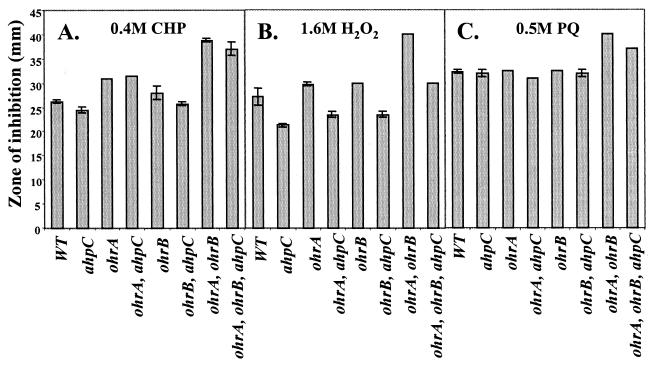
Disk diffusion assays were used to determine if OhrA and OhrB protect cells against ROS and to determine if these functions are redundant with AhpCF. Mutation of ohrA, but not ohrB or ahpC, leads to significantly increased sensitivity to CHP (Fig. 1A) and tert-butyl hydroperoxide (data not shown). The ohrA ohrB double mutant displays much greater sensitivity to CHP than either single mutant, suggesting that both proteins are involved in CHP detoxification and that lack of one can be partially compensated for by the presence of the other. In contrast, AhpCF does not appear to play a significant role in CHP resistance, a finding consistent with our previous studies. In all four strains containing an ahpC mutation, resistance to CHP is not significantly altered relative to the control strain (Fig. 1A). Thus, even in the absence of both OhrA and OhrB, AhpCF still does not play a measurable role in CHP resistance. These strains all lack AhpCF function since, as reported previously (7), mutation of ahpC leads to derepression of catalase and a consequent increase in H2O2 resistance (Fig. 1B). In addition to greatly increased sensitivity to CHP, the ohrA ohrB double mutant also displays a striking sensitivity to both H2O2 (Fig. 1B) and the superoxide-generating compound, paraquat (Fig. 1C).
FIG. 1.
Roles of OhrA, OhrB, and AhpCF in protection against ROS. The sensitivity of each indicated strain was measured as a zone of growth inhibition in a disk diffusion assay. Filters contained either 0.4 M CHP (A), 1.6 M H2O2 (B), or 0.5 M paraquat (C). The data shown are representative of three experiments. The error bars indicate the standard deviations from duplicate samples. PQ, paraquat.
Transcriptional regulation of ohrA and ohrB.
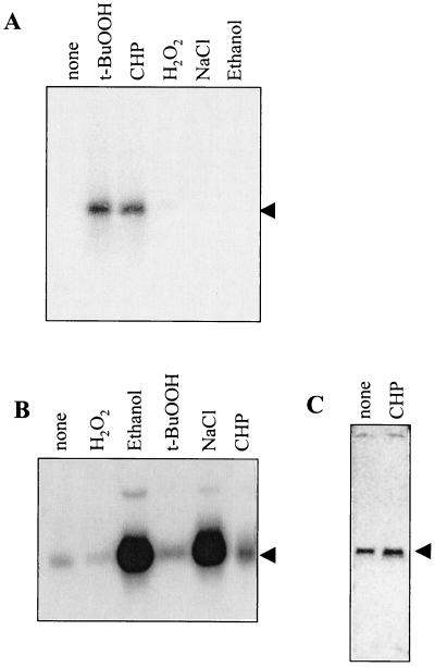
Northern blot analysis of CU1065 RNA isolated after exposure to various stresses demonstrates that ohrA is strongly induced by tert-butyl hydroperoxide and CHP, but not by H2O2, ethanol, or salt (Fig. 2A). In contrast, ohrB is strongly induced by ethanol or salt (Fig. 2B), a result consistent with the data of Volker et al. (38). It is also weakly inducible by tert-butyl hydroperoxide and CHP (Fig. 2B).
FIG. 2.
Northern analysis ohr region genes. Expression of ohrA (A), ohrB (B), and ohrR (C) was measured using 10 μg of total RNA from each sample separated on a 1% formaldehyde gel. RNA was transferred to a nylon membrane and hybridized with a radiolabeled DNA fragment containing the coding region of each gene. Arrows indicate the major transcript of each gene. Cells were either uninduced (none) or were treated with 100 μM CHP, 100 μM tert-butyl hydroperoxide (t-BuOOH), 100 μM H2O2, 4% ethanol, or 4% NaCl for 15 min as indicated.
The regulation of ohrA by organic peroxides was also confirmed in primer extension experiments. A major ohrA transcript was found in cells induced with tert-butyl hydroperoxide and corresponds to a candidate ςA-dependent promoter (Fig. 3). This inducible transcript corresponds to the transcript previously described for the ohrA gene (38). The constitutive signal corresponding to an apparent start site further upstream may be due to readthrough transcripts from the upstream proBA operon: this signal may result from reverse transcriptase pausing or termination at the base of the proBA terminator stem-loop. Readthrough from this upstream operon is consistent with the observation that ohrA expression is enhanced in minimal medium (38).
FIG. 3.
Primer extension analysis of the ohrA promoter. Cells were grown and treated as described for Fig. 2 prior to RNA isolation. The major alkyl peroxide responsive transcriptional start point for the ohrA gene corresponds to position −27 relative to the start codon, in agreement with previously published start site mapping data (38). The origin of the larger band is not clear, but may be due to readthrough transcription from the upstream proAB operon.
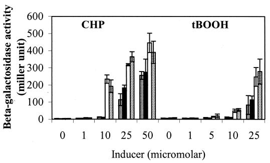
The induction of ohrA by organic peroxides was also confirmed using transcriptional reporter fusions (Table 2 and Fig. 4). With the pMUTIN derived transcriptional fusion, ohrA-lacZ expression can be induced ∼100-fold by either CHP or tert-butyl hydroperoxide (Fig. 4). Similar regulation is also seen when a 219-bp region containing the ohrA promoter is used to generate a lacZ fusion inserted ectopically in SPβ (Table 2). This suggests that all necessary cis-regulatory elements are present within this DNA fragment.
TABLE 2.
β-Galactosidase activity of ohrA and ohrR transcription fusion in wild-type and ohrR backgrounds
| Strain | Genotype
|
Mean
β-Galactosidase activity ± SD (Miller unit)a
|
||
|---|---|---|---|---|
| Mutation | Reporter | Uninduced | 100 μM CHP | |
| HB2012 | None | ohrA | 3.44 ± 0.09 | 90.47 ± 1.35 |
| HB2014 | ohrR | ohrA | 513.04 ± 19.58 | 520.31 ± 16.43 |
| HB2011 | None | ohrR | 3.65 ± 0.19 | 3.73 ± 0.12 |
| HB2013 | ohrR | ohrR | 4.27 ± 0.14 | 4.46 ± 0.24 |
| HB2031 | None | ohrA* | 128.55 ± 4.12 | ND |
| HB2044 | ohrR | ohrA* | 230.37 ± 31.22 | ND |
ND, not done.
FIG. 4.
Effect of an ahpC mutation on induction of ohrA by organic hydroperoxides. β-Galactosidase activities were assayed in various mutants (bars: gray, ohrA, HB574; black, ohrA ohrB, HB2003; white, ohrA ahpC, HB2008; cross-hatched, ohrA ohrB ahpC, HB2010). Cells were grown to mid-log phase, and various concentrations of CHP or tert-butyl hydroperoxide (tBOOH) were added to the cultures for 15 min at 37°C with shaking. The data shown are representative of triplicate determinations.
Although AhpCF, at the levels present under these growth conditions, does not contribute significantly to protection against the killing action of CHP (Fig. 1A) or tert-butyl hydroperoxide (data not shown), AhpCF can reduce these compounds in vivo. This is apparent since the ohrA promoter can be induced by CHP and tert-butyl hydroperoxide at lower concentrations in strains carrying an ahpC mutation (Fig. 4). Note that these experiments were performed using the pMUTIN derived ohrA-lacZ fusion, so all strains are also mutant for ohrA.
OhrR is a repressor of ohrA.
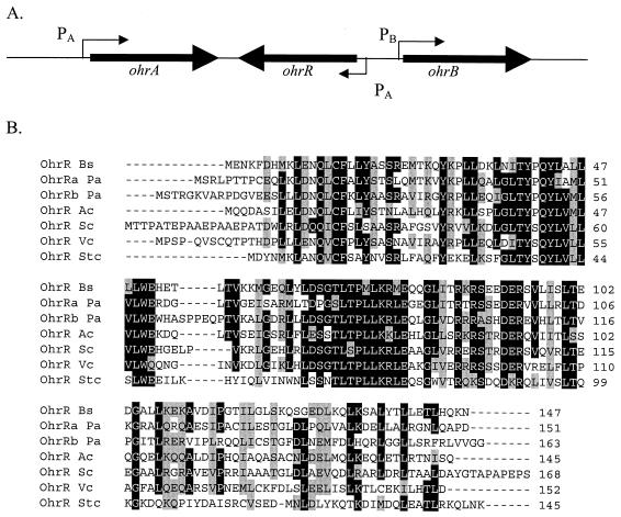
The ohrA and ohrB genes are transcribed in the same direction and are separated by ohrR (formerly ykmA), which is transcribed in the opposite direction and encodes a member of the MarR family of transcriptional repressors (Fig. 5). This proximity makes OhrR a good candidate for a regulator of ohrA and/or ohrB. In addition, an OhrR family member is known to repress ohr expression in X. campestris (S.M., unpublished data).
FIG. 5.
ohrR encodes a MarR-like repressor of ohrA. (A) Schematic of the ohrA ohrR ohrB region. PA indicates a ςA-dependent promoter element; PB indicates a ςB-dependent promoter. (B) alignment of OhrR with other closely related MarR family members. The abbreviations used are as follows (strain; GenBank accession number): OhrR Bs (B. subtilis; E69857), OhrRa Pa (P. aeruginosa PAO1; D83290), OhrRb Pa (P. aeruginosa PAO1; G83292), OhrR Ac (Acinetobacter sp. strain ADP1; CAA70318), OhrR Sc (Streptomyces coelicolor; CAB87337); OhrR Vc (Vibrio cholerae group O1 strain N16961; B82389), and OhrR Stc (Staphylococcus sciuri strain ATCC 29062). The amino acid sequences were aligned (using CLUSTALW) and conserved residues highlighted using the BoxShade utility.
To determine if OhrR is a transcriptional regulator of ohrA and/or ohrB, β-galactosidase activity was measured in wild-type (HB2012) and ohrR mutant (HB2014) cells harboring an ohrA-cat-lacZ transcriptional fusion carried at SPβ (Table 2). The >100-fold upregulation of ohrA in the ohrR mutant was also confirmed in strains constructed using the pMUTIN integrational vector (which are additionally mutant for ohrA). The β-galactosidase activity in cells harboring ohrA-lacZ and an ohrR mutation (HB2001) was very high (∼2,500 U) compared to cells harboring ohrA-lacZ alone (HB574) (∼6 U). In contrast, mutation of ohrR did not greatly affect the level of expression of the ohrB-lacZ fusion, which is very low in growing cells (1 to 2 U). These data demonstrate that mutation of ohrR is sufficient for derepression of ohrA, but not ohrB.
There is no significant increase in ohrR-cat-lacZ activity in ohrR versus wild-type cells (Table 2), suggesting that OhrR is not autoregulated. Moreover, expression of the ohrR-cat-lacZ fusion did not respond to CHP treatment (Table 2), a finding consistent with the slight response to CHP (1.3-fold induction) observed in the Northern analysis of ohrR mRNA (Fig. 2C).
Putative binding site of OhrR.
Inspection of the ohrA promoter region reveals possible binding motifs for OhrR. The ohrA promoter region contains one perfect inverted repeat (TACAATT-AATTGTA) and an adjacent imperfect repeat with three mismatches (Fig. 6A). Alternatively, this region may be viewed as an 11-bp direct repeat.
FIG. 6.
Genetic identification of sequences required for OhrR-mediated repression. The perfect inverted repeat is indicated in capital letters with matching bases identified by a vertical line. (A) In the ohrA promoter, there are two adjacent inverted repeats. The first is imperfect; the second is a perfect inverted repeat (thick arrows). This region also contains two 11-bp direct repeats (thin arrows). The −10 and −35 regions are shown in boldface. (B) The sequence of the mutant promoter region (ohrA∗) is shown with a dashed line to indicate the 15-bp deletion. A new −10 element is created by the deletion. (C) A related, imperfect inverted repeat is found overlapping the ohrR promoter region.
To determine if these sequence motifs are important for OhrR-mediated repression, we selected for mutant strains that were derepressed for ohrA-cat-lacZ expression and characterized the resulting cis-acting mutants. Two independent mutants (ohrA∗) contained the identical 15-bp deletion (Fig. 6B). These mutations likely arose from unequal crossing over between the two 11-bp direct-repeat elements noted above. Remarkably, this deletion also removes the native −10 element of the ohrA promoter but replaces this region with another sequence that closely matches the −10 consensus, thereby likely generating a new ςA-dependent promoter.
To determine if this altered promoter retains sequences that bind OhrR, the ohrA∗-cat-lacZ fusion from one representative strain (HB2031) was transduced into the ohrR mutant to generate strain HB2044. Comparison of β-galactosidase activity in the wild-type and ohrR mutant cells indicates that OhrR still exerts a small, but reproducible, repressive effect on this promoter (Table 2). This result is consistent with models in which OhrR binds to the inverted repeat sequences noted above and suggests that the imperfect inverted repeat, which is retained in the mutant promoter region, may be sufficient for mediating some repression by OhrR.
DISCUSSION
Cells have evolved numerous overlapping mechanisms to protect against the ravages of ROS (35, 36). In the case of organic hydroperoxides, the best-studied defensive enzyme is alkyl hydroperoxide reductase, encoded by the ahpCF operon. However, bacterial cells contain additional activities that are important in protection against organic peroxides, including other peroxiredoxins and, as described here, members of the Ohr family. The role of Ohr in defense against oxidative stress was first described in X. campestris pv. phaseoli (27), and recent results indicate a similar function in Pseudomonas aeruginosa (28). Ohr proteins are not obviously homologous to known peroxidases, but it is reasonable to speculate that these proteins may enzymatically detoxify peroxides. Although Ohr expression is clearly regulated, the mechanisms controlling Ohr expression have yet to be described.
We have shown that the both OhrA and OhrB contribute to organic hydroperoxide resistance. Unlike PerR regulated genes, which can be induced by either organic hydroperoxides or H2O2 (7, 8, 12, 13), ohrA responds specifically to organic hydroperoxides, and this regulation requires OhrR. Consistent with previous studies, ohrB expression responds to heat, ethanol, and salt stress as part of the ςB-dependent general stress response (Fig. 2A) (38). However, OhrB also has a role in organic hydroperoxide resistance, as shown by the increased CHP sensitivity of the ohrA ohrB double mutant (Fig. 1).
The relationship between the Ohr proteins and AhpCF is complex. Interestingly, only ohrA is under the control of OhrR. It is possible that OhrA plays the primary protective role when cells are exposed to organic hydroperoxides and OhrB is involved in detoxification of organic hydroperoxides produced during general stress. It is also possible that OhrA, OhrB, and the Ahp/TSA family members have distinct, albeit overlapping, substrate selectivities. Introduction of an ahpC mutation into the ohrA, ohrB, or ohrA ohrB strains did not increase sensitivity to organic hydroperoxides (Fig. 1), suggesting that AhpCF does not play a major role in protecting cells against the killing action of these organic hydroperoxides. The lack of a protective role for AhpCF in the present studies may result from the use of logarithmically growing cells (in which ahpCF is expressed at a low level) and the use of defined organic peroxides as the stressor. AhpCF and other genes repressed by PerR are known to be induced upon entry into stationary phase, upon starvation for iron and manganese, or in response to peroxides (7, 8, 14). In stationary-phase cells or under conditions in which both H2O2 and organic peroxides are generated, AhpCF levels would be elevated and could thereby contribute to oxidative defenses. Indeed, perR mutant cells have elevated resistance to CHP that depends on the ahpC gene (8). It is curious that AhpCF overproduction (in a perR mutant) leads to a CHP-resistant phenotype, whereas OhrA overproduction (in an ohrR mutant) does not, although OhrA is now sufficiently abundant as to be visible by Coomassie blue staining of whole-cell lysates (data not shown). Similarly, Ohr overproduction in X. campestris did not increase resistance to organic hydroperoxides (27).
The presence of two Ohr paralogs with distinct regulation is reminiscent of other genes involved in oxidative defense in B. subtilis. The katA gene is induced by ROS by virtue of its regulation by PerR, while the katB and katX genes are part of the ςB regulon (4, 5, 8, 17, 30). Similarly, PerR represses expression of the Dps homolog encoded by mrgA (12), while a second Dps homolog encoded by the dps gene is regulated by ςB (2).
Our genetic analysis defines a 15-bp region required for OhrR-mediated repression of the ohrA gene. This region includes a perfect inverted repeat, TACAATT-AATTGTA, which likely defines the OhrR binding site. Related imperfect inverted repeat sequences (three mismatches) are found in the ohrA and the ohrR promoter regions. Analysis of the ohrA∗ mutant suggests that an imperfect inverted repeat element may still allow some residual regulation by OhrR (Table 2). However, the imperfect inverted repeat overlapping the ohrR promoter does not appear to mediate repression, since we found no evidence for ohrR autoregulation (Table 2).
OhrA and OhrB are representative of a large family of conserved proteins found throughout the Bacterial domain (3). Our data lend further support to the suggestion that these proteins function in protecting cells against organic peroxides. Moreover, since ohr homologs are often found closely associated with an ohrR-like gene (3), the mechanism of regulation described here may also be conserved. Thus, OhrR is a novel type of organic peroxide-sensing transcription factor and represents a third regulator (together with PerR and ςB) involved in oxidative stress responses in B. subtilis.
ACKNOWLEDGMENTS
This study is based upon work supported by the National Science Foundation under grant MCB-9630411 (to J.D.H.), a grant from the Chulabhorn Research Institute to the Laboratory of Biotechnology, grants to S.M. from the Thai Research Fund (BRG/10/2543), and a career development award (RCF 01-40-0005) from NSTDA.
REFERENCES
- 1.Antelmann H, Engelmann S, Schmid R, Hecker M. General and oxidative stress responses in Bacillus subtilis: cloning, expression, and mutation of the alkyl hydroperoxide reductase operon. J Bacteriol. 1996;178:6571–6578. doi: 10.1128/jb.178.22.6571-6578.1996. [DOI] [PMC free article] [PubMed] [Google Scholar]
- 2.Antelmann H, Engelmann S, Schmid R, Sorokin A, Lapidus A, Hecker M. Expression of a stress- and starvation-induced dps/pexB-homologous gene is controlled by the alternative sigma factor sigmaB in Bacillus subtilis , J. Bacteriol. 1997;179:7251–7256. doi: 10.1128/jb.179.23.7251-7256.1997. [DOI] [PMC free article] [PubMed] [Google Scholar]
- 3.Atichartpongkul, S., S. Lopraset, P. Vattanaviboon, W. Whangsuk, J. D. Helmann, and S. Mongkolsuk. Bacterial Ohr and OsmC paralogs define two protein families with distinct functions and patterns of expression. Microbiology, in press. [DOI] [PubMed]
- 4.Bagyan I, Casillas-Martinez L, Setlow P. The katX gene, which codes for the catalase in spores of Bacillus subtilis, is a forespore-specific gene controlled by sigmaF, and KatX is essential for hydrogen peroxide resistance of the germinating spore. J Bacteriol. 1998;180:2057–2062. doi: 10.1128/jb.180.8.2057-2062.1998. [DOI] [PMC free article] [PubMed] [Google Scholar]
- 5.Bol D K, Yasbin R E. Analysis of the dual regulatory mechanisms controlling expression of the vegetative catalase gene of Bacillus subtilis. J. Bacteriol. 1994;176:6744–6748. doi: 10.1128/jb.176.21.6744-6748.1994. [DOI] [PMC free article] [PubMed] [Google Scholar]
- 6.Bouvier J, Gordia S, Kampmann G, Lange R, Hengge-Aronis R, Gutierrez C. Interplay between global regulators of Escherichia coli: effect of RpoS, Lrp and H-NS on transcription of the gene osmC. Mol. Microbiol. 1998;28:971–980. doi: 10.1046/j.1365-2958.1998.00855.x. [DOI] [PubMed] [Google Scholar]
- 7.Bsat N, Chen L, Helmann J D. Mutation of the Bacillus subtilis alkyl hydroperoxide reductase (ahpCF) operon reveals compensatory interactions among hydrogen peroxide stress genes. J Bacteriol. 1996;178:6579–6586. doi: 10.1128/jb.178.22.6579-6586.1996. [DOI] [PMC free article] [PubMed] [Google Scholar]
- 8.Bsat N, Herbig A, Casillas-Martinez L, Setlow P, Helmann J D. Bacillus subtiliscontains multiple Fur homologues: identification of the iron uptake (Fur) and peroxide regulon (PerR) repressors. Mol Microbiol. 1998;29:189–198. doi: 10.1046/j.1365-2958.1998.00921.x. [DOI] [PubMed] [Google Scholar]
- 9.Carmel-Harel O, Storz G. Roles of the glutathione- and thioredoxin-dependent reduction systems in the Escherichia coli and Saccharomyces cerevisiaeresponses to oxidative stress. Annu Rev Microbiol. 2000;54:439–461. doi: 10.1146/annurev.micro.54.1.439. [DOI] [PubMed] [Google Scholar]
- 10.Cha M K, Kim H K, Kim I H. Mutation and mutagenesis of thiol peroxidase of Escherichia coliand a new type of thiol peroxidase family. J Bacteriol. 1996;178:5610–5614. doi: 10.1128/jb.178.19.5610-5614.1996. [DOI] [PMC free article] [PubMed] [Google Scholar]
- 11.Chae H Z, Robison K, Poole L B, Church G, Storz G, Rhee S G. Cloning and sequencing of thiol-specific antioxidant from mammalian brain: alkyl hydroperoxide reductase and thiol-specific antioxidant define a large family of antioxidant enzymes. Proc Natl Acad Sci USA. 1994;91:7017–7021. doi: 10.1073/pnas.91.15.7017. [DOI] [PMC free article] [PubMed] [Google Scholar]
- 12.Chen L, Helmann J D. Bacillus subtilisMrgA is a Dps(PexB) homologue: evidence for metalloregulation of an oxidative-stress gene. Mol Microbiol. 1995;18:295–300. doi: 10.1111/j.1365-2958.1995.mmi_18020295.x. [DOI] [PubMed] [Google Scholar]
- 13.Chen L, Keramati L, Helmann J D. Coordinate regulation of Bacillus subtilisperoxide stress genes by hydrogen peroxide and metal ions. Proc Natl Acad Sci USA. 1995;92:8190–8194. doi: 10.1073/pnas.92.18.8190. [DOI] [PMC free article] [PubMed] [Google Scholar]
- 14.Chen L, Xie Q W, Nathan C. Alkyl hydroperoxide reductase subunit C (AhpC) protects bacterial and human cells against reactive nitrogen intermediates. Mol Cell. 1998;1:795–805. doi: 10.1016/s1097-2765(00)80079-9. [DOI] [PubMed] [Google Scholar]
- 15.Dowds B C. The oxidative stress response in Bacillus subtilis. FEMS Microbiol Lett. 1994;124:255–263. doi: 10.1111/j.1574-6968.1994.tb07294.x. [DOI] [PubMed] [Google Scholar]
- 16.Ellis H R, Poole L B. Roles for the two cysteine residues of AhpC in catalysis of peroxide reduction by alkyl hydroperoxide reductase from Salmonella typhimurium. Biochemistry. 1997;36:13349–13356. doi: 10.1021/bi9713658. [DOI] [PubMed] [Google Scholar]
- 17.Engelmann S, Lindner C, Hecker M. Cloning, nucleotide sequence, and regulation of katE encoding a sigma B-dependent catalase in Bacillus subtilis. J Bacteriol. 1995;177:5598–5605. doi: 10.1128/jb.177.19.5598-5605.1995. [DOI] [PMC free article] [PubMed] [Google Scholar]
- 18.Gordia S, Gutierrez C. Growth-phase-dependent expression of the osmotically inducible gene osmC of Escherichia coliK-12. Mol Microbiol. 1996;19:729–736. doi: 10.1046/j.1365-2958.1996.418945.x. [DOI] [PubMed] [Google Scholar]
- 19.Guerout-Fleury A M, Shazand K, Frandsen N, Stragier P. Antibiotic-resistance cassettes for Bacillus subtilis. Gene. 1995;167:335–336. doi: 10.1016/0378-1119(95)00652-4. [DOI] [PubMed] [Google Scholar]
- 20.Gutierrez C, Devedjian J C. Osmotic induction of gene osmC expression in Escherichia coliK12. J Mol Biol. 1991;220:959–973. doi: 10.1016/0022-2836(91)90366-e. [DOI] [PubMed] [Google Scholar]
- 21.Henriques A O, Melsen L R, Moran C P., Jr Involvement of superoxide dismutase in spore coat assembly in Bacillus subtilis. J Bacteriol. 1998;180:2285–2291. doi: 10.1128/jb.180.9.2285-2291.1998. [DOI] [PMC free article] [PubMed] [Google Scholar]
- 22.Inaoka T, Matsumura Y, Tsuchido T. Molecular cloning and nucleotide sequence of the superoxide dismutase gene and characterization of its product from Bacillus subtilis. J. Bacteriol. 1998;180:3697–3703. doi: 10.1128/jb.180.14.3697-3703.1998. [DOI] [PMC free article] [PubMed] [Google Scholar]
- 23.Inaoka T, Matsumura Y, Tsuchido T. SodA and manganese are essential for resistance to oxidative stress in growing and sporulating cells of Bacillus subtilis. J. Bacteriol. 1999;181:1939–1943. doi: 10.1128/jb.181.6.1939-1943.1999. [DOI] [PMC free article] [PubMed] [Google Scholar]
- 24.Jacobson F S, Morgan R W, Christman M F, Ames B N. An alkyl hydroperoxide reductase from Salmonella typhimuriuminvolved in the defense of DNA against oxidative damage. Purification and properties. J Biol Chem. 1989;264:1488–1496. [PubMed] [Google Scholar]
- 25.Li Calzi M, Poole L B. Requirement for the two AhpF cystine disulfide centers in catalysis of peroxide reduction by alkyl hydroperoxide reductase. Biochemistry. 1997;36:13357–13364. doi: 10.1021/bi9713660. [DOI] [PubMed] [Google Scholar]
- 26.Miller J H. Experiments in molecular genetics. Cold Spring Harbor, N.Y: Cold Spring Harbor Laboratory; 1972. [Google Scholar]
- 27.Mongkolsuk S, Praituan W, Loprasert S, Fuangthong M, Chamnongpol S. Identification and characterization of a new organic hydroperoxide resistance (ohr) gene with a novel pattern of oxidative stress regulation from Xanthomonas campestrispv. phaseoli. J. Bacteriol. 1998;180:2636–2643. doi: 10.1128/jb.180.10.2636-2643.1998. [DOI] [PMC free article] [PubMed] [Google Scholar]
- 28.Ochsner U, Hassett D J, Vasil M L. Genetic and physiological characterization of ohr, encoding a protein involved in organic hydroperoxide resistance in Pseudomonas aeruginosa. J Bacteriol. 2001;183:773–778. doi: 10.1128/JB.183.2.773-778.2001. [DOI] [PMC free article] [PubMed] [Google Scholar]
- 29.Park S G, Cha M K, Jeong W, Kim I H. Distinct physiological functions of thiol peroxidase isoenzymes in Saccharomyces cerevisiae. J Biol Chem. 2000;275:5723–5732. doi: 10.1074/jbc.275.8.5723. [DOI] [PubMed] [Google Scholar]
- 30.Petersohn A, Engelmann S, Setlow P, Hecker M. The katX gene of Bacillus subtilisis under dual control of sigmaB and sigmaF. Mol Gen Genet. 1999;262:173–179. doi: 10.1007/s004380051072. [DOI] [PubMed] [Google Scholar]
- 31.Poole L B. Flavin-dependent alkyl hydroperoxide reductase from Salmonella typhimurium. 2. Cystine disulfides involved in catalysis of peroxide reduction. Biochemistry. 1996;35:65–75. doi: 10.1021/bi951888k. [DOI] [PubMed] [Google Scholar]
- 32.Poole L B, Ellis H R. Flavin-dependent alkyl hydroperoxide reductase from Salmonella typhimurium. 1. Purification and enzymatic activities of overexpressed AhpF and AhpC proteins. Biochemistry. 1996;35:56–64. doi: 10.1021/bi951887s. [DOI] [PubMed] [Google Scholar]
- 33.Rhee S G, Kang S W, Netto L E, Seo M S, Stadtman E R. A family of novel peroxidases, peroxiredoxins. Biofactors. 1999;10:207–209. doi: 10.1002/biof.5520100218. [DOI] [PubMed] [Google Scholar]
- 34.Slack F J, Mueller J P, Sonenshein A L. Mutations that relieve nutritional repression of the Bacillus subtilisdipeptide permease operon. J Bacteriol. 1993;175:4605–4614. doi: 10.1128/jb.175.15.4605-4614.1993. [DOI] [PMC free article] [PubMed] [Google Scholar]
- 35.Storz G, Imlay J A. Oxidative stress. Curr Opin Microbiol. 1999;2:188–194. doi: 10.1016/s1369-5274(99)80033-2. [DOI] [PubMed] [Google Scholar]
- 36.Storz G, Zheng M. Oxidative stress. In: Storz G, Hengge-Aronis R, editors. Bacterial stress responses. Washington, D.C.: ASM Press; 2000. pp. 47–59. [Google Scholar]
- 37.Vander Horn P B, Zahler S A. Cloning and nucleotide sequence of the leucyl-tRNA synthetase gene of Bacillus subtilis. J. Bacteriol. 1992;174:3928–3935. doi: 10.1128/jb.174.12.3928-3935.1992. [DOI] [PMC free article] [PubMed] [Google Scholar]
- 38.Volker U, Andersen K K, Antelmann H, Devine K M, Hecker M. One of two osmC homologs in Bacillus subtilisis part of the sigmaB-dependent general stress regulon. J Bacteriol. 1998;180:4212–4218. doi: 10.1128/jb.180.16.4212-4218.1998. [DOI] [PMC free article] [PubMed] [Google Scholar]
- 39.Youngman P. Use of transposons and integrational vectors for mutagenesis and construction of gene fusions in Bacillus species. In: Harwood C R A C, editor. Molecular biological methods for Bacillus. Chichester, England: John Wiley & Sons; 1990. pp. 221–266. [Google Scholar]
- 40.Zuber P, Losick R. Role of AbrB in Spo0A- and Spo0B-dependent utilization of a sporulation promoter in Bacillus subtilis. J. Bacteriol. 1987;169:2223–2230. doi: 10.1128/jb.169.5.2223-2230.1987. [DOI] [PMC free article] [PubMed] [Google Scholar]


