PLOS ONE logoLink to PLOS ONE
. 2022 Oct 4;17(10):e0275315. doi: 10.1371/journal.pone.0275315

The intervertebral disc during growth: Signal intensity changes on magnetic resonance imaging and their relevance to low back pain

Teija Lund 1,‡,*, Dietrich Schlenzka 2,, Martina Lohman 3,#, Leena Ristolainen 2,#, Hannu Kautiainen 4,#, Erkko Klemetti 2,, Kalevi Österman 2,
Editor: Alejandro A Espinoza Orías5
PMCID: PMC9531821  PMID: 36194584

Abstract

Life-time prevalence of low back pain (LBP) in children and adolescents varies from 7% to 72%. Disc changes on magnetic resonance imaging (MRI) have been reported in up to 44% of children with earliest observations around pre-puberty. In this longitudinal cohort study, our objective was to determine the natural history of disc changes from childhood to early adulthood, and the possible association of these changes to LBP. Healthy 8-year-old schoolchildren were recruited for this longitudinal study consisting of a semi-structured interview, a clinical examination, and an MRI investigation at the age of 8–9 (Y8), 11–12 (Y12) and 18–19 (Y19) years. The interview inquired about LBP without trauma. T2-weighted sagittal MRI of the lumbar spine was acquired. Life-long prevalence of LBP was determined, and the disc signal intensity (SI) at the three lowest lumbar levels was assessed both visually using the Schneiderman classification (Bright-Speckled-Dark), and digitally using the disc to cerebrospinal fluid -SI ratio. Possible associations between SI changes and LBP were analyzed. Ninety-four of 208 eligible children were included at Y8 in 1994, 13 and 23 participants were lost to follow-up at Y12 and Y19, respectively. Prevalence of LBP increased after the pubertal growth spurt reaching 54% at Y19. On MRI, 18%, 10% and 38% of participants had disc SI changes at Y8, Y12 and Y19, respectively. No significant associations between self-reported LBP and either qualitative or quantitative disc SI changes were observed at any age. Life-time prevalence of LBP reached 54% by early adulthood. Disc SI changes on MRI traditionally labeled as degenerative were seen earlier than previously reported. Changes in disc SI were not associated with the presence of LBP in childhood, adolescence or early adulthood.

Introduction

In recent years, low back pain (LBP) in children and adolescents has been recognized as a major public health concern. What was once deemed a rare occurrence is now acknowledged a common condition with possible repercussions into adult life.

Varying prevalence of LBP in children and adolescents has been reported depending on study design, definition of LBP, recall period, and age of study participants. In a systematic literature review, the lifetime prevalence of LBP in adolescence ranged from 7 to 72% [1]. Although most children and adolescents report no LBP or low probability of LBP, 16 to 37% suffer occasional bouts, and up to 10% report repeated episodes [2]. The prevalence of LBP increases with age [1, 310] reaching adult levels by the end of puberty [5]. A clear association has been suggested between puberty and back pain [11] at least partly explained by the growth spurt [12]. Childhood LBP seems to be a significant risk factor for LBP in adulthood [1315] with reported odds ratios from 3.5 to 4 [14, 15].

The gradual loss of water content associated with “aging” of the intervertebral disc is manifested by a reduced signal intensity (SI) of the nucleus pulposus on T2-weighted magnetic resonance imaging (MRI). Several studies have reported an association between disc degeneration (DD) and LBP in adults [1618]. Although signs of DD on MRI are common in asymptomatic individuals as well [17, 18], a meta-analysis in an adult population concluded that they are more prevalent in the LBP population [19]. In pioneering research on adolescents, about one third of 15-year-olds were found to have at least one degenerated disc on MRI [20, 21]. While no significant difference in the prevalence of MRI findings between adolescents with or without LBP was noticed in the earliest studies [20], those with DD at an early age seemed to be at greater risk of having recurrent LBP in the future [21, 22]. In a more recent study, 35% of 13- to 20-year-old subjects presented with DD on MRI, and almost one third of them had multilevel involvement [23]. Nearly two out of three 12-14-year-old school children demonstrated some degree of DD on MRI in a cross-sectional study of 439 subjects [24]. A recent meta-analysis established a pooled prevalence of DD on MRI in 44% and 22% of adolescents with or without LBP, respectively [25].

Previous research has shown that degenerative changes of lumbar intervertebral discs are common in adolescents after the pubertal growth spurt. Little is known about the onset and development of these changes during growth, or their relevance to the clinical symptom of LBP. In the present study, our primary objectives were 1) to explore the SI changes in lumbar intervertebral discs on MRI during growth in a group of healthy school children, and 2) to investigate whether these changes were correlated to the clinical symptom of LBP.

Methods

Subject recruitment and study flow (Fig 1)

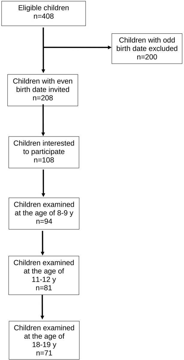
Fig 1. Flow diagram of the study.

Fig 1

In 1994, we aimed to recruit a cohort of 100 school children for a longitudinal follow-up study on the natural history of lumbar intervertebral discs in healthy children. Six elementary schools from a total of 71 were randomly chosen from the urban capital area of Helsinki. All 2nd graders with an even birth date were invited to participate via a letter to their parents. Using this criterium, 208 out of 408 children were eligible; of them 108 were interested in participating. The baseline examination was performed at the age of 8–9 years (Y8) with follow-up examinations at the ages of 11–12 years (Y12) and 18–19 years (Y19). All time points included a semi-structured interview, a clinical examination, and an MRI investigation of the lumbar spine.

This study was conducted according to the Declaration of Helsinki for research on human participants. The ethical approval was granted by the Ethics Committee of the Invalid Foundation on January 22, 1993; the study was registered to the Research Institute of the Invalid Foundation (later Research Institute Orton) on April 7, 1993. The study protocol was approved by the authorities of the Helsinki school district on August 30, 1993. A written informed consent from the parents of each child was obtained before commencement of the study.

Semi-structured interview

In the semi-structured interview, the participants were asked whether they had ever had LBP without associated trauma. At Y19, the interview included more detailed information about the LBP (last week/last month/last year/earlier) and recorded a possible contact to a physician for the LBP symptom. To precisely localize the anatomical area of interest the participants were shown a body map highlighting the lumbar area. At Y19, the participants were asked whether they smoked, and if yes, how many cigarettes per day for how long.

Clinical examination

The subject’s height and weight were measured with a stadiometer and a balance-beam scale, respectively. Body Mass Index (BMI) for Y8 and Y12 was defined using the ISO-BMI formula taking into account the child´s age and gender; for Y19 we used the standard formula of BMI calculation for adults (weight in kilograms divided by the square of the height in meters). The clinical examination focused on identifying signs of scoliosis, functional leg length inequality, hamstring tightness, and reflex abnormalities.

MRI investigation

At Y8 and Y12, the MRI investigation was obtained with a high-field 1.0T scanner (Siemens Magnetom, Siemens, Erlangen, Germany) using a dedicated spine coil. Only T2-weighted sagittal images were acquired with the following imaging parameters: TR 2500 ms, TE 80/15 ms, FOV 260, image matrix 256x256, slice thickness 4.0 mm, slice interval 4.4 mm, acq 1. At Y19, the MRI investigation was performed with a high-field 1.5T MRI scanner and a dedicated spine coil (Siemens Symphony, Siemens, Erlangen, Germany). The imaging parameters were as follows: TR 4630 ms, TE 107ms, FOV 280, image matrix 384x288, slice thickness 4.0 mm, slice interval 4.4 mm, acq.2.

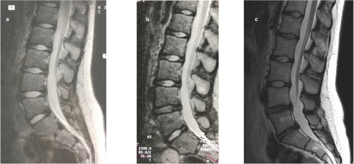
The SI of the three lowest intervertebral discs (L3/L4, L4/L5 and L5/S1) was assessed both qualitatively and quantitatively from sagittal T2-weighted midline images. For the qualitative visual evaluation, a modified Schneiderman classification [26] was used. The discs were categorized as Bright with a preserved SI, Speckled with a heterogeneously decreased SI, or Dark with a diffuse loss of SI (Fig 2). A musculoskeletal radiologist (third author) and a spine surgeon (first author) independently graded the discs without any knowledge of the subject´s clinical characteristics. In case of discrepancy, the assessment of the third evaluator (second author) was used for consensus. This turned out to be necessary in 38 of the 732 discs (5.2%). The inter-rater agreement (Scott/Fleiss agreement coefficients with ordinal weights) for the three intervertebral levels combined at Y19 (with the highest prevalence of disc changes) ranged from 0.70 to 0.89 describing a substantial to almost perfect agreement [27].

Fig 2. The evolution of disc changes throughout the study period in one study participant.

Fig 2

The MRI images illustrate the visual assessment of the signal intensity (SI) of the intervertebral disc using the Schneiderman classification. At Y8, all lumbar discs presented with a Bright nucleus pulposus (a). At Y12, the L4/L5 disc was graded Speckled (b), and at Y19, both L4/L5 and L5/S1 discs were graded Dark (c).

The SI of the intervertebral disc was assessed quantitatively by a computerized method with a region of interest (ROI) marked digitally from each nucleus pulposus. The ROI in each individual disc was marked using a freehand technique according to the estimated area of the nucleus pulposus. As an internal reference the SI of the adjacent cerebrospinal fluid (CSF) was used for a disc to CSF -SI ratio [28]. For the ROI of the CSF at every level, the area in the anterior dural sac immediately posterior to the disc was chosen to exclude the effect of the nerve roots. The measurements were performed by a musculoskeletal radiologist (third author) and a physician (sixth author) trained with the measurement technique.

Data analysis

The descriptive statistics are presented as means with standard deviations (SD) or as counts with percentages. Repeated measures were analysed using generalising estimating equations (GEE) models with the unstructured correlation structure. Generalized estimating equations were developed as an extension of the general linear model (e.g., OLS regression analysis) to analyze longitudinal and other correlated data. GEE models take into account the correlation between repeated measurements in the same subject; models do not require complete data and can be fit even when individuals do not have observations at all time points. In case of violation of the assumptions (e.g., non-normality) for continuous variables, a bootstrap-type method or Monte Carlo p values (small number of observations) for categorical variables were used. The normality of variables was evaluated graphically and by using the Shapiro–Wilk W test. No adjustment was made for multiple testing. Stata 17.0 (StataCorp LP; College Station, Texas, USA) statistical package was used for the analysis.

Results

Of the 108 children expressing interest in the study, 94 (46 females and 48 males) eventually participated. Two children did not want to go through the MRI investigation at Y8 but were included in the analysis with clinical data and MRI investigations at the two later time points. At Y12 and Y19, 81 and 71 study subjects participated in the interview, clinical examination and MRI resulting in 86% and 76% follow-up, respectively.

Table 1 gives a more detailed description of our study participants. The only statistically significant difference between sexes was seen at Y19 when males were significantly taller and weighted more than females (p<0.001). The growth for females between Y8 and Y12 was 21 cm (95% CI: 18 to 23; p<0.001) for a relative growth of 1.16 (95% CI: 1.13 to 1.18); and between Y12 and Y19 13 cm (95% CI: 10 to 17; p<0.001) for a relative growth of 1.09 (95% CI: 1.07–1.11). For males the growth between Y8 and Y12 was 19 cm (95% CI: 15 to 22, p<0.001) for a relative growth of 1.14 (95% CI: 1.11 to 1.16), and between Y12 and Y19 28 cm (95% CI: 25 to 31, p<0.001) for a relative growth of 1.18 (95% CI: 1.16 to 1.21). Between Y12 and Y19 the relative growth of males was significantly more than that of females (p<0.001).

Table 1. Description of study participants.

Y8 (N = 94) Y12 (N = 81) Y19 (N = 71)
Mean age (SD), y 8.5(0.4) 11.9 (0.5) 19.3 (0.6)
Female: Male, N 46:48 39:42 35:36
Mean height (SD), cm
Female 131 (6) 152 (8) 165 (7)
Male 133 (7) 152 (8) 180* (7)
Mean weight (SD), kg
Female 28.5 (6.0) 42.0 (9.9) 60.8 (12.0)
Male 28.8 (5.5) 43.0 (10.7) 73.7* (17.2)
Mean BMI (SD)
Female 16.4 (2.5) 18.1 (3.3) 22.3 (4.3)
Male 16.1 (2.2) 18.5 (3.4) 22.8 (5.1)
LBP, % (95% CI)
All** 6 (2 to 13) 13 (7 to 23) 54 (42 to 66)
Female 9 (2 to 21) 8 (2 to 20) 56 (38 to 73)
Male 4 (1 to 15) 19 (9 to 34) 53 (35 to 70)

*p<0.001 according to sex

**p<0.001 for all participants according to age

Occurrence of LBP

By the age of 19, 54% of the participants had experienced LBP without associated trauma. The increase in occurrence of LBP with age for the whole study population was statistically significant (p<0.001). Table 1 for the occurrence of LBP at different study time points.

Visual assessment of disc changes and their correlation to LBP

In general, the L3/L4 disc remained stable throughout the study period; while some changes were noticed at the L4/L5 disc, most of the changes developed in the L5/S1 disc. Progression of at least one grade at L4/L5 and L5/S1 was demonstrated in 12% and 20% of participants, respectively, from Y8 to Y19. Specifically, at Y8, 21 of the 276 discs (7.6%) were graded as Speckled in 17 participants (18%); four participants had two-level involvement. Nine discs (3.7%) demonstrated Speckled pattern at Y12 in eight participants with two-level involvement in one participant. By Y19, 37 of the 213 discs (17.4%) demonstrated changes with 11 discs (5.2%) graded as Dark. Disc changes were noticed in 27 participants (38%) with two-level involvement in eight participants and one participant with a three-level involvement. Two participants had Dark discs both at L4/L5 and L5/S1 levels.

No association was found between visual assessment of the SI (Bright—Speckled—Dark) and LBP when the most degenerated disc at any level was selected for analysis (Table 2).

Table 2. Association of the visual assessment of the most degenerated disc to self-reported LBP.

No LBP N (%) LBP N (%) p-value
Schneiderman score
Y8 0.91
Bright 70 (81) 5 (83)
Speckled 16 (19) 1 (17)
Dark 0 0
Y12 0.93
Bright 63 (90) 10 (91)
Speckled 7 (10) 1 (9)
Dark 0 0
Y19 0.98
Bright 20 (61) 24 (63)
Speckled 10 (30) 8 (21)
Dark 3 (9) 6 (16)

Disc to cerebrospinal fluid -SI ratio and its association to LBP

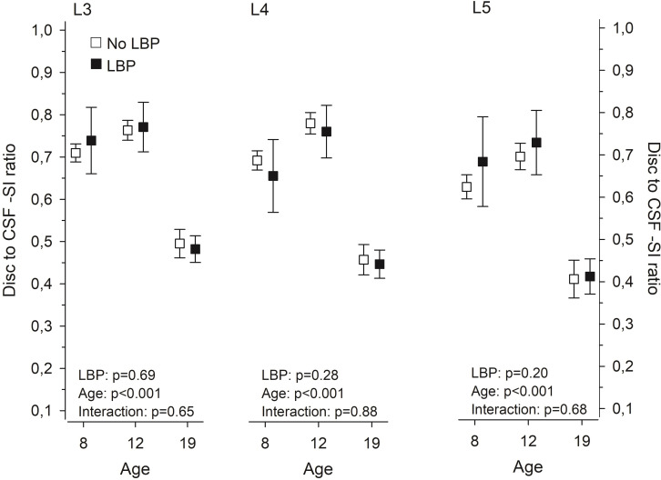
In the disc to CSF -SI ratio, no significant changes were noticed between Y8 and Y12, whereas the ratio markedly decreased by Y19 (Fig 3). No statistically significant difference in the disc to CSF -SI ratio between participants with or without LBP was noticed at any level as illustrated in Fig 3.

Fig 3. Computerized measurement of the disc signal intensity (SI) to cerebrospinal fluid SI–ratio at 8, 12, and 19 years in subjects with or without LBP.

Fig 3

Means with whiskers representing 95 per cent confidence intervals.

At Y19, BMI showed a statistically significant association to the disc to CSF -SI ratio at the L5/S1 level (p = 0.035); no other statistically significant associations emerged at any of the disc levels at the different study time points (Table 3).

Table 3. The Spearman correlation between BMI and Disc to CSF -SI ratio.

Y8 Y12 Y19
(N = 92) (N = 81) (N = 71)
r (95% CI) r (95% CI) r (95% CI)
L3/L4 0.16 (-0.05 to 0.28) -0.03 (-0.25 to 0.19) 0.05 (-0.19 to 0.28)
L4/L5 0.00 (-0.21 to 0.21) -0.02 (-0.24 to 0.20) -0.04 (-0.27 to 0.20)
L5/S1 -0.14 (-0.33 to 0.07) -0.06 (-0.27 to 0.17) -0.25* (-0.46 to -0.02)

* p = 0.035

Discussion

In the present study, we describe the natural history of lumbar intervertebral discs (L3/L4, L4/L5 and L5/S1) from childhood to early adulthood and examine the association of changes in the disc SI to the clinical symptom of LBP. At the age of 8–9 years, 18% of the participants presented with MRI findings that have traditionally been considered early signs of degeneration. At the age of 18–19 years, 17% of the discs demonstrated SI changes in 38% of the participants. By this time 54% of the participants had experienced LBP without associated trauma. The disc SI changes did not associate with the presence of LBP in childhood, adolescence or early adulthood.

The strength of the present study lies in its design; to our knowledge, this is the first longitudinal study assessing the natural history of lumbar intervertebral discs with MRI from childhood to early adulthood. However, several limitations need to be considered when interpreting our results.

Healthy school children with an even birth date were recruited to this study. The rationale was to have children with an odd birth date as possible controls with no history of repeated inquiries about LBP. Of the 208 eligible participants, 108 expressed interest in the study. It is entirely possible that a family history of LBP affected their decision to participate. In a population-based twin study, Hestbaek et al. showed a significant genetic influence on the development of early LBP [29]. While some research has shown significant association between parental and child back pain [30, 31] with children potentially modeling the symptoms of their parents [30], others have found no evidence of learned pain behavior in children [32]. Based on a recent systematic review, up to 47% of children and adolescents report either occasional or recurrent LBP [2]. The prevalence estimates surpass 50% at the age of 18 to 20 years [5]. The prevalence of LBP in the present study, 54% by the age of 18–19 years, corresponds with previous studies on larger cohorts reporting lifetime prevalence from 40% to 79% by adolescence and early adulthood [3237].

One could speculate that repeated inquiries about the presence of LBP will result in increased reporting. We tried to avoid this by keeping the discussion of LBP limited. The participants and their parents were informed that the main purpose of the study was to investigate the growth of the lumbar spine in children (“Healthy spine in a growing child”). Moreover, self-reported LBP should be regarded with a degree of uncertainty. When studying children and adolescents, particularly about lifetime prevalence of LBP, memory decay may influence the results. Although high level of forgetfulness of previous LBP has been shown in children [3], this appears less significant for recurrent or more severe episodes [38]. It may well be that our participants had forgotten about previous less severe episodes, as at Y19 84% of participants with LBP reported pain either last week or last month. The pain intensity was not formally evaluated, but only seven participants (18% of those reporting LBP) had visited a physician suggesting moderate LBP with limited effect on the activities of daily life.

Ninety-four (94) children comprised our initial study group. At Y12, 13 participants were lost to follow-up. Four of them had mild MRI changes at Y8, and one had reported LBP without disc changes. At Y19, 23 participants were lost to follow-up. Only four of them had disc changes in the first and/or second MRI investigation; one of them had reported LBP at Y12. Three additional participants lost to follow-up had reported LBP at Y12 without disc changes. Most of the participants lost to follow-up at Y19 had not experienced LBP or demonstrated disc changes at the previous examinations.

Our study covered the evolution of MRI technology; the first two MRI investigations were performed with a 1.0T scanner and the last one with a 1.5T scanner. Some evidence suggests that the field strength of the MRI equipment does not have a significant effect on the assessment of spinal morphology [39]. Our main interest was the SI of the intervertebral disc at the three lowest lumbar levels. While SI can be assessed qualitatively and quantitatively, it is dependent on a variety of technical and patient-related factors, e.g., the distance of the object of interest (in our case the intervertebral disc) from the surface coil. As the absolute contrast and SI on each MRI slice is determined by the brightest pixel, non-standardized SI measurements would introduce a significant error when comparing different study participants and time points. To minimize the effect of confounding factors and to compare the SI within and between participants at different time points, we used the SI of the adjacent CSF as a reference for a relative SI. The adjacent CSF has the advantage of being close to the intervertebral disc and having a relatively constant SI [40, 41]. The disc to CSF -SI ratio has proven sensitive to early disc changes in young subjects [28]. All MRI investigations were performed in the morning to prevent possible diurnal variation of the SI.

No grading system for early intervertebral disc changes in children and adolescents has been introduced. We used the Schneiderman classification [26], albeit slightly modified as we did not expect advanced disc changes in our young study participants. The computerized disc to CSF -SI ratio remained relatively stable between Y8 and Y12 with a marked decrease at Y19 reflecting the results of the visual assessment.

At the age of 8–9 years, 18% of our participants presented with mild disc changes (Speckled) on T2-weighted MRI-images. By the age of 18–19 years 38% of the participants had disc changes (Speckled or Dark). A recent systematic review and meta-analysis of abnormalities in the pediatric spine on MRI demonstrated a 22% (95%CI 9%-38%) pooled prevalence of DD in children without LBP and 44% (95%CI 23%-65%) in children with LBP [25]. In a prospective cross-sectional study of 439 schoolchildren, Kjaer et al found DD on MRI in approximately one third of their 12-14-year-old participants [24]. Our results are in line with these previous studies.

In the present study, we did not find an association between disc SI changes and self-reported LBP. This is contrary to a systematic review reporting higher prevalence of DD in children with LBP [25]. It is noteworthy that the studies included in the meta-analysis were performed in a hospital setting, possibly implying a more severe symptom state and overestimation of the difference between children with and without LBP. The earliest MRI studies from the 1990s suggested that young subjects with early degenerative changes are more prone to LBP in the future [21, 22, 42]. In a cross-sectional MRI study on young adults, moderately degenerated discs were likely to be associated with more severe clinical symptoms compared to mildly degenerated discs [18]. However, DD was also found in one third of asymptomatic subjects. In the present study, no association between the most degenerated disc (defined by the Schneiderman classification) regardless of disc level and self-reported LBP was found. In a cross-sectional study of 439 12-14-year-old adolescents, most disc-related findings were only weakly associated with LBP; statistically significant associations for boys emerged in the upper and for girls in the lower lumbar levels [24]. In our study, only the three lowest lumbar levels were analyzed as significant disc changes in the upper lumbar spine in this group of young participants were unlikely.

Only T2-weighted sagittal images were obtained to reduce the scanning time in our young participants. Thus, our analysis was limited to changes in the SI of the intervertebral discs, which might have caused us to overlook other morphological changes related to LBP. For example, a more significant association between disc bulge and LBP has been suggested in younger adults compared to older subjects [19]. DD, however, is the most common structural abnormality in the pediatric spine on MRI [25]. and thus of special interest clinically.

For a clinically meaningful analysis of the evolution of disc height we would have needed a standing lumbar spine x-ray at each of the study time points. This would have exposed our participants to unnecessary radiation and was not included in the study design. Moreover, the validity of absolute disc height, alone or in combination with other measures, as an indicator of early DD has been questioned [43]. Pfirrmann et al., in their MRI investigation of 70 asymptomatic adults, concluded that the association of DD and disc height was stronger in older individuals compared to younger subjects [44].

For the disc to CSF -SI ratio, higher BMI at Y19 correlated statistically significantly with lower relative SI at the L5/S1 level, although the correlation was only fair. High BMI at 16 years of age has previously been shown to be associated with lumbar DD among young males [45]. In another population-based cross-sectional study, overweight or obese adolescents and young adults had more severe DD than underweight or normal-weight individuals [23]. Compared to these previous findings, our results are more in line with those of van den Heuvel et al. who found no association between increased BMI and disc SI in their 9-year-old subjects [46].

We did not perform a formal power analysis to define the number of participants. Due to financial constraints we had to restrict our study population to approximately 100 children and 300 MRI investigations; 94 children formed the initial study group, 13 and 23 participants were lost to follow-up at Y12 and Y19, respectively. Thus, it is entirely possible that due to a small number of participants our study did not have enough power to detect possible associations between disc SI changes and LBP.

Conclusions

Our study adds to the existing evidence in providing data on the natural history of intervertebral disc morphology in a group of healthy subjects from childhood to early adulthood. The prevalence of LBP increased significantly with age reaching 54% by the age of 18–19 years. Some mild disc SI changes on MRI were seen at the age of 8–9 years in 18% of our participants; after the growth spurt 38% of our 18-19-year-old participants demonstrated disc SI changes. In this small study population, these disc SI changes did not have an association with LBP. If adolescents and young adults complain of LBP without symptoms and signs of specific etiology, it is unlikely that an MRI investigation will benefit the diagnostic workup or therapy, and it may eventually lead to poorer health outcomes due to unfounded conviction that incidental and innocuous findings on MRI are the cause of pain.

Supporting information

S1 File. Supporting data set.

(XLSX)

Data Availability

All relevant data are within the article and its Supporting Information files.

Funding Statement

This research (official recipient: DS) was supported by The Research Institute of the Invalid Foundation (later Research Institute Orton) through grants by the Ministry of Social Affairs and Health in Finland (Project Identification Number A2500/465), and by the Siviä Kosti Foundation of the Invalid Foundation in Helsinki, Finland. The funders did not play any role in the study design, data collection and analysis, decision to publish, or preparation of the manuscript.

References

  • 1.Jeffries LJ, Milanese SF, Grimmer-Somers KA. Epidemiology of adolescent spinal pain: A systematic overview of the research literature. Spine. 2007;32: 2630–2637. doi: 10.1097/BRS.0b013e318158d70b [DOI] [PubMed] [Google Scholar]
  • 2.Junge T, Wedderkopp N, Boyle E, Kjaer P. The natural course of low back pain from childhood to young adulthood—a systematic review. Chiropr Man Therap. 2019;27: 10. doi: 10.1186/s12998-018-0231-x [DOI] [PMC free article] [PubMed] [Google Scholar]
  • 3.Burton AK, Clarke RD, McClune TD, Tillotson KM. The natural history of low back pain in adolescents. Spine. 1996;21: 2323–2328. doi: 10.1097/00007632-199610150-00004 [DOI] [PubMed] [Google Scholar]
  • 4.Taimela S, Kujala UM, Salminen JJ, Viljanen T. The prevalence of low back pain among children and adolescents. A nationwide, cohort-based questionnaire survey in Finland. Spine. 1997;22: 1132–1136. doi: 10.1097/00007632-199705150-00013 [DOI] [PubMed] [Google Scholar]
  • 5.Leboeuf-Yde C, Ohm Kyvik K. At what age does low back pain become a common problem? A study of 29424 individuals aged 12–41 years. Spine. 1998;23: 228–234. doi: 10.1097/00007632-199801150-00015 [DOI] [PubMed] [Google Scholar]
  • 6.Jones GT, Watson KD, Silman AJ, Symmons DPM, Macfarlane GJ. Predictors of low back pain in British schoolchildren: a population-based prospective cohort study. Pediatrics. 2003;111: 822–828. doi: 10.1542/peds.111.4.822 [DOI] [PubMed] [Google Scholar]
  • 7.Hestbaek L, Leboeuf-Yde C, Ohm Kyvik K, Vach W, Russell MB, Skadhauge, et al. Comorbidity with low back pain. A cross-sectional population-based survey of 12- to 22-year-olds. Spine. 2004;29: 1483–1491. doi: 10.1097/01.brs.0000129230.52977.86 [DOI] [PubMed] [Google Scholar]
  • 8.Sato T, Ito T, Hirano T, Morita O, Kikuchi R, Endo N, et al. Low back pain in childhood and adolescence: a cross-sectional study in Nigata City. Eur Spine J. 2008;17: 1441–1447. doi: 10.1007/s00586-008-0788-5 [DOI] [PMC free article] [PubMed] [Google Scholar]
  • 9.Kjaer P, Wedderkopp N, Korsholm L, Leboeuf-Yde C. Prevalence and tracking of back pain from childhood to adolescence. BMC Musculoskelet Disord. 2011;12: 98. doi: 10.1186/1471-2474-12-98 [DOI] [PMC free article] [PubMed] [Google Scholar]
  • 10.Joergensen AC, Hestbaek L, Andersen PK, Nybo Andersen A-M. Epidemiology of spinal pain in children: a study within the Danish National Birth Cohort. Eur J Pediatr. 2019;178: 695–706. doi: 10.1007/s00431-019-03326-7 [DOI] [PMC free article] [PubMed] [Google Scholar]
  • 11.Lardon A, Leboeuf-Yde C, Le Scanff C, Wedderkopp N. Is puberty a risk factor for back pain in the young? a systematic critical literature review. Chiropr Man Therap. 2014;22: 27. doi: 10.1186/s12998-014-0027-6 [DOI] [PMC free article] [PubMed] [Google Scholar]
  • 12.Wedderkopp N, Andersen LB, Froberg K, Leboeuf-Yde C. Back pain reporting in young girls appears to be puberty-related. BMC Musculoskelet Disord. 2005;6: 52. doi: 10.1186/1471-2474-6-52 [DOI] [PMC free article] [PubMed] [Google Scholar]
  • 13.Harreby M, Neergaard K, Hesselsøe G, Kjer J. Are radiologic changes in the thoracic and lumbar spine of adolescents risk factors for low back pain in adults? A 25-year prospective cohort study of 640 school children. Spine. 1995;20: 2298–2320. doi: 10.1097/00007632-199511000-00007 [DOI] [PubMed] [Google Scholar]
  • 14.Hestbaek L, Leboeuf-Yde C, Ohm Kyvik K. Is comorbidity in adolescence a predictor for adult low back pain? A prospective study of a young population. BMC Musculoskelet Disord. 2006;7: 29. doi: 10.1186/1471-2474-7-29 [DOI] [PMC free article] [PubMed] [Google Scholar]
  • 15.Hestbaek L, Leboeuf-Yde C, Ohm Kyvik K, Manniche C. The course of low back pain from adolescence to adulthood: eight-year follow-up of 9600 twins. Spine. 2006;31: 468–472. doi: 10.1097/01.brs.0000199958.04073.d9 [DOI] [PubMed] [Google Scholar]
  • 16.Luoma K, Riihimäki H, Luukkonen R, Raininko R, Viikari-Juntura E, Lamminen A. Low back pain in relation to lumbar disc degeneration. Spine. 2000;25: 487–492. doi: 10.1097/00007632-200002150-00016 [DOI] [PubMed] [Google Scholar]
  • 17.Cheung KMC, Karppinen J, Chan D, Ho DWH, Song Y-Q, Sham P, et al. Prevalence and pattern of lumbar magnetic resonance imaging changes in a population study of one thousand forty-three individuals. Spine. 2009;34: 934–940. doi: 10.1097/BRS.0b013e3181a01b3f [DOI] [PubMed] [Google Scholar]
  • 18.Takatalo J, Karppinen J, Niinimäki J, Taimela S, Näyhä S, Mutanen P, et al. Does lumbar disc degeneration on magnetic resonance imaging associate with low back symptom severity in young Finnish adults? Spine. 2011;36: 2180–2189. doi: 10.1097/BRS.0b013e3182077122 [DOI] [PubMed] [Google Scholar]
  • 19.Brinjikji W, Diehn FE, Jarvik JG, Carr CM, Kallmes DF, Murad MH, et al. MRI findings of disc degeneration are more prevalent in adults with low back pain than in asymptomatic controls: A systematic review and meta-analysis. Am J Neuroradiol. 2015;36: 2394–2399. doi: 10.3174/ajnr.A4498 [DOI] [PMC free article] [PubMed] [Google Scholar]
  • 20.Tertti MO, Salminen JJ, Paajanen HEK, Terho PH, Kormano MJ, et al. Low-back pain and disk degeneration in children: a case-control MR imaging study. Radiology. 1991;180: 503–507. doi: 10.1148/radiology.180.2.1829844 [DOI] [PubMed] [Google Scholar]
  • 21.Salminen JJ, Erkintalo MO, Pentti J, Oksanen A, Kormano MJ. Recurrent low back pain and early disc degeneration in the young. Spine. 1999;24: 1316–1321. doi: 10.1097/00007632-199907010-00008 [DOI] [PubMed] [Google Scholar]
  • 22.Salminen JJ, Erkintalo M, Laine M, Pentti J. Low back pain in the young. A prospective three-year follow-up study of subjects with and without low back pain. Spine. 1995;20: 2101–2108. doi: 10.1097/00007632-199510000-00006 [DOI] [PubMed] [Google Scholar]
  • 23.Samartzis D, Karppinen J, Mok F, Fong DYT, Luk KDK, Cheung KMC. A population-based study of juvenile disc degeneration and its association with overweight and obesity, low back pain and diminished functional status. J Bone Joint Surg Am. 2011;93: 662–670. doi: 10.2106/JBJS.I.01568 [DOI] [PubMed] [Google Scholar]
  • 24.Kjaer P, Leboeuf-Yde C, Solgaard Sorensen J, Bendix T. An epidemiologic study of MRI and low back pain in 13-year-old children. Spine. 2005;30: 798–806. doi: 10.1097/01.brs.0000157424.72598.ec [DOI] [PubMed] [Google Scholar]
  • 25.van den Heuvel MM, Oei EHG, Bierma-Zeinstra SMA, van Middelkoop M. The prevalence of abnormalities in the pediatric spine on MRI. A systematic review and meta-analysis. Spine. 2020;45: E1185–E1196. doi: 10.1097/BRS.0000000000003527 [DOI] [PubMed] [Google Scholar]
  • 26.Schneiderman G, Flannigan B, Kingston S, Thomas J, Dillin WH, Watkins RG. Magnetic resonance imaging in the diagnosis of disc degeneration: correlation with discography. Spine. 1987;12: 276–281. doi: 10.1097/00007632-198704000-00016 [DOI] [PubMed] [Google Scholar]
  • 27.Landis JR, Koch GG. The measurement of observer agreement for categorical data. Biometrics. 1977;33: 159–174 [PubMed] [Google Scholar]
  • 28.Videman T, Nummi P, Battie MC, Gill K. Digital assessment of MRI for lumbar disc desiccation. A comparison of digital versus subjective assessments and digital intensity profiles versus discogram and macro anatomic findings. Spine. 1994;19: 192–198. doi: 10.1097/00007632-199401001-00014 [DOI] [PubMed] [Google Scholar]
  • 29.Hestbaek L, Iachine IA, Leboeuf-Yde C, Ohm Kyvik K, Manniche C. Heredity of low back pain in a young population: a classical twin study. Twin Res. 2004;7: 16–26. doi: 10.1375/13690520460741408 [DOI] [PubMed] [Google Scholar]
  • 30.Grøholt E-K, Stigum H, Nordhagen R, Köhler L. Recurrent pain in children, socio-economic factors and accumulation in families. Eur J Epidemiol. 2003;18: 965–975. doi: 10.1023/a:1025889912964 [DOI] [PubMed] [Google Scholar]
  • 31.Balagué F, Bibbo E, Mélot C, Szpalski M, Gunzburg R, Keller TS. The association between isoinertial trunk muscle performance and low back pain in male adolescents. Eur Spine J. 2010;19: 624–632. doi: 10.1007/s00586-009-1168-5 [DOI] [PMC free article] [PubMed] [Google Scholar]
  • 32.Mustard CA, Kalcevich C, Frank JW, Boyle M. Childhood and early adult predictors of risk of incident back pain: Ontario Child Health Study 2001 follow-up. Am J Epidemiol. 2005;162: 779–786. doi: 10.1093/aje/kwi271 [DOI] [PubMed] [Google Scholar]
  • 33.Balagué F, Skovron ML, Nordin M, Dutoit G, Pol LR, Waldburger M. Low back pain in schoolchildren. A study of familial and psychological factors. Spine. 1995;20: 1265–1270. doi: 10.1097/00007632-199506000-00012 [DOI] [PubMed] [Google Scholar]
  • 34.Harreby M, Nygaard B, Jessen T, Larsen E, Storr-Paulsen A, Lindahl A, et al. Risk factors for low back pain in a cohort of 1389 Danish school children: an epidemiologic study. Eur Spine J. 1999;8: 444–450. doi: 10.1007/s005860050203 [DOI] [PMC free article] [PubMed] [Google Scholar]
  • 35.Kovacs FM, Gestoso M, Gil Del Real MT, Lopéz J, Mufraggi N, Méndez JI. Risk factors for non-specific low back pain in schoolchildren and their parents: a population based study. Pain. 2003;103: 259–268. doi: 10.1016/S0304-3959(02)00454-2 [DOI] [PubMed] [Google Scholar]
  • 36.Jones MA, Stratton G, Reilly T, Unnithan VB. A school-based survey of recurrent non-specific low-back pain prevalence and consequences in children. Health Educ Res. 2004;19: 284–289. doi: 10.1093/her/cyg025 [DOI] [PubMed] [Google Scholar]
  • 37.Poussa MS, Heliövaara MM, Seitsamo JT, Könönen MH, Hurmerinta K, Nissinen MJ. Anthropometric measurements and growth as predictors of low-back pain: a cohort study of children followed up from the age of 11 to 22 years. Eur Spine J. 2005;14: 595–598. doi: 10.1007/s00586-004-0872-4 [DOI] [PMC free article] [PubMed] [Google Scholar]
  • 38.Milanese S, Grimmer-Somers K. What is adolescent low back pain? Current definitions used to define the adolescent with low back pain. J Pain Research. 2010;3: 57–66. doi: 10.2147/jpr.s10025 [DOI] [PMC free article] [PubMed] [Google Scholar]
  • 39.Maubon AJ, Ferru J-M, Berger V, Soulage MC, DeGraef M, Aubas P, et al. Effect of field strength on MR images: Comparison of the same subject at 0.5, 1.0 and 1.5 T. RadioGraphics. 1999;19: 1057–1067. doi: 10.1148/radiographics.19.4.g99jl281057 [DOI] [PubMed] [Google Scholar]
  • 40.Luoma K, Raininko R, Nummi P, Luukkonen R. Is the signal intensity of cerebrospinal fluid constant? Intensity measurements with high and low field magnetic resonance imagers. Magn Reson Imagaging. 1993;11: 549–555. doi: 10.1016/0730-725x(93)90474-r [DOI] [PubMed] [Google Scholar]
  • 41.Luoma EK, Raininko R, Nummi PJ, Luukkonen R, Manninen HI, Riihimäki HA. Suitability of cerebrospinal fluid as a signal-intensity reference on MRI: evaluation of signal-intensity variations in the lumbosacral dural sac. Neuroradiology. 1997;39: 728–732. doi: 10.1007/s002340050496 [DOI] [PubMed] [Google Scholar]
  • 42.Paajanen H, Erkintalo M, Parkkola R, Salminen J, Kormano M. Age-dependent correlation of low-back pain and lumbar disc degeneration. Arch Orthop Trauma Surg. 1997;116: 106–107. doi: 10.1007/BF00434112 [DOI] [PubMed] [Google Scholar]
  • 43.Luoma K, Vehmas T, Riihimäki H, Raininko R. Disc height and signal intensity of the nucleus pulposus on magnetic resonance imaging as indicators of lumbar disc degeneration. Spine. 2001;26:680–686. doi: 10.1097/00007632-200103150-00026 [DOI] [PubMed] [Google Scholar]
  • 44.Pfirrmann CWA, Metzdorf A, Elfering A, Hodler J, Boos N. Effect of aging and degeneration on disc volume and shape: A quantitative study in asymptomatic volunteers. J Orthop Res. 2006;24:1086–1094. doi: 10.1002/jor.20113 [DOI] [PubMed] [Google Scholar]
  • 45.Takatalo J, Karppinen J, Taimela S, Niinimäki J, Laitinen J, Blanco Sequeiros R, et al. Body mass index is associated with lumbar disc degeneration in young Finnish males: subsample of Northern Finland birth cohort study 1986. BMC Musculoskelet Disord. 2013;14:87. doi: 10.1186/1471-2474-14-87 [DOI] [PMC free article] [PubMed] [Google Scholar]
  • 46.van den Heuvel MM, Oei EHG, Renkens JJM, Bierma-Zeinstra SMA, van Middelkoop M. Structural spinal abnormalities on MRI and associations with weight status in a general pediatric population. Spine J 2021;21:465–476. doi: 10.1016/j.spinee.2020.10.003 [DOI] [PubMed] [Google Scholar]

Decision Letter 0

Alejandro A Espinoza Orías

17 Mar 2022

PONE-D-22-02909The intervertebral disc during growth: changes on magnetic resonance imaging and their relevance to low back painPLOS ONE

Dear Dr. Lund,

Thank you for submitting your manuscript to PLOS ONE. After careful consideration, we feel that it has merit but does not fully meet PLOS ONE’s publication criteria as it currently stands. Therefore, we invite you to submit a revised version of the manuscript that addresses the points raised during the review process.

The reviewer panel is of the impression that the study does contribute to the field, especially by providing new data on natural progression of the intervertebral disc conditions in a young cohort. However, there are a few important statistical and methodological issues that need to be addressed in detail before the manuscript can be accepted for publication. Namely, a proper power of study, a detailed statistical evaluation of low back pain prevalence, consistency in MRI data, and the inclusion of clinical and morphological spinal parameters. Please refer to the reviewer comments for more details on these questions.

Please submit your revised manuscript by May 01 2022 11:59PM. If you will need more time than this to complete your revisions, please reply to this message or contact the journal office at plosone@plos.org. When you're ready to submit your revision, log on to https://www.editorialmanager.com/pone/ and select the 'Submissions Needing Revision' folder to locate your manuscript file.

Please include the following items when submitting your revised manuscript:

  • A rebuttal letter that responds to each point raised by the academic editor and reviewer(s). You should upload this letter as a separate file labeled 'Response to Reviewers'.

  • A marked-up copy of your manuscript that highlights changes made to the original version. You should upload this as a separate file labeled 'Revised Manuscript with Track Changes'.

  • An unmarked version of your revised paper without tracked changes. You should upload this as a separate file labeled 'Manuscript'.

If you would like to make changes to your financial disclosure, please include your updated statement in your cover letter. Guidelines for resubmitting your figure files are available below the reviewer comments at the end of this letter.

If applicable, we recommend that you deposit your laboratory protocols in protocols.io to enhance the reproducibility of your results. Protocols.io assigns your protocol its own identifier (DOI) so that it can be cited independently in the future. For instructions see: https://journals.plos.org/plosone/s/submission-guidelines#loc-laboratory-protocols. Additionally, PLOS ONE offers an option for publishing peer-reviewed Lab Protocol articles, which describe protocols hosted on protocols.io. Read more information on sharing protocols at https://plos.org/protocols?utm_medium=editorial-email&utm_source=authorletters&utm_campaign=protocols.

We look forward to receiving your revised manuscript.

Kind regards,

Alejandro A. Espinoza Orías, PhD

Academic Editor

PLOS ONE

Journal Requirements:

When submitting your revision, we need you to address these additional requirements.

1. Please ensure that your manuscript meets PLOS ONE's style requirements, including those for file naming. The PLOS ONE style templates can be found at 

https://journals.plos.org/plosone/s/file?id=wjVg/PLOSOne_formatting_sample_main_body.pdf and 

https://journals.plos.org/plosone/s/file?id=ba62/PLOSOne_formatting_sample_title_authors_affiliations.pdf.

2. We note that the grant information you provided in the ‘Funding Information’ and ‘Financial Disclosure’ sections do not match. 

When you resubmit, please ensure that you provide the correct grant numbers for the awards you received for your study in the ‘Funding Information’ section.

3. We noted in your submission details that a portion of your manuscript may have been presented or published elsewhere. [Our long-term follow-up (currently under review for publication) describes a subset (approx 50%) of the original study group. The analysis approaches the data from a different perspective; no text or figures in the present manuscript have been taken from the manuscript on the long-term results.] Please clarify whether this publication was peer-reviewed and formally published. If this work was previously peer-reviewed and published, in the cover letter please provide the reason that this work does not constitute dual publication and should be included in the current manuscript.

4. In your Data Availability statement, you have not specified where the minimal data set underlying the results described in your manuscript can be found. PLOS defines a study's minimal data set as the underlying data used to reach the conclusions drawn in the manuscript and any additional data required to replicate the reported study findings in their entirety. All PLOS journals require that the minimal data set be made fully available. For more information about our data policy, please see http://journals.plos.org/plosone/s/data-availability.

Upon re-submitting your revised manuscript, please upload your study’s minimal underlying data set as either Supporting Information files or to a stable, public repository and include the relevant URLs, DOIs, or accession numbers within your revised cover letter. For a list of acceptable repositories, please see http://journals.plos.org/plosone/s/data-availability#loc-recommended-repositories. Any potentially identifying patient information must be fully anonymized.

Important: If there are ethical or legal restrictions to sharing your data publicly, please explain these restrictions in detail. Please see our guidelines for more information on what we consider unacceptable restrictions to publicly sharing data: http://journals.plos.org/plosone/s/data-availability#loc-unacceptable-data-access-restrictions. Note that it is not acceptable for the authors to be the sole named individuals responsible for ensuring data access.

We will update your Data Availability statement to reflect the information you provide in your cover letter.

[Note: HTML markup is below. Please do not edit.]

Reviewers' comments:

Reviewer's Responses to Questions

Comments to the Author

1. Is the manuscript technically sound, and do the data support the conclusions?

The manuscript must describe a technically sound piece of scientific research with data that supports the conclusions. Experiments must have been conducted rigorously, with appropriate controls, replication, and sample sizes. The conclusions must be drawn appropriately based on the data presented.

Reviewer #1: Partly

Reviewer #2: Yes

**********

2. Has the statistical analysis been performed appropriately and rigorously?

Reviewer #1: No

Reviewer #2: Yes

**********

3. Have the authors made all data underlying the findings in their manuscript fully available?

The PLOS Data policy requires authors to make all data underlying the findings described in their manuscript fully available without restriction, with rare exception (please refer to the Data Availability Statement in the manuscript PDF file). The data should be provided as part of the manuscript or its supporting information, or deposited to a public repository. For example, in addition to summary statistics, the data points behind means, medians and variance measures should be available. If there are restrictions on publicly sharing data—e.g. participant privacy or use of data from a third party—those must be specified.

Reviewer #1: No

Reviewer #2: Yes

**********

4. Is the manuscript presented in an intelligible fashion and written in standard English?

PLOS ONE does not copyedit accepted manuscripts, so the language in submitted articles must be clear, correct, and unambiguous. Any typographical or grammatical errors should be corrected at revision, so please note any specific errors here.

Reviewer #1: Yes

Reviewer #2: Yes

**********

5. Review Comments to the Author

Please use the space provided to explain your answers to the questions above. You may also include additional comments for the author, including concerns about dual publication, research ethics, or publication ethics. (Please upload your review as an attachment if it exceeds 20,000 characters)

Reviewer #1: “In this longitudinal cohort study [of at least 71 children (8-19 yr)], the objective was to determine the natural history of disc changes from childhood to early adulthood, and the possible association of these changes to LBP.” Outcomes included a survey for life-long back pain and an MRI-based classification of the disc and csf. Prevalence of LBP increased with aging but was not associated to disc hydration intensity. Some key structural measures that are possible were not determined and some key statistical metrics are either not included or explicitly stated.

Conceptual comments

None.

Major technical comments:

• Two different MRIs were used in the study, where the first two scans were of Y9 and T12 and the second scanner was used for Y19. The assumption is made that the relative change between disc and CSF would account for machine/parameter/calibration differences but this assumes a linear relationship and may not be the case. Disc/csf ratios of Y9 or Y12 on the latter scanner (1.5T) must be corroborated to those in the former (1.0T)? Otherwise, the age-related effect is due to use of a different scanner and parameter differences.

• References >43 are missing.

• “The most significant increase occurred after Y12 with 54% of participants having experienced LBP. . .” but the prevalence of LBP was not compared statistically. A Chi-square or something similar is necessary to draw this conclusion with respect to age or degree intensity and is necessary for all of the nominal data in the manuscript.

• What is the disc height, disc concavity and/or spinal curvature (Cobb angle)? Are these correlated to LBP? Do they change with age?

• Does disc morphology, intensity changes or LBP with aging remain following normalization to BMI or smoking?

• The sample size for the following statement appears unclear: “The only significant finding was that all participants with a dark disc at L4/L5 level at Y19 (n=4) reported LBP (p=0.048).” In Table 2, there were n=6 participants with LBP. Secondly, what was the control group to determine this finding: Y8 or Y12 of the same participants, incidence of LBP in dark disc at Y19?

• How did incidence of LBP in an individual relate to disc morphology and hydration?

• “The prevalence of LBP increased significantly after the pubertal growth spurt reaching 54% by the age of 18-19 years.” This statement is not supported by any particular data and requires a comparison showing that the height change from 12-19 was greater than 12-9 in this cohort. Y19 smoked while all others did not. Any number of other reasons may have engendered LBP.

• Include LBP incidence per child.

Minor comments

• “At the age of 8-9 years, 18% of the participants presented with MRI findings that have traditionally been considered early signs of degeneration.” What outcome is this and from where in Table 2 is this data? A more specific statement is needed that explains that the “traditional” metric is disc intensity distribution for all children irrespective of the LBP incidence.

• What was the level of activity of the participants, e.g., sports, etc.? Many people were confined indoors because of COVID19. When were these latter data collected?

Reviewer #2: The manuscript details a very interesting study of children over a decade of life attempting to investigate relationships between low back pain and structural changes within the disc observed on MRI. The longitudinal nature of the investigation is impressive, and this data will certainly be a great contribution to the field as studies of this nature are lacking. Please see below for specific comments and suggestions:

1. Abstract – claiming that disc changes on MRI “are not associated with the presence of LBP” is somewhat misleading – the authors state that there was a significant finding that participants with a “dark” disc at L45 at Y19 reported back pain.

2. Methods - For the signal intensity characterization on MRI – what were the dimensions of the ROIs used? Or were they scaled to the size of the disc?

3. Table 1 – were there statistically significant differences in demographics at any age?

4. Were the children all from families with similar socioeconomic status? Studies have suggested socioeconomic status may be a factor in the risk for development of back pain.

5. Results – Prevalence of Back Pain – Was smoking associated with an increased prevalence of back pain? Was back pain more prevalent in males versus females at any age group? Given this large and unique data set, these points I think would be of interest to readers.

6. MRI Results – it seems data from only L3-S1 was utilized. Is there a reason the L1-2 and L2-L3 discs were excluded from analysis?

7. Results, Figure 2 – labels need to be added to this Figure, are these scans from the same patient at each time point? There does not appear to be a figure legend included.

**********

6. PLOS authors have the option to publish the peer review history of their article (what does this mean?). If published, this will include your full peer review and any attached files.

If you choose “no”, your identity will remain anonymous but your review may still be made public.

Do you want your identity to be public for this peer review? For information about this choice, including consent withdrawal, please see our Privacy Policy.

Reviewer #1: No

Reviewer #2: No

[NOTE: If reviewer comments were submitted as an attachment file, they will be attached to this email and accessible via the submission site. Please log into your account, locate the manuscript record, and check for the action link "View Attachments". If this link does not appear, there are no attachment files.]

While revising your submission, please upload your figure files to the Preflight Analysis and Conversion Engine (PACE) digital diagnostic tool, https://pacev2.apexcovantage.com/. PACE helps ensure that figures meet PLOS requirements. To use PACE, you must first register as a user. Registration is free. Then, login and navigate to the UPLOAD tab, where you will find detailed instructions on how to use the tool. If you encounter any issues or have any questions when using PACE, please email PLOS at figures@plos.org. Please note that Supporting Information files do not need this step.

PLoS One. 2022 Oct 4;17(10):e0275315. doi: 10.1371/journal.pone.0275315.r002

Author response to Decision Letter 0


6 Apr 2022

We appreciate the opportunity to re-submit our revised manuscript to PLOS ONE and are grateful for the thorough and insightful comments of your reviewers. We have addressed their comments in this response letter and made changes in the manuscript accordingly. We believe these changes have made our manuscript and our message stronger and clearer.

Editor´s comments:

We have addressed the reviewers’ comments regarding a statistical evaluation of LBP prevalence, consistency in MRI data, and the inclusion of additional clinical and morphological spinal parameters in our response below.

We did not perform a formal power analysis. When we started the study, MRI investigations were still relatively rare and significantly more expensive than nowadays. Our funding allowed us to perform three consecutive MRIs on around 100 participants (300 MRIs) which defined the size of the original study group. Although the power of our study may not be enough for any definitive conclusions, we believe the study is still unique in its design and scope.

Journal requirements:

1. We have re-checked that our manuscript meets the PLOS ONE requirements.

2. We have revised the funding information. The grants received do not have an official grant number; they are listed using a specific project number (A2500/465 for the current research project).

3. The long-term follow-up consisting of a sub-population of the original study population (reported here) has been accepted for publication in the European Spine Journal (peer-reviewed and published online before print). We have included the online version of the manuscript to this response letter as a supplemental file for your review.

4. We have included a supplemental file with a data set of the results described in the present manuscript. We continue to analyze the data and intend to report additional results in the future.

Reviewer´s comments:

Reviewer #1:

1. This is a valid concern and a possible source of error. Our study spanned the evolution of MRI technology with the first two MRI studies performed with a 1.0T scanner and the last one with a 1.5T scanner. The contrast on MRI depends on the field strength and imaging parameters; all our MRI studies were performed using a high-field scanner. Using different scanners introduces a possibility of error but standardizing the SI measurements with a specific internal reference (the adjacent CSF) in each study subject for all disc levels separately minimizes the risk. The same method has been used in the few longitudinal studies published to date using MRI scanners with different field strengths.

The brightest pixel determines the contrast and SI on each MRI slice. Thus, standardization to a tissue or liquid (such as CSF) that is homogeneous e.g., biologically and regarding the relaxation time, is mandatory. Our reference was measured separately for all disc levels on each study participant to ensure maximum accuracy. We acknowledge that non-standardized SI measurements would have introduced a significant error when comparing different study participants and time points.

In the revised manuscript, we have described our measurement technique for the relative signal intensity in more detail; moreover, we have strengthened the discussion on this topic when discussing the limitations of our study.

2. We thank the reviewer for pointing out an unfortunate mix-up in our reference list. We have addressed this, and the reference list is now correct.

3. This is an important remark by the reviewer. We have added information on the prevalence of LBP at each study time point to Table 1.

4. The decrease in disc SI on MRI is the most common finding in children and adolescents with increasing age. We decided to concentrate on this parameter in our primary analysis. No universally accepted grading system for early intervertebral disc changes in children and adolescents has been established. Our initial study design was based on the Schneiderman classification introduced in 1987; the Schneiderman classification focuses on the disc SI. We are currently in the process of re-assessing the MRIs using the Pfirrmann classification (introduced in 2001) that considers not only the disc SI but also the disc height. However, we did not expect major disc height reduction in our young study population. We did not evaluate disc concavity but recognize the wide variability of disc shapes seen in real life; however, no classification has addressed this phenomenon making its analysis difficult. The effect of spinal curvature (lumbar lordosis) on disc degeneration is an interesting question. We only had supine MRI images and thus could not evaluate the development of lumbar lordosis during growth. For a reliable and meaningful measure of lumbar lordosis we would have needed standing x-rays from our study participants in each study time point. When planning the study, we did not expect the ethical committee to accept this, and thus did not include it in our study protocol.

5. In the present manuscript, we decided to concentrate on the disc SI and its association to LBP; this decision was made consciously to keep the reporting concise and the message clear. We hope that this decision is acceptable. We have information on the BMI and smoking and are currently in the process of analyzing the data in relation to disc SI and LBP. We hope to report our findings in further manuscripts. In the present manuscript, we have deleted the information on smoking from Table 1.

6. We have revised Table 2 such that the message comes across clearer. The revised Results section also has a more detailed description of the disc findings at the three study time points. At ages 8 and 12, no dark discs were found in any of the study subjects. At age 19, dark disc at L4/L5 was seen in 4 participants and at L5/S1 in 7 participants. Two participants had dark discs both at L4/L5 and L5/S1 levels leaving 9 participants with dark discs at age 19. The only significant finding was that the 4 participants with a dark disc at L4/L5 level reported LBP.

7. We have added the results of our analysis of the relative change in height to the results section. The results confirm that by age 12 girls had already entered the growth spurt while with boys more growth occurred after the age of 12 years. We did not have a control group as the study design was a longitudinal observational study. We agree with the reviewer that there are many factors that might affect both the changes in disc SI and the occurrence of LBP. We are in the process of analyzing the effect of e.g., BMI, smoking, physical activity level, family socio-economic status, family history of LBP, and hope to report our results in the future.

8. We have included the prevalence of LBP in Table 1. The supplemental file on the data set reported herein gives detailed information about LBP at an individual level.

9. A more detailed description has been added to the Results section.

10. We will analyze the effect of activity level both to disc SI and LBP. We started our study in 1994-1995 so the final study time point was before the COVID era.

Reviewer #2:

1. We thank the reviewer for this comment. It is true that this finding was borderline significant. We did not want to put too much emphasis on it as it was only 4 participants, but in this revised manuscript, we have added this information to the abstract and the body of the text as well.

2. The dimensions of the ROIs for the disc SI measurement were scaled according to the size of the nucleus pulposus of each disc. We have added a more detailed description of the measurement method to the revised manuscript.

3. We have added (Table 1) a statistical analysis of growth between the different time points in girls and boys separately. Otherwise, the data on Table 1 is related to growth (a biological phenomenon) and as such are not suited for statistical analysis. Moreover, the Results section now includes a more detailed description of the relative growth indicating that by Y12 girls were already in the growth spurt phase while for boys this started later.

4. Our national education system is based on public schools, and thus majority of children from all socioeconomic classes attend the public school system. All six schools involved in the present study were part of the national school system. We have detailed information on the parental education and family income levels and intend to analyze this data in relation to LBP.

5. We agree with the reviewer that the data set is unique and hope to publish our results on the effect of gender, smoking, BMI, sports activities, family history of LBP etc. on both the disc SI and the occurrence of LBP. In the present manuscript, we decided to concentrate on the development of disc SI from childhood to early adulthood with special emphasis on self-reported LBP. We hope that this decision is acceptable.

6. In the original study protocol, we decided to concentrate on the three lowest disc levels, as “degeneration” usually starts at the lowest lumbar levels, and we did not expect any major changes at the upper lumbar levels in our young study population. In the ongoing analysis, using the more recent Pfirrmann classification we have analyzed the SI of all the lumbar discs. The preliminary analysis corroborates our original hypothesis of disc changes occurring in the lowest lumbar levels up to early adulthood.

7. We have delineated the legend for Figure 2 more clearly compared to the original manuscript. The labels have been added accordingly.

Attachment

Submitted filename: Response to reviewers_PONE.docx

Decision Letter 1

Alejandro A Espinoza Orías

20 Jun 2022

PONE-D-22-02909R1The intervertebral disc during growth: signal intensity changes on magnetic resonance imaging and their relevance to low back painPLOS ONE

Dear Dr. Lund,

Thank you for submitting your manuscript to PLOS ONE. After careful consideration, we feel that it has merit but does not fully meet PLOS ONE’s publication criteria as it currently stands. Therefore, we invite you to submit a revised version of the manuscript that addresses the points raised during the review process. The  reviewer panel was of the opinion that the statistics were not treated properly in the manuscript and that only minor superficial corrections were made to the initial submission. For example, the cohort of only four subjects with a dark disc needs more scrutiny and statistical support since it is a very small sample size. Please refer to the detailed comments by the reviewers for these and other questions. Adding a statistician to the author team to address these data treatment/analysis issues would be beneficial in this case.

Please submit your revised manuscript by Jul 16 2022 11:59PM. If you will need more time than this to complete your revisions, please reply to this message or contact the journal office at plosone@plos.org. When you're ready to submit your revision, log on to https://www.editorialmanager.com/pone/ and select the 'Submissions Needing Revision' folder to locate your manuscript file.

Please include the following items when submitting your revised manuscript:

  • A rebuttal letter that responds to each point raised by the academic editor and reviewer(s). You should upload this letter as a separate file labeled 'Response to Reviewers'.

  • A marked-up copy of your manuscript that highlights changes made to the original version. You should upload this as a separate file labeled 'Revised Manuscript with Track Changes'.

  • An unmarked version of your revised paper without tracked changes. You should upload this as a separate file labeled 'Manuscript'.

If you would like to make changes to your financial disclosure, please include your updated statement in your cover letter. Guidelines for resubmitting your figure files are available below the reviewer comments at the end of this letter.

If applicable, we recommend that you deposit your laboratory protocols in protocols.io to enhance the reproducibility of your results. Protocols.io assigns your protocol its own identifier (DOI) so that it can be cited independently in the future. For instructions see: https://journals.plos.org/plosone/s/submission-guidelines#loc-laboratory-protocols. Additionally, PLOS ONE offers an option for publishing peer-reviewed Lab Protocol articles, which describe protocols hosted on protocols.io. Read more information on sharing protocols at https://plos.org/protocols?utm_medium=editorial-email&utm_source=authorletters&utm_campaign=protocols.

We look forward to receiving your revised manuscript.

Kind regards,

Alejandro A. Espinoza Orías, PhD

Academic Editor

PLOS ONE

[Note: HTML markup is below. Please do not edit.]

Reviewers' comments:

Reviewer's Responses to Questions

Comments to the Author

1. If the authors have adequately addressed your comments raised in a previous round of review and you feel that this manuscript is now acceptable for publication, you may indicate that here to bypass the “Comments to the Author” section, enter your conflict of interest statement in the “Confidential to Editor” section, and submit your "Accept" recommendation.

Reviewer #1: (No Response)

Reviewer #2: All comments have been addressed

Reviewer #3: (No Response)

**********

2. Is the manuscript technically sound, and do the data support the conclusions?

The manuscript must describe a technically sound piece of scientific research with data that supports the conclusions. Experiments must have been conducted rigorously, with appropriate controls, replication, and sample sizes. The conclusions must be drawn appropriately based on the data presented.

Reviewer #1: No

Reviewer #2: Yes

Reviewer #3: (No Response)

**********

3. Has the statistical analysis been performed appropriately and rigorously?

Reviewer #1: No

Reviewer #2: Yes

Reviewer #3: (No Response)

**********

4. Have the authors made all data underlying the findings in their manuscript fully available?

The PLOS Data policy requires authors to make all data underlying the findings described in their manuscript fully available without restriction, with rare exception (please refer to the Data Availability Statement in the manuscript PDF file). The data should be provided as part of the manuscript or its supporting information, or deposited to a public repository. For example, in addition to summary statistics, the data points behind means, medians and variance measures should be available. If there are restrictions on publicly sharing data—e.g. participant privacy or use of data from a third party—those must be specified.

Reviewer #1: Yes

Reviewer #2: Yes

Reviewer #3: (No Response)

**********

5. Is the manuscript presented in an intelligible fashion and written in standard English?

PLOS ONE does not copyedit accepted manuscripts, so the language in submitted articles must be clear, correct, and unambiguous. Any typographical or grammatical errors should be corrected at revision, so please note any specific errors here.

Reviewer #1: Yes

Reviewer #2: Yes

Reviewer #3: (No Response)

**********

6. Review Comments to the Author

Please use the space provided to explain your answers to the questions above. You may also include additional comments for the author, including concerns about dual publication, research ethics, or publication ethics. (Please upload your review as an attachment if it exceeds 20,000 characters)

Reviewer #1: Overall, the authors have not addressed the comments or concerns of Reviewer 1.

There are no statistical comparisons listed in the tables.

A chi-square can determine whether age influenced the incidence of LBP (for nominal data) and this was not included.

The authors did not calculate disc height and therefore, could not respond to the subsequent questions.

The authors chose to ignore the potential influence of BMI on SI even after the reviewer asked for clarification.

“The only significant finding was that the 4 participants with a dark disc at L4/L5 level reported LBP.” This statement is not clearer because the comparison is not stated. There are three ages and three types of intensity. What is compared to what to draw this conclusion?

From Response: “We have added the results of our analysis of the relative change in height to the results section. The results confirm that by age 12 girls had already entered the growth spurt while with boys more growth occurred after the age of 12 years.” From manuscript: lines 203-209 show that the relative growth between males and females was similar and they were not compared statistically.

Separately, how can a growth of 19 cm have a p>0.001? It is likely less than 0.001.

Reviewer #2: (No Response)

Reviewer #3: Authors conduct a longitudinal cohort study to determine the history of disc changes from age 8 to 19 years old and evaluate the association between these changes and self-reported low back pain (LBP). They analyzed 208 participants and observe 54% prevalence of LBP at year 19. Also they observed the association between dark disc at L4/L5 level at year 19 and LBP.

1. Abstract/line 224. “all four participants with a dark disc at…” only four participants in this analysis? The sample size is too small. Please comment on the generalizability of this result. Also, what statistical test was performed here for sample size of 4? Is it suitable for such a small sample?

2. Line 332. Please clearly report the power analysis results so that readers know how to evaluate these insignificant results.

**********

7. PLOS authors have the option to publish the peer review history of their article (what does this mean?). If published, this will include your full peer review and any attached files.

If you choose “no”, your identity will remain anonymous but your review may still be made public.

Do you want your identity to be public for this peer review? For information about this choice, including consent withdrawal, please see our Privacy Policy.

Reviewer #1: No

Reviewer #2: No

Reviewer #3: No

[NOTE: If reviewer comments were submitted as an attachment file, they will be attached to this email and accessible via the submission site. Please log into your account, locate the manuscript record, and check for the action link "View Attachments". If this link does not appear, there are no attachment files.]

While revising your submission, please upload your figure files to the Preflight Analysis and Conversion Engine (PACE) digital diagnostic tool, https://pacev2.apexcovantage.com/. PACE helps ensure that figures meet PLOS requirements. To use PACE, you must first register as a user. Registration is free. Then, login and navigate to the UPLOAD tab, where you will find detailed instructions on how to use the tool. If you encounter any issues or have any questions when using PACE, please email PLOS at figures@plos.org. Please note that Supporting Information files do not need this step.

PLoS One. 2022 Oct 4;17(10):e0275315. doi: 10.1371/journal.pone.0275315.r004

Author response to Decision Letter 1


31 Aug 2022

Re: PONE-D-22-02909R1 (The intervertebral disc during growth: signal intensity changes on magnetic resonance imaging and their relevance to low back pain)

Dear Dr. Espinoza Orias

We would like to take this opportunity to thank you and your reviewer´s for your continued commitment to make our manuscript better. Please see below our detailed responses to your and your reviewer´s comments.

Author response to the editor

Editor´s comment:

Adding a statistician to the author team to address these data treatment/analysis issues would be beneficial in this case.

Author response:

We have had an experienced professional medical statistician in our author team from the beginning (Dr Hannu Kautiainen). PubMed gives him a list of more than 850 peer-reviewed publications as a co-author. It would have been impossible to analyze and handle the vast amount of complex data without him.

Author response to reviewers

Reviewer 1:

Reviewer comment:

Overall, the authors have not addressed the comments or concerns of Reviewer 1.

Author response:

We regret that our revisions to the abovementioned manuscript did not meet the standards of Reviewer 1. We have now carefully gone through the comments and responded to them to the best of our ability. We hope that this re-revision will address the reviewer´s concerns regarding our manuscript. Our initial plan for publication of the results was to concentrate on the association between changes in disc signal intensity and LBP first and then to elaborate on the association of e.g., demographics with disc signal intensity and LBP. We have now reconsidered our previous decision and included additional data in this re-revision of our manuscript.

Author action:

Please see below our detailed responses and the actions we have taken based on the comments of Reviewer 1.

Reviewer comment:

There are no statistical comparisons listed in the tables.

Author response:

The information in Table 1 describing the growth of the participants is not susceptible to statistical analysis. We have analyzed our baseline demographic data at each study time point separately for females and males; the results of this analysis have been included in the manuscript and in Table 1. Statistical analysis of the data in Table 2, please see below.

Author action:

Table 1

Both in the manuscript and in Table 1, we have added information on the statistically significant differences between males and females throughout the study period.

Results (2nd chapter) in the re-revised manuscript state the following: The only statistically significant difference between sexes was seen at Y19 when males were significantly taller and weighted more than females (p<0.001).

In Table 1, the statistically significant findings between females and males have been highlighted with an asterisk (*).

Regarding the statistical analysis of the occurrence of low back pain with age (Table 1), please see the separate comment below.

Table 2

To avoid the problems of multiple testing, we analyzed the data presented in Table 2 according to the most degenerated disc regardless of the intervertebral level. With this analysis the previous statistically borderline significant (p=0.048) association between a dark disc at the L4/L5 level and LBP in four study subjects disappeared. We have revised the manuscript accordingly:

We have deleted all information on the above-mentioned finding from the abstract and the text proper. Specifically, in the abstract, the conclusion now states: Changes in disc SI were not associated with the presence of LBP in childhood, adolescence or early adulthood. In the Results, we deleted the following statement: “When the disc levels were considered separately, the only significant finding was that all participants with a Dark disc at L4/L5 level at Y19 (n=4) reported LBP (p=0.048).” In the Discussion, the following statement was deleted: “The only statistically significant association between disc degeneration and LBP emerged in the four participants with a dark disc at L4/L5 level at the age of 19 years who all reported LBP.” In the revised manuscript, we stated “The most significant increase in both LBP and disc changes occurred after the pubertal growth spurt. The only statistically significant association between disc degeneration and LBP emerged in the four participants with a dark disc at L4/L5 level at the age of 19 years who all reported LBP.” This statement has now been modified as follows (Discussion, 1st chapter): The disc SI changes did not associate with the presence of LBP in childhood, adolescence or early adulthood. In the Conclusions, we deleted the following statement: “The only significant relationship between LBP and disc SI changes was noticed in the four participants with a dark disc at L4/L5 level at the age of 19 years.” The Conclusions now state: In this small study population, these disc SI changes did not have an association with LBP.

Based on the abovementioned analysis, we have significantly simplified Table 2; the p values are included in the Table.

Table 2. Association of the visual assessment of the most degenerated disc to self-reported LBP

No LBP

N (%) LBP

N (%) p-value

Schneiderman score

Y8 0.91

Bright 70 (81) 5 (83)

Speckled 16 (19) 1 (17)

Dark 0 0

Y12 0.93

Bright 63 (90) 10 (91)

Speckled 7 (10) 1 (9)

Dark 0 0

Y19 0.98

Bright 20 (61) 24 (63)

Speckled 10 (30) 8 (21)

Dark 3 (9) 6 (16)

Reviewer comment:

A chi-square can determine whether age influenced the incidence of LBP (for nominal data) and this was not included.

Author response: In the previous version of the manuscript, we only reported the confidence intervals.

Author action:

We have now included the statistical analysis of the influence of age on the incidence of LBP both in the manuscript and in Table 1. The analysis was performed using the Monte Carlo p values for the whole study group, i.e., females and males together. The increased occurrence of LBP with age was found to be statistically significant.

In the manuscript, the information is given as follows (Results, chapter “Occurrence of LBP): By the age of 19, 54% of the participants had experienced LBP without associated trauma. The increase in occurrence of LBP with age for the whole study population was statistically significant (p<0.001). Table 1 for the occurrence of LBP at different study time points.

In Table 1, this statistical significance is illustrated by **.

Reviewer comment:

The authors did not calculate disc height and therefore, could not respond to the subsequent questions.

Author response:

In the first revision round, Reviewer 1 addressed this issue as follows: Are these correlated to LBP? Do they change with age? The association of disc height to low back pain is an interesting line of research but was not our primary focus when we designed our study. Clinically meaningful evaluation of disc height from supine MRI images is limited; to achieve this goal we would have needed standing x-rays from our study participants at each study time point. This would have exposed our participants to radiation and was thus not included in the study protocol. Further, in the additional reference # 44 (Luoma K, Vehmas T, Riihimäki H, Raininko R. Disc height and signal intensity of the nucleus pulposus on magnetic resonance imaging as indicators of lumbar disc degeneration. Spine. 2001;26:680-686. doi 10.1097/00007632-200103150-00026), the validity of absolute disc height, alone or combined with other measures, as an indicator of early disc degeneration is questioned.

We have continued to analyze our MRI data using the Pfirrmann classification (Pfirrmann CWA, Metzdorf A, Zanetti M, Hodler J, Boos N. Magnetic resonance classification of lumbar intervertebral disc degeneration. Spine. 2001;26:1873-1878. doi 10.1097/00007632-200109010-00011) which considers not only the signal intensity of the nucleus pulposus but also the disc height. In the present manuscript, we report the results based on the original study design with assessment of disc signal intensity using the Schneiderman classification. We are currently analyzing the results using the Pfirrmann classification.

Author action:

Lack of information about the disc height has been added to the Discussion as a further limitation of our study (Discussion, 10th and especially 11th chapter): Only T2-weighted sagittal images were obtained to reduce the scanning time in our young participants. Thus, our analysis was limited to changes in the SI of the intervertebral discs, which might have caused us to overlook other morphological changes related to LBP. For example, a more significant association between disc bulge and LBP has been suggested in younger adults compared to older subjects [19]. DD, however, is the most common structural abnormality in the pediatric spine on MRI [25] and thus of special interest clinically.

For a clinically meaningful analysis of the evolution of disc height we would have needed a standing lumbar spine x-ray at each of the study time points. This would have exposed our participants to unnecessary radiation and was not included in the study design. Moreover, the validity of absolute disc height, alone or in combination with other measures, as an indicator of early disc degeneration has been questioned [44]. Pfirrmann et al., in their MRI investigation of 70 asymptomatic adults, concluded that the association of disc degeneration and disc height was stronger in older individuals compared to younger subjects [45].

Reviewer comment:

The authors chose to ignore the potential influence of BMI on SI even after the reviewer asked for clarification.

Author response:

Our initial plan for publication of the results was to first concentrate on the association between changes in disc signal intensity and LBP and then elaborate on the association of e.g., BMI and other demographics with disc signal intensity and LBP. In this re-revised manuscript, we have now included data on the influence of BMI on the disc to CSF -SI ratio.

Author action:

We have addressed the influence of BMI on the computerized quantitative SI (i.e. the ratio between disc and CSF SI) as follows (In Results, under “Disc to cerebrospinal fluid -SI ratio and its association to LBP”): “At Y19, BMI showed a statistically significant association to the disc to CSF -SI ratio at the L5/S1 level (p=0.035); no other statistically significant associations emerged at any of the disc levels at the different study time points (Table 3).” The data on BMI is illustrated in the new Table 3.

Table 3. The Spearman correlation between BMI and Disc to CSF -SI ratio

Y8

(N=92)

r (95% CI) Y12

(N=81)

r (95% CI) Y19

(N=71)

r (95% CI)

L3/L4 0.16 (-0.05 to 0.28) -0.03(-0.25 to 0.19) 0.05 (-0.19 to 0.28)

L4/L5 -0.00 (-0.21 to 0.21) -0.02 (-0.24 to 0.20) -0.04 (-0.27 to 0.20)

L5/S1 -0.14 (-0.33 to 0.07) -0.06 (-0.27 to 0.17) -0.25* (-0.46 to -0.02)

p=0.035

In the Discussion, we have included the following chapter to compare our results with previous literature: For the disc to CSF -SI ratio, higher BMI at Y19 correlated statistically significantly with lower relative SI at the L5/S1 level, although the correlation was only fair. High BMI at 16 years of age has previously been shown to be associated with lumbar DD among young males [46]. In another population-based cross-sectional study, overweight or obese adolescents and young adults had more severe DD than underweight or normal-weight individuals [23]. Compared to these previous findings, our results are more in line with those of van den Heuvel et al. who found no association between increased BMI and disc SI in their 9-year-old subjects [47].

Reviewer comment:

“The only significant finding was that the 4 participants with a dark disc at L4/L5 level reported LBP.” This statement is not clearer because the comparison is not stated. There are three ages and three types of intensity. What is compared to what to draw this conclusion?

Author response:

We have further analyzed the data presented in Table 1. To avoid the problems of multiple testing, we performed the analysis using the most degenerated disc regardless of intervertebral level. After this analysis the previous statistically borderline significant (p=0.048) association between a dark disc at the L4/L5 level and LBP in four study subjects disappeared.

Author action:

We have made the necessary changes to this manuscript (please see above the discussion under statistical significance in the Tables).

Reviewer comment:

From Response: “We have added the results of our analysis of the relative change in height to the results section. The results confirm that by age 12 girls had already entered the growth spurt while with boys more growth occurred after the age of 12 years.” From manuscript: lines 203-209 show that the relative growth between males and females was similar and they were not compared statistically.

Author response: Thank you for this comment. In the revised manuscript, we only reported the confidence intervals.

Author action: We have added the following statement to the Results (2nd chapter): Between Y12 and Y19 the relative growth of males was significantly more than that of females (p<0.001).

Reviewer comment:

Separately, how can a growth of 19 cm have a p>0.001? It is likely less than 0.001.

Author response: We apologize for this unfortunate typo and thank the reviewer for pointing it out.

Author action: We have corrected the p-value to p<0.001.

Reviewer 2: (No Response)

Reviewer 3: Authors conduct a longitudinal cohort study to determine the history of disc changes from age 8 to 19 years old and evaluate the association between these changes and self-reported low back pain (LBP). They analyzed 208 participants and observe 54% prevalence of LBP at year 19. Also they observed the association between dark disc at L4/L5 level at year 19 and LBP.

Reviewer comment:

1. Abstract/line 224. “all four participants with a dark disc at…” only four participants in this analysis? The sample size is too small. Please comment on the generalizability of this result. Also, what statistical test was performed here for sample size of 4? Is it suitable for such a small sample?

Author response:

As a response to the comment of Reviewer 1 regarding the association of disc signal intensity changes and LBP, we did further statistical analysis based on data in Table 2. To avoid the problems of multiple testing, we analyzed the data anew according to the most degenerated disc regardless of intervertebral level. With this analysis the previous statistically borderline significant (p=0.048) association between a dark disc at the L4/L5 level and LBP in four study subjects disappeared.

Author action:

We have deleted all information on the above-mentioned finding from the abstract and the text proper. Specifically, in the abstract, the conclusion now states: Changes in disc SI were not associated with the presence of LBP in childhood, adolescence, or early adulthood. In the Results, we deleted the following statement: “When the disc levels were considered separately, the only significant finding was that all participants with a Dark disc at L4/L5 level at Y19 (n=4) reported LBP (p=0.048).” In the Discussion, the following statement was deleted: “The only statistically significant association between disc degeneration and LBP emerged in the four participants with a dark disc at L4/L5 level at the age of 19 years who all reported LBP.” In the revised manuscript, we stated “The most significant increase in both LBP and disc changes occurred after the pubertal growth spurt. The only statistically significant association between disc degeneration and LBP emerged in the four participants with a dark disc at L4/L5 level at the age of 19 years who all reported LBP.” This statement has now been modified as follows (Discussion, 1st chapter): The disc SI changes did not associate with the presence of LBP in childhood, adolescence or early adulthood. In the Conclusions, we deleted the following statement: “The only significant relationship between LBP and disc SI changes was noticed in the four participants with a dark disc at L4/L5 level at the age of 19 years.” The Conclusions now state: In this small study population, these disc SI changes did not have an association with LBP. Based on the abovementioned analysis, we have significantly simplified Table 2.

Reviewer comment:

Please clearly report the power analysis results so that readers know how to evaluate these insignificant results.

Author response:

We did not perform a formal power analysis. When we started our study, lumbar spine MRI was still relatively rare and thus significantly more expensive than today. Financial constraints (funding) limited the number of participants to approximately 100, i.e. 300 MRI investigations. The last chapter of our manuscript highlighted this issue.

Author action:

We have stated more clearly that this is a small study population. In Discussion, this has been emphasized as follows (last chapter): Thus, it is entirely possible that due to a small number of participants our study did not have enough power to detect possible associations between disc SI changes and LBP. Further, the Conclusions now emphasize: In this small study population, these disc SI changes did not have an association with LBP.

Yours sincerely,

Teija Lund, MD, PhD

Attachment

Submitted filename: Re-re-revision.docx

Decision Letter 2

Alejandro A Espinoza Orías

14 Sep 2022

The intervertebral disc during growth: signal intensity changes on magnetic resonance imaging and their relevance to low back pain

PONE-D-22-02909R2

Dear Dr. Lund,

We’re pleased to inform you that your manuscript has been judged scientifically suitable for publication and will be formally accepted for publication once it meets all outstanding technical requirements.

Within one week, you’ll receive an e-mail detailing the required amendments. When these have been addressed, you’ll receive a formal acceptance letter and your manuscript will be scheduled for publication.

An invoice for payment will follow shortly after the formal acceptance. To ensure an efficient process, please log into Editorial Manager at http://www.editorialmanager.com/pone/, click the 'Update My Information' link at the top of the page, and double check that your user information is up-to-date. If you have any billing related questions, please contact our Author Billing department directly at authorbilling@plos.org.

If your institution or institutions have a press office, please notify them about your upcoming paper to help maximize its impact. If they’ll be preparing press materials, please inform our press team as soon as possible -- no later than 48 hours after receiving the formal acceptance. Your manuscript will remain under strict press embargo until 2 pm Eastern Time on the date of publication. For more information, please contact onepress@plos.org.

Kind regards,

Alejandro A. Espinoza Orías, PhD

Academic Editor

PLOS ONE

Additional Editor Comments (optional):

Reviewers' comments:

Reviewer's Responses to Questions

Comments to the Author

1. If the authors have adequately addressed your comments raised in a previous round of review and you feel that this manuscript is now acceptable for publication, you may indicate that here to bypass the “Comments to the Author” section, enter your conflict of interest statement in the “Confidential to Editor” section, and submit your "Accept" recommendation.

Reviewer #3: (No Response)

**********

2. Is the manuscript technically sound, and do the data support the conclusions?

The manuscript must describe a technically sound piece of scientific research with data that supports the conclusions. Experiments must have been conducted rigorously, with appropriate controls, replication, and sample sizes. The conclusions must be drawn appropriately based on the data presented.

Reviewer #3: (No Response)

**********

3. Has the statistical analysis been performed appropriately and rigorously?

Reviewer #3: (No Response)

**********

4. Have the authors made all data underlying the findings in their manuscript fully available?

The PLOS Data policy requires authors to make all data underlying the findings described in their manuscript fully available without restriction, with rare exception (please refer to the Data Availability Statement in the manuscript PDF file). The data should be provided as part of the manuscript or its supporting information, or deposited to a public repository. For example, in addition to summary statistics, the data points behind means, medians and variance measures should be available. If there are restrictions on publicly sharing data—e.g. participant privacy or use of data from a third party—those must be specified.

Reviewer #3: (No Response)

**********

5. Is the manuscript presented in an intelligible fashion and written in standard English?

PLOS ONE does not copyedit accepted manuscripts, so the language in submitted articles must be clear, correct, and unambiguous. Any typographical or grammatical errors should be corrected at revision, so please note any specific errors here.

Reviewer #3: (No Response)

**********

6. Review Comments to the Author

Please use the space provided to explain your answers to the questions above. You may also include additional comments for the author, including concerns about dual publication, research ethics, or publication ethics. (Please upload your review as an attachment if it exceeds 20,000 characters)

Reviewer #3: (No Response)

**********

7. PLOS authors have the option to publish the peer review history of their article (what does this mean?). If published, this will include your full peer review and any attached files.

If you choose “no”, your identity will remain anonymous but your review may still be made public.

Do you want your identity to be public for this peer review? For information about this choice, including consent withdrawal, please see our Privacy Policy.

Reviewer #3: No

**********

Acceptance letter

Alejandro A Espinoza Orías

20 Sep 2022

PONE-D-22-02909R2

The intervertebral disc during growth: signal intensity changes on magnetic resonance imaging and their relevance to low back pain

Dear Dr. Lund:

I'm pleased to inform you that your manuscript has been deemed suitable for publication in PLOS ONE. Congratulations! Your manuscript is now with our production department.

If your institution or institutions have a press office, please let them know about your upcoming paper now to help maximize its impact. If they'll be preparing press materials, please inform our press team within the next 48 hours. Your manuscript will remain under strict press embargo until 2 pm Eastern Time on the date of publication. For more information please contact onepress@plos.org.

If we can help with anything else, please email us at plosone@plos.org.

Thank you for submitting your work to PLOS ONE and supporting open access.

Kind regards,

PLOS ONE Editorial Office Staff

on behalf of

Dr. Alejandro A. Espinoza Orías

Academic Editor

PLOS ONE

Associated Data

    This section collects any data citations, data availability statements, or supplementary materials included in this article.

    Supplementary Materials

    S1 File. Supporting data set.

    (XLSX)

    Attachment

    Submitted filename: Response to reviewers_PONE.docx

    Attachment

    Submitted filename: Re-re-revision.docx

    Data Availability Statement

    All relevant data are within the article and its Supporting Information files.


    Articles from PLoS ONE are provided here courtesy of PLOS

    RESOURCES