Abstract
Chronic obstructive pulmonary disease (COPD) is a complex and heterogeneous disease. Not all patients with COPD respond to available drugs. Identifying respondents to therapy is critical to delivering the most appropriate treatment and avoiding unnecessary medication. Recognition of individual patients’ dominant characteristics by phenotype is a useful tool to better understand their disease and tailor treatment accordingly. To look for a suitable phenotype, it is important to understand what makes COPD complex and heterogeneous. The pathology of COPD includes small airway disease and/or emphysema. Thus, COPD is not a single disease entity. In addition, there are two types (panlobular and centrilobular) of emphysema in COPD. The coexistence of different pathological subtypes could be the reason for the complexity and heterogeneity of COPD. Thus, it is necessary to look for the phenotype based on the difference in the underlying pathology. Review of the literature has shown that clinical manifestation and therapeutic response to pharmacological therapy are different depending on the presence of computed tomography–defined airway wall thickening in COPD patients. Defining the phenotype of COPD based on the underlying pathology is encouraging as most clinical manifestations can be distinguished by the presence of increased airway wall thickness. Pharmacological therapy has shown significant effect on COPD with airway wall thickening. However, it has limited use in COPD without an airway disease. The phenotype of COPD based on the underlying pathology can be a useful tool to better understand the disease and adjust treatment accordingly.
Keywords: Chronic Obstructive Pulmonary Disease, Phenotype, Small Airway Disease, Emphysema, Panlobular Emphysema, Centrilobular Emphysema
Introduction
Chronic obstructive pulmonary disease (COPD) is defined as a common, preventable, and treatable condition. It is characterized by persistent respiratory symptoms and airflow limitation resulting from abnormalities in the airways and/or alveoli [1]. Such abnormalities are usually caused by significant exposure to harmful particles or gases. COPD is a leading cause of morbidity and mortality worldwide. It is a significant economic and social burden. In 2017, around 300 million cases of COPD were reported worldwide [2], with around 3.2 million deaths related to COPD, placing the disease seventh in the global list of causes of disability [3] and third among the world’s leading causes of death [4]. COPD is also a leading cause of disability and death in the United States. In 2018, 16.4 million adults reported a diagnosis of COPD, corresponding to 6.6% of adults, with those over 65 years of age having the highest rate of illness [5]. In the Republic of Korea, the prevalence of COPD in those over the age of 40 was estimated to be 13.4% in 2015 by the Korea National Health and Nutrition Examination Survey using spirometry [6]. In those over 65 years of age, its prevalence was 28.1%.
Patients with COPD show large clinical, functional, radiological, cellular and molecular variabilities of the phenotype. The course of the disease and response to pharmacological treatment show equally large variabilities [7]. Therefore, COPD is a very complex and heterogeneous disease. Not all patients with COPD respond to available drugs [8]. Identifying respondents is critical to delivering the most appropriate treatment and avoiding unnecessary medication [9].
Recognition of individual patients’ predominant characteristics by phenotype is a useful tool for better understanding their disease and adjusting treatment accordingly. The phenotype in COPD has been defined as a single or a combination of disease features that describe differences between individuals in terms of clinically meaningful outcomes (symptoms, exacerbations, response to therapy, disease progression, or death) [10]. The phenotype should be able to classify patients into subgroups with prognostic value and determine the most appropriate therapy to obtain the better result. As the field of phenotyping of COPD is not advanced enough to understand the mechanism behind each clinical presentation, there is an urgent need to search for an identifiable phenotype of COPD. To achieve successful phenotyping of COPD, it is necessary to understand why COPD is a complex and heterogeneous disease.
The pathology of COPD includes small airway disease and/or emphysema. Thus, COPD is not a single disease entity. In addition, there are two types of emphysema in COPD, panlobular emphysema and centrilobular emphysema. Coexistence of different pathological subtypes of small airway disease and/or emphysema could be the fundamental cause of the complexity and heterogeneity of COPD. Thus, it is necessary to look for the phenotype based on the difference in the underlying pathology. To achieve this goal, it is necessary to discuss the pathophysiological difference between small airway disease and emphysema as well as differences in clinical manifestation and response to pharmacological therapy in these two distinct subtypes of COPD.
Pathophysiological Difference between Small Airway Disease and Emphysema
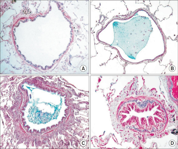
Pathological characteristics of COPD include inflammation of the small airway (small airway disease) and destruction of the lung parenchyma (emphysema). Small airway disease contributes to airflow limitation by narrowing and obliterating the airway lumen (Figure 1C, D) compared to normal small airway (Figure 1A, B) and by actively narrowing the airway through an increased smooth muscle [11]. Emphysema, on the other hand, helps restrict airflow by reducing the elastic recoil pressure available to drive air out of the lungs by destroying the parenchyma and decreasing tethering of small airways through low elastic load applied to the airways. In addition, destruction of alveolar attachments can lead to narrowing and premature closure of airways.
Fig. 1.

Figures of small airway, cited from Hogg JC [11] (Lancet 2004;364:709-21) with permission from Elsevier Inc. (A) Normal small airway. (B) Small airway containing plug of mucus with relatively few cells. (C) Acutely inflamed airway with thickened wall, in which the lumen is partly filled with an inflammatory exudate of mucus and cells. (D) Airway surrounded by connective tissue, which appears as if it might restrict normal enlargement of the lumen and unfolding of the epithelial lining.
Difference between Panlobular and Centrilobular Emphysema
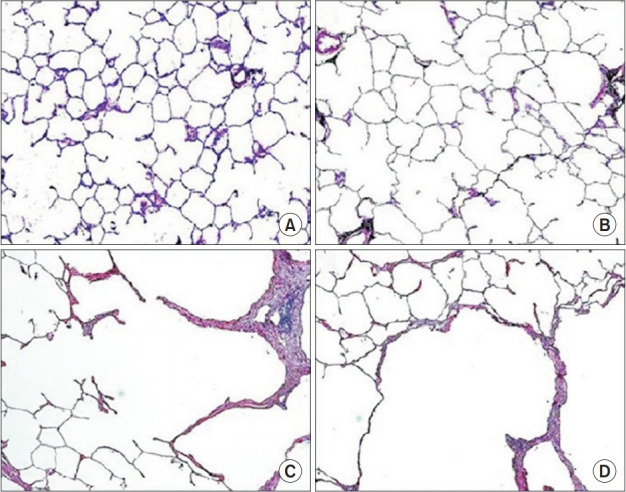
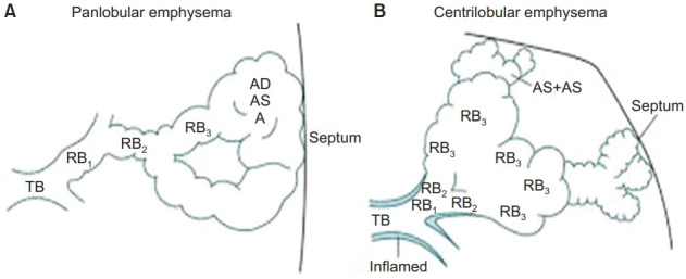
Two main types of emphysema have been recognized in COPD: (1) panlobular emphysema (PLE) commonly associated with α 1 -antitrypsin deficiency and also smoking; (2) centrilobular emphysema (CLE) known as smoker’s emphysema. Paraseptal emphysema is not associated with increased symptoms or reduced lung function [12]. PLE shows diffuse even alveolar enlargement (Figure 2B) comapared to non-smoking control lung (Figure 2A). Therefore, PLE is associated with uniform damage of air space, affecting primarily the lower lobes of the lungs. CLE shows an uneven pattern of lung destruction with thickened wall of the terminal bronchiole (Figure 2C) [13], affecting mainly the upper lobes. A schematic representation of the gross pathological difference between these two emphysemas is shown in Figure 3 [14].
Fig. 2.

Representative microscopic images of the parenchyma of the lungs, cited from author’s prior publication [13] (Kim WD et al. Respirology 2013;18:688-96). (A) Image of a non-smoking control lung. (B) Image of a panlobular emphysema lung. (C) Image of a centrilobular emphysema lung with thickened wall of terminal bronchiole. (D) Image of a mixed panlobular and centrilobular emphysema lung with thickened wall of terminal bronchiole.
Fig. 3.

A schematic representation of gross pathological difference between panlobular and centrilobular emphysemas, cited from Thurlbeck WM and Wright JL [14] (Thurlbeck’s chronic airflow obstruction) with permission from Wright JL. (A) Panlobular emphsema shows enlargement and destruction of the air spaces that uniformly affecting the acinus. (B) Centrilobular emphysema selectively and dominantly affects the respiratory bronchiole with inflamed terminal bronchiole. TB: terminal bronchiole; RB: respiratory bronchiole; AD: alveolar duct; AS: alveolar sac.
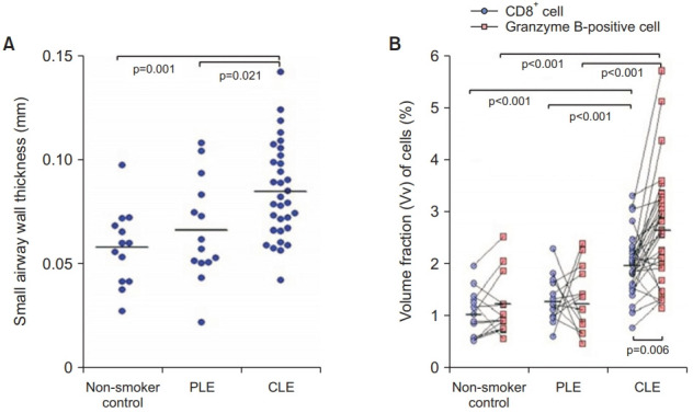
The original description of CLE was obtained through an autopsy study, showing that all CLE lesions had a feeding bronchiole lined with abnormal epithelium, accompanied by varying degrees of thickening of the airway wall and narrowing of the lumen [15]. In addition to this observational study, objective and quantitative morphometric measurements of small airway pathological scores in surgically resected lung specimens indicated that CLE had a higher degree of small airway abnormalities than PLE, mainly due to higher muscle score and fibrosis [16]. Direct microscopic measurements of thicknesses of small airway walls in resected lung samples also showed that thicknesses of small airway walls in CLE lungs were greater than those of non-smoker control lungs [17]. There was a significant correlation between the small airway wall thickness and the degree of airflow limitation in these CLE lungs. Further morphometric analysis of resected lung samples showed significantly increased wall thicknesses of small airways in CLE compared to those of non-smoker control or PLE lungs (Figure 4A) [13]. Additional histologic analysis of cryosections cut from the frozen tissue blocks of isolated COPD lungs showed that CLE lungs had thickened airway walls compared to control lungs [18].
Fig. 4.

Thickness of small airway walls and volume fraction (Vv) of cells in small airway walls, cited from author’s prior publication [13] (Kim WD et al. Respirology 2013;18:688-96). (A) Small airway wall thickness is greater in CLE than in nonsmoker control of PLE lungs. (B) The Vv of CD8+ and granzyme B-positive cells in small airway walls is greater in CLE than in non-smoker control or PLE lungs. PLE: Panlobular emphysema; CLE: centrilobular emphysema.
Immunohistochemical studies have shown that the volume fraction of CD8 + and granzyme B-positive cells in small airway walls (Figure 4B) [13] and the number of these cells on the alveolar walls [19] in CLE lungs are greater than those in non-smoker control or PLE lungs, suggesting a difference in cellular background between these two emphysemas. In addition, mast cells were more dominant in smooth muscles of small airways and alveolar walls of CLE compared to PLE [20]. Blood interleukin (IL)-6, matrix metalloproteinase-7, and tumor necrosis factor α were associated with emphysema, while IL-6, IL-13, IL-2 receptor, interferon γ, and C-reactive protein were associated with bronchial thickening in smokers based on quantitative computed tomography (QCT), suggesting different inflammatory biomarker patterns in these COPD subtypes [21]. QCT is the method of quantifying the presence and percentage of low attenuation areas of emphysema and airway wall thickness of segmental and subsegmental airways of lungs [22].
The fact that CLE shows uneven lung destruction with airway thickening and the preferential distribution in the upper lobes is consistent with the concept of airborne disease [16]. On the other hand, the diffuse uniform destruction of lungs without airway involvement and the preferential distribution in the lower lobes where blood flow is greater than that in the upper lobes suggest that PLE may arise from a blood-mediated mechanism of protease-antiprotease imbalance [17].
Reported relative frequencies of PLE and CLE are variable. Of 19 series of random cases from mostly autopsy or necropsy studies, CLE was considerably more common than PLE in 10 studies, PLE was more common than CLE in 4 studies, and PLE and CLE were considered equal in five studies [23]. QCT examination of COPD patients showed 174 airway-predominant and 75 emphysema-predominant COPD [24]. A study of the visual assessment of chest computed tomography (CT) scans of COPD patients found 63 predominant PLE and 55 predominant CLE [25].
Relationship between Small Airway Disease and Centrilobular Emphysema
It was reported that young smokers who died suddenly outside the hospital had definite abnormalities in the peripheral airways [26]. The authors hypothesized that these lesions might be precursors of severe anatomical lesions in smokers. A MicroCT study of resected lung samples also showed that narrowing of terminal bronchioles preceded the occurrence of centrilobular emphysematous destruction [18]. In lung tissues, remodeling of terminal and transitional bronchioles not affected by emphysema provides further evidence that disease of small airways precedes emphysematous lesions [27]. Therefore, it is believed that the pathogenesis of CLE begins with inflammation, remodeling, and destruction of small airways with subsequent spread into the peribronchiolar alveolar wall tissue and destruction of the center of the lobule [28]. Thus, the pathology of COPD can be redefined as small airway disease with CLE and PLE.
Differential Diagnosis between Panlobular and Centrilobular Emphysema
Differential diagnosis between PLE and small airway disease with CLE is not easy because the emphysema is shared by the two. Chest CT scans are becoming routine for smokers with COPD to check for lung cancer and to evaluate pulmonary nodules. Fortunately, improved CT-based imaging of lungs can be used to differentiate emphysematous phenotypes in a less invasive way [29]. QCT is useful to identify and sequentially assess the extent of emphysematous lung destruction and changes in airway walls in patients with COPD [30]. It has been shown that CT measurements of the thickening and narrowing of the relatively large airways can serve as a surrogate for pathological changes in small airways that are not measurable on conventional CT [31]. It is now possible to non-invasively assess the relationship of QCT-defined thickening of airways or emphysema with clinically relevant outcomes.
Different Clinical Manifestation of COPD Depending on the Presence of Increased Airway Wall Thickness
1. Clinical presentations
In a study with 463 COPD patients, the CT-measured airway wall thickness was significantly related to morning cough, chronic cough, and wheezing [32]. In 100 male smokers, smokers with chronic respiratory symptoms such as cough, excessive mucus, dyspnea, and wheezing had thicker CT-measured bronchial walls than those without symptoms [33]. In 56 COPD patients, CT-measured thicker walls were associated with clinical features that might represent a bronchitic phenotype (Medical Research Council bronchitis score, frequent exacerbations, total St. George’s score, and body mass index [BMI]), independent of emphysema [34]. BMI was negatively correlated with the degree of emphysema. One study of 3,171 current or former smokers found that patients with confluent or advanced destructive emphysema, equivalent to PLE, had lower BMI than those with mild CLE [35]. In 1,200 patients with COPD, relative influence of CT-defined airway disease was greater for SGRQ (St. George’s Respiratory Questionnaire) scores and relative influence of emphysema was greater for BODE (Body mass index, airflow Obstruction, Dyspnea and Exercise capacity) index [36].
2. Acute bronchodilator responsiveness
Preoperative methacholine challenge was compared to morphologic and cellular inflammatory features of airways in the surgically resected lungs of CLE and PLE [37]. The reactivity of airways was significantly higher in CLE than in PLE. And the reactivity of airways was determined by the degree of pathological abnormality in small airways. Small airway morphometry was performed in resected lungs of 67 patients with advanced emphysema undergoing lung volume reduction surgery [38]. The group with reversibility to bronchodilator had increased smooth muscle mass in small airways than the irreversible group. Of 2,355 bronchodilator-negative COPD patients and 1,306 positive patients, bronchodilator-responsive patients had CT evidence of thicker airways than bronchodilator-unresponsive patients [39].
3. Exacerbation of COPD
In a study of 1,002 COPD patients with QCT measurements of emphysema and airway disease, the multivariate model showed that an increase in segmental bronchial wall thickness was associated with a higher frequency of exacerbations [24]. Of 167 patients with COPD, patients with mild emphysema and severe airway changes had significantly more frequent exacerbations than patients with moderate emphysema with mild airway changes [40].
4. Progression of COPD
In 131 COPD patients with follow-up study over 3.7 years, rapid fall in forced expiratory volume in 1 second (FEV1) was more strongly influenced by emphysema-dominant phenotype than by airway-dominant phenotype on CT [41]. In 1,184 smoker and non-smoker participants in a 6-year longitudinal study, the total airway count on CT, possibly reflecting airway-related disease changes, was independently associated with a decrease in lung function [42].
On the other hand, chest CT scans of 116 cigarette smokers showed that PLE was more frequent in individuals with Global Initiative for Obstructive Lung Disease (GOLD) stage 4 [25]. The authors of the study suggested that some people with predominant PLE might have a higher genetic susceptibility to emphysema or a faster disease progression. This finding is in line with the report of 3,171 ever-smokers, showing that confluent and advanced destructive emphysema, equivalent to PLE, were more common in GOLD stage 4 [35].
Although COPD is generally considered progressive, this disease often remains stable [28]. In more than half of 2,163 COPD patients, the rate of decline in FEV1 over a 3-year period was not greater than that in people without lung disease [43]. A study to calculate the rate of lung function decline over a 5-year period showed that patients with a rapid decline had a lower proportion of regulatory T cells in the bronchoalveolar lavage fluid than patients with non-rapid decline [44]. The authors suggested that the inability to upregulate regulatory T cells (i.e., the inability to suppress the inflammatory response after smoking) could lead to a more rapid decline in lung function. A cross-sectional study of surgically resected lung specimens showed that the number of alveolar granzyme B-positive cells was higher in CLE lungs compared to that in non-smoker control and PLE lungs [19]. This number of alveolar granzyme B-positive cells was positively correlated with FEV1 in CLE. Another study also showed a positive correlation between the volume fraction of granzyme B-positive cells in small airways and FEV1 in CLE lungs [13]. These results mean that CLE lungs with mild airflow limitation have more granzyme B-positive cells both in small airways and on alveolar walls than CLE lungs with severe airflow limitation. The authors of the study postulated that the degree of lung destruction and thus the progression of airflow limitation in CLE might be determined by the individual amount of available granzyme B-positive cells, which are thought to represent an activated state of cells with a regulatory function [19].
5. Mortality
In 609 patients with severe emphysema, increased mortality was independently associated with greater lower-lung zone emphysema [45]. An 8-year mortality study in 947 ever-smokers showed that CT-measured airway thickness did not predict mortality, whereas emphysema was a strong independent predictor of mortality [46].
6. Association of COPD with cardiovascular disease
Sixty COPD patients underwent a right heart catheterization and chest CT examination [47]. Airway wall thickness was an independent predictor associated with an increase in mean pulmonary artery pressure. In contrast to quantification of emphysema, CT measurement of airway remodeling was correlated with mean pulmonary artery pressure. Chest CT was performed for emphysematous lesions, airway lesions, and epicardial adipose tissue (EAT) in 180 smokers [48]. The EAT area was independently related to the wall thickness of the airways. EAT has been shown to be a non-invasive marker that could predict cardiovascular disease (CVD) progression [49]. The result suggests a mechanistic link between airway-predominant COPD and CVD.
7. Association of COPD with low bone mineral density
When X-ray absorptiometry measurements of bone mineral density were performed for 190 current and former smokers, quantitative emphysema, but not CT-measured airway wall thickness index, was inversely associated with bone mineral density [50]. Emphysema was a strong, independent predictor of low bone mineral density. In 3,321 current and former smokers, emphysema was associated with both low volumetric bone mineral density and vertebral fractures [51]. Airway disease was associated with higher bone density. In 75 patients with emphysema-predominant and 174 with airway-predominant COPD on CT, osteoporosis was significantly more common in emphysema-predominant COPD subjects [24].
8. Association of COPD with lung cancer
When CT scans of 279 participants diagnosed with lung cancer were analyzed, the emphysema index was most closely related to lung cancer [52]. Airway dimensions were not associated with lung cancer. In 947 ever-smokers who were followed up for 10 years, base- line emphysema on CT remained a significant predictor of lung cancer incidence [53]. Airway wall thickness did not independently predict cancer.
9. Association of COPD with diabetes mellitus
Of 75 patients with emphysema-predominant and 174 with airway-predominant COPD on CT, diabetes was more common in patients with airway-predominant cases [24]. Of 4,197 COPD subjects, non-emphysematous COPD (defined by airflow limitation with a lack of emphysema on chest CT) was associated with an increased risk of diabetes [54].
Summary of different clinical manifestation
In summary, chronic respiratory symptoms, greater influence for SGRQ score, positive bronchodilator responsiveness, exacerbation, cardiovascular disease, and diabetes mellitus were associated with COPD patients having thickened airway walls. On the other hand, lower BMI, greater influence for BODE index, rapid progression, mortality, low bone mineral density, and lung cancer were associated with COPD without airway wall thickening (Table 1).
Table 1.
Different clinical manifestation of COPD depending on airway wall thickening
| COPD with airway wall thickening | COPD without airway wall thickening |
|---|---|
| Chronic respiratory symptoms [32-34] | Lower BMI [34,35] |
| Greater influence for SGRQ score [36] | Greater influence for BODE index [36] |
| Positive bronchodilator response [37-39] | Rapid progression [25,41] |
| Exacerbation [24,40] | Mortality [46] |
| Cardiovascular disease [47,48] | Low bone mineral density [24,50,51] |
| Diabetes mellitus [24,54] | Lung cancer [52,53] |
COPD: chronic obstructive pulmonary disease; BMI: body mass index; SGRQ: St. George’s Respiratory Questionnaire; BODE: Body mass index, airflow Obstruction, Dyspnea and Exercise capacity.
Different Response to Pharmacological Therapy Depending on the Presence of Increased Airway Wall Thickness
Lung samples were examined in 35 COPD patients within 12 months of administration of isoproterenol. Patients with an increased bronchial gland-bronchial wall ratio (Reid index) showed a significantly greater improvement in FEV1 after bronchodilator therapy compared to patients with a normal Reid index [55]. This index of the ratio of gland thickness to wall thickness measured between cartilage and epithelial basement membrane was introduced as a measure of chronic bronchitis [56]. In 85 COPD patients, increase in FEV1 in response to treatment with inhaled corticosteroid for 2–3 months was significantly higher in emphysema with bronchial wall thickening on high-resolution computed tomography than in emphysema without airway thickening [57]. When 226 patients received combination of inhaled long-acting beta-agonist and corticosteroid for 3 months, internal perimeter of 10 mm measured by integral-based half-band method (Pi10-IBHB), reflecting the severity of small airway disease on CT, was the only independent variable predicting an increase in FEV1, suggesting that COPD with predominant airway disease would be more treatable than COPD with predominant emphysema [58]. When 60 COPD patients were randomized to receive bronchodilator or bronchodilator with corticosteroid for 16 weeks, airway wall thickening and airway narrowing on CT were decreased after treatment with combination of bronchodilator and corticosteroid [59]. It was found that changes in airway dimensions were proportional to the improvement in FEV1.
When 254 COPD patients were randomly assigned to inhaled corticosteroid or placebo and followed up with annual CT for 2–4 years, there was no significant difference in the annual decrease in FEV1 between corticosteroid and placebo [60]. Long-term inhalation of corticosteroid showed a non-significant trend in reducing the progression of emphysema from annual CT scans. When 165 COPD patients received inhalation of a long-acting beta-agonist and corticosteroid for three months, CT-defined emphysema-dominant patients showed no improvement in FEV1 or dyspnea after three months of treatment [61].
The difference in response to pharmacological therapy is summarized in Table 2.
Table 2.
Different response of COPD to pharmacological therapy
| Favorable response to therapy | Poor response to therapy |
|---|---|
| Increased Reid index [55] | Emphysema on CT [60] |
| Emphysema with bronchial wall thickening on HRCT [57] | CT-defined emphysema-dominant |
| Higher Pi10-IBHB on CT [58] | COPD [61] |
| Airway wall thickening on CT [59] |
COPD: chronic obstructive pulmonary disease; Reid index: bronchial gland-bronchial wall ratio; CT: computed tomography; HRCT: high-resolution computed tomography; Pi10-IBHB: internal perimeter of 10 mm measured by integral-based half-band method.
Conclusion
Phenotyping of COPD based on the underlying subtype of pathology is encouraging since most clinical manifestations can be distinguished by the presence of increased airway wall thickness on CT. Although further studies with large numbers of subjects are desirable, available data indicated that pharmacological therapy has a significant effect in COPD patients with increased airway wall thickness. However, it has limited benefit for COPD patients without airway thickening.
The phenotype of COPD based on the CT-defined underlying pathology was able to describe differences in clinically meaningful outcomes between patients. It could also classify patients into subgroups with prognostic value of responsiveness to pharmacological therapy. This phenotype can be a useful tool to better understand the disease and adjust treatment accordingly.
Footnotes
Conflicts of Interest
No potential conflict of interest relevant to this article was reported.
Funding
This work was supported by a grant (No. F01-2004-000-10180-0) from the Korea Science and Engineering Foundation, Republic of Korea.
REFERENCES
- 1.Global Initiative for Chronic Obstructive Lung Disease . Fontana: Global Initiative for Chronic Obstructive Lung Disease; 2021. Global strategy for the diagnosis, management, and prevention of chronic obstructive pulmonary disease, 2022 report [Internet] [cited 2021 Dec 19]. Available from: https://goldcopd.org. [Google Scholar]
- 2.GBD 2017 Disease and Injury Incidence and Prevalence Collaborators Global, regional, and national incidence, prevalence, and years lived with disability for 354 diseases and injuries for 195 countries and territories, 1990–2017: a systematic analysis for the Global Burden of Disease Study 2017. Lancet. 2018;392:1789–858. doi: 10.1016/S0140-6736(18)32279-7. [DOI] [PMC free article] [PubMed] [Google Scholar]
- 3.GBD 2017 Causes of Death Collaborators Global, regional, and national age-sex-specific mortality for 282 causes of death in 195 countries and territories, 1980-2017: a systematic analysis for the Global Burden of Disease Study 2017. Lancet. 2018;392:1736–88. doi: 10.1016/S0140-6736(18)32203-7. [DOI] [PMC free article] [PubMed] [Google Scholar]
- 4.World Health Organization . Geneva: World Health Organization; 2019. Burden of COPD [Internet] [cited 2021 Dec 19]. Available from: https://www.who.int/respiratory/copd/burden/en/ [Google Scholar]
- 5.Chicago: American Lung Association; 2021. Understanding COPD trends [Internet] [cited 2021 Dec 19]. Available from: http://www.lung.org/blog/understanding-copd-trends. [Google Scholar]
- 6.Hwang YI, Park YB, Yoo KH. Recent trends in the prevalence of chronic obstructive pulmonary disease in Korea. Tuberc Respir Dis. 2017;80:226–9. doi: 10.4046/trd.2017.80.3.226. [DOI] [PMC free article] [PubMed] [Google Scholar]
- 7.Montuschi P, Malerba M, Santini G, Miravitlles M. Pharmacological treatment of chronic obstructive pulmonary disease: from evidence-based medicine to phenotyping. Drug Discov Today. 2014;19:1928–35. doi: 10.1016/j.drudis.2014.08.004. [DOI] [PubMed] [Google Scholar]
- 8.Agusti A. The path to personalised medicine in COPD. Thorax. 2014;69:857–64. doi: 10.1136/thoraxjnl-2014-205507. [DOI] [PubMed] [Google Scholar]
- 9.Miravitlles M, Soler-Cataluna JJ, Calle M, Soriano JB. Treatment of COPD by clinical phenotypes: putting old evidence into clinical practice. Eur Respir J. 2013;41:1252–6. doi: 10.1183/09031936.00118912. [DOI] [PubMed] [Google Scholar]
- 10.Han MK, Agusti A, Calverley PM, Celli BR, Criner G, Curtis JL, et al. Chronic obstructive pulmonary disease phenotypes: the future of COPD. Am J Respir Crit Care Med. 2010;182:598–604. doi: 10.1164/rccm.200912-1843CC. [DOI] [PMC free article] [PubMed] [Google Scholar]
- 11.Hogg JC. Pathophysiology of airflow limitation in chronic obstructive pulmonary disease. Lancet. 2004;364:709–21. doi: 10.1016/S0140-6736(04)16900-6. [DOI] [PubMed] [Google Scholar]
- 12.Smith BM, Austin JH, Newell JD, Jr, D’Souza BM, Rozenshtein A, Hoffman EA, et al. Pulmonary emphysema subtypes on computed tomography: the MESA COPD study. Am J Med. 2014;127:94. doi: 10.1016/j.amjmed.2013.09.020. [DOI] [PMC free article] [PubMed] [Google Scholar]
- 13.Kim WD, Chi HS, Choe KH, Oh YM, Lee SD, Kim KR, et al. A possible role for CD8 + and non-CD8 + cell granzyme B in early small airway wall remodelling in centrilobular emphysema. Respirology. 2013;18:688–96. doi: 10.1111/resp.12069. [DOI] [PubMed] [Google Scholar]
- 14.Thurlbeck WM, Wright JL. In: Thurlbeck’s chronic airflow obstruction. 2nd ed. Thurlbeck WM, Wright JL, editors. Hamilton: B.C. Decker Inc; 1999. Emphysema: classification, morphology, and associations; pp. 85–137. [Google Scholar]
- 15.Leopold JG, Gough J. The centrilobular form of hypertrophic emphysema and its relation to chronic bronchitis. Thorax. 1957;12:219–35. doi: 10.1136/thx.12.3.219. [DOI] [PMC free article] [PubMed] [Google Scholar]
- 16.Kim WD, Eidelman DH, Izquierdo JL, Ghezzo H, Saetta MP, Cosio MG. Centrilobular and panlobular emphysema in smokers. Two distinct morphologic and functional entities. Am Rev Respir Dis. 1991;144:1385–90. doi: 10.1164/ajrccm/144.6.1385. [DOI] [PubMed] [Google Scholar]
- 17.Kim WD, Ling SH, Coxson HO, English JC, Yee J, Levy RD, et al. The association between small airway obstruction and emphysema phenotypes in COPD. Chest. 2007;131:1372–8. doi: 10.1378/chest.06-2194. [DOI] [PubMed] [Google Scholar]
- 18.McDonough JE, Yuan R, Suzuki M, Seyednejad N, Elliott WM, Sanchez PG, et al. Small-airway obstruction and emphysema in chronic obstructive pulmonary disease. N Engl J Med. 2011;365:1567–75. doi: 10.1056/NEJMoa1106955. [DOI] [PMC free article] [PubMed] [Google Scholar]
- 19.Kim WD, Chi HS, Choe KH, Kim WS, Hogg JC, Sin DD. The role of granzyme B containing cells in the progression of chronic obstructive pulmonary disease. Tuberc Respir Dis. 2020;83(Suppl 1):S25–33. doi: 10.4046/trd.2020.0089. [DOI] [PMC free article] [PubMed] [Google Scholar]
- 20.Ballarin A, Bazzan E, Zenteno RH, Turato G, Baraldo S, Zanovello D, et al. Mast cell infiltration discriminates between histopathological phenotypes of chronic obstructive pulmonary disease. Am J Respir Crit Care Med. 2012;186:233–9. doi: 10.1164/rccm.201112-2142OC. [DOI] [PubMed] [Google Scholar]
- 21.Bon JM, Leader JK, Weissfeld JL, Coxson HO, Zheng B, Branch RA, et al. The influence of radiographic phenotype and smoking status on peripheral blood biomarker patterns in chronic obstructive pulmonary disease. PLoS One. 2009;4:e6865. doi: 10.1371/journal.pone.0006865. [DOI] [PMC free article] [PubMed] [Google Scholar]
- 22.Lynch DA, Al-Qaisi MA. Quantitative computed tomography in chronic obstructive pulmonary disease. J Thorac Imaging. 2013;28:284–90. doi: 10.1097/RTI.0b013e318298733c. [DOI] [PMC free article] [PubMed] [Google Scholar]
- 23.Thurlbeck WM, Wright JL. In: Thurlbeck’s chronic airflow obstruction. 2nd ed. Thurlbeck WM, Wright JL, editors. Hamilton: B.C. Decker Inc; 1999. Chapter 5. Emphysema: etiology, pathogenesis, animal models, and epidemiology; pp. 145–87. [Google Scholar]
- 24.Han MK, Kazerooni EA, Lynch DA, Liu LX, Murray S, Curtis JL, et al. Chronic obstructive pulmonary disease exacerbations in the COPDGene study: associated radiologic phenotypes. Radiology. 2011;261:274–82. doi: 10.1148/radiol.11110173. [DOI] [PMC free article] [PubMed] [Google Scholar]
- 25.Sverzellati N, Lynch DA, Pistolesi M, Kauczor HU, Grenier PA, Wilson C, et al. Physiologic and quantitative computed tomography differences between centrilobular and panlobular emphysema in COPD. Chronic Obstr Pulm Dis. 2014;1:125–32. doi: 10.15326/jcopdf.1.1.2014.0114. [DOI] [PMC free article] [PubMed] [Google Scholar]
- 26.Niewoehner DE, Kleinerman J, Rice DB. Pathologic changes in the peripheral airways of young cigarette smokers. N Engl J Med. 1974;291:755–8. doi: 10.1056/NEJM197410102911503. [DOI] [PubMed] [Google Scholar]
- 27.Higham A, Quinn AM, Cancado JED, Singh D. The pathology of small airways disease in COPD: historical aspects and future directions. Respir Res. 2019;20:49. doi: 10.1186/s12931-019-1017-y. [DOI] [PMC free article] [PubMed] [Google Scholar]
- 28.Singh D. Small airway disease in patients with chronic obstructive pulmonary disease. Tuberc Respir Dis. 2017;80:317–24. doi: 10.4046/trd.2017.0080. [DOI] [PMC free article] [PubMed] [Google Scholar]
- 29.Foster WL, Jr, Gimenez EI, Roubidoux MA, Sherrier RH, Shannon RH, Roggli VL, et al. The emphysemas: radiologic-pathologic correlations. Radiographics. 1993;13:311–28. doi: 10.1148/radiographics.13.2.8460222. [DOI] [PubMed] [Google Scholar]
- 30.Lynch DA, Austin JH, Hogg JC, Grenier PA, Kauczor HU, Bankier AA, et al. CT-definable subtypes of chronic obstructive pulmonary disease: a statement of the Fleischner Society. Radiology. 2015;277:192–205. doi: 10.1148/radiol.2015141579. [DOI] [PMC free article] [PubMed] [Google Scholar]
- 31.Nakano Y, Wong JC, de Jong PA, Buzatu L, Nagao T, Coxson HO, et al. The prediction of small airway dimensions using computed tomography. Am J Respir Crit Care Med. 2005;171:142–6. doi: 10.1164/rccm.200407-874OC. [DOI] [PubMed] [Google Scholar]
- 32.Grydeland TB, Dirksen A, Coxson HO, Eagan TM, Thorsen E, Pillai SG, et al. Quantitative computed tomography measures of emphysema and airway wall thickness are related to respiratory symptoms. Am J Respir Crit Care Med. 2010;181:353–9. doi: 10.1164/rccm.200907-1008OC. [DOI] [PubMed] [Google Scholar]
- 33.Xie X, Dijkstra AE, Vonk JM, Oudkerk M, Vliegenthart R, Groen HJ. Chronic respiratory symptoms associated with airway wall thickening measured by thin-slice low-dose CT. AJR Am J Roentgenol. 2014;203:W383–90. doi: 10.2214/AJR.13.11536. [DOI] [PubMed] [Google Scholar]
- 34.Mair G, Maclay J, Miller JJ, McAllister D, Connell M, Murchison JT, et al. Airway dimensions in COPD: relationships with clinical variables. Respir Med. 2010;104:1683–90. doi: 10.1016/j.rmed.2010.04.021. [DOI] [PubMed] [Google Scholar]
- 35.Lynch DA, Moore CM, Wilson C, Nevrekar D, Jennermann T, Humphries SM, et al. CT-based visual classification of emphysema: association with mortality in the COPD-Gene study. Radiology. 2018;288:859–66. doi: 10.1148/radiol.2018172294. [DOI] [PMC free article] [PubMed] [Google Scholar]
- 36.Martinez CH, Chen YH, Westgate PM, Liu LX, Murray S, Curtis JL, et al. Relationship between quantitative CT metrics and health status and BODE in chronic obstructive pulmonary disease. Thorax. 2012;67:399–406. doi: 10.1136/thoraxjnl-2011-201185. [DOI] [PMC free article] [PubMed] [Google Scholar]
- 37.Finkelstein R, Ma HD, Ghezzo H, Whittaker K, Fraser RS, Cosio MG. Morphometry of small airways in smokers and its relationship to emphysema type and hyperresponsiveness. Am J Respir Crit Care Med. 1995;152:267–76. doi: 10.1164/ajrccm.152.1.7599834. [DOI] [PubMed] [Google Scholar]
- 38.Kim V, Pechulis RM, Abuel-Haija M, Solomides CC, Gaughan JP, Criner GJ. Small airway pathology and bronchoreversibility in advanced emphysema. COPD. 2010;7:93–101. doi: 10.3109/15412551003631691. [DOI] [PubMed] [Google Scholar]
- 39.Kim V, Desai P, Newell JD, Make BJ, Washko GR, Silverman EK, et al. Airway wall thickness is increased in COPD patients with bronchodilator responsiveness. Respir Res. 2014;15:84. doi: 10.1186/s12931-014-0084-3. [DOI] [PMC free article] [PubMed] [Google Scholar]
- 40.Karayama M, Inui N, Yasui H, Kono M, Hozumi H, Suzuki Y, et al. Clinical features of three-dimensional computed tomography-based radiologic phenotypes of chronic obstructive pulmonary disease. Int J Chron Obstruct Pulmon Dis. 2019;14:1333–42. doi: 10.2147/COPD.S207267. [DOI] [PMC free article] [PubMed] [Google Scholar]
- 41.Tanabe N, Muro S, Tanaka S, Sato S, Oguma T, Kiyokawa H, et al. Emphysema distribution and annual changes in pulmonary function in male patients with chronic obstructive pulmonary disease. Respir Res. 2012;13:31. doi: 10.1186/1465-9921-13-31. [DOI] [PMC free article] [PubMed] [Google Scholar]
- 42.Kirby M, Tanabe N, Tan WC, Zhou G, Obeidat M, Hague CJ, et al. Total airway count on computed tomography and the risk of chronic obstructive pulmonary disease progression: findings from a population-based study. Am J Respir Crit Care Med. 2018;197:56–65. doi: 10.1164/rccm.201704-0692OC. [DOI] [PubMed] [Google Scholar]
- 43.Vestbo J, Edwards LD, Scanlon PD, Yates JC, Agusti A, Bakke P, et al. Changes in forced expiratory volume in 1 second over time in COPD. N Engl J Med. 2011;365:1184–92. doi: 10.1056/NEJMoa1105482. [DOI] [PubMed] [Google Scholar]
- 44.Eriksson Strom J, Pourazar J, Linder R, Blomberg A, Lindberg A, Bucht A, et al. Airway regulatory T cells are decreased in COPD with a rapid decline in lung function. Respir Res. 2020;21:330. doi: 10.1186/s12931-020-01593-9. [DOI] [PMC free article] [PubMed] [Google Scholar]
- 45.Martinez FJ, Foster G, Curtis JL, Criner G, Weinmann G, Fishman A, et al. Predictors of mortality in patients with emphysema and severe airflow obstruction. Am J Respir Crit Care Med. 2006;173:1326–34. doi: 10.1164/rccm.200510-1677OC. [DOI] [PMC free article] [PubMed] [Google Scholar]
- 46.Johannessen A, Skorge TD, Bottai M, Grydeland TB, Nilsen RM, Coxson H, et al. Mortality by level of emphysema and airway wall thickness. Am J Respir Crit Care Med. 2013;187:602–8. doi: 10.1164/rccm.201209-1722OC. [DOI] [PubMed] [Google Scholar]
- 47.Dournes G, Laurent F, Coste F, Dromer C, Blanchard E, Picard F, et al. Computed tomographic measurement of airway remodeling and emphysema in advanced chronic obstructive pulmonary disease. Correlation with pulmonary hypertension. Am J Respir Crit Care Med. 2015;191:63–70. doi: 10.1164/rccm.201408-1423OC. [DOI] [PubMed] [Google Scholar]
- 48.Higami Y, Ogawa E, Ryujin Y, Goto K, Seto R, Wada H, et al. Increased epicardial adipose tissue is associated with the airway dominant phenotype of chronic obstructive pulmonary disease. PLoS One. 2016;11:e0148794. doi: 10.1371/journal.pone.0148794. [DOI] [PMC free article] [PubMed] [Google Scholar]
- 49.Talman AH, Psaltis PJ, Cameron JD, Meredith IT, Seneviratne SK, Wong DT. Epicardial adipose tissue: far more than a fat depot. Cardiovasc Diagn Ther. 2014;4:416–29. doi: 10.3978/j.issn.2223-3652.2014.11.05. [DOI] [PMC free article] [PubMed] [Google Scholar]
- 50.Bon J, Fuhrman CR, Weissfeld JL, Duncan SR, Branch RA, Chang CC, et al. Radiographic emphysema predicts low bone mineral density in a tobacco-exposed cohort. Am J Respir Crit Care Med. 2011;183:885–90. doi: 10.1164/rccm.201004-0666OC. [DOI] [PMC free article] [PubMed] [Google Scholar]
- 51.Jaramillo JD, Wilson C, Stinson DS, Lynch DA, Bowler RP, Lutz S, et al. Reduced bone density and vertebral fractures in smokers: men and COPD patients at increased risk. Ann Am Thorac Soc. 2015;12:648–56. doi: 10.1513/AnnalsATS.201412-591OC. [DOI] [PMC free article] [PubMed] [Google Scholar]
- 52.Gierada DS, Guniganti P, Newman BJ, Dransfield MT, Kvale PA, Lynch DA, et al. Quantitative CT assessment of emphysema and airways in relation to lung cancer risk. Radiology. 2011;261:950–9. doi: 10.1148/radiol.11110542. [DOI] [PMC free article] [PubMed] [Google Scholar]
- 53.Aamli Gagnat A, Gjerdevik M, Gallefoss F, Coxson HO, Gulsvik A, Bakke P. Incidence of non-pulmonary cancer and lung cancer by amount of emphysema and airway wall thickness: a community-based cohort. Eur Respir J. 2017;49:1601162. doi: 10.1183/13993003.01162-2016. [DOI] [PubMed] [Google Scholar]
- 54.Hersh CP, Make BJ, Lynch DA, Barr RG, Bowler RP, Calverley PM, et al. Non-emphysematous chronic obstructive pulmonary disease is associated with diabetes mellitus. BMC Pulm Med. 2014;14:164. doi: 10.1186/1471-2466-14-164. [DOI] [PMC free article] [PubMed] [Google Scholar]
- 55.Boushy SF. The use of expiratory forced flows for deter-mining response to bronchodilator therapy. Chest. 1972;62:534–41. doi: 10.1378/chest.62.5.534. [DOI] [PubMed] [Google Scholar]
- 56.Reid L. Measurement of the bronchial mucous gland layer: a diagnostic yardstick in chronic bronchitis. Thorax. 1960;15:132–41. doi: 10.1136/thx.15.2.132. [DOI] [PMC free article] [PubMed] [Google Scholar]
- 57.Kitaguchi Y, Fujimoto K, Kubo K, Honda T. Characteristics of COPD phenotypes classified according to the findings of HRCT. Respir Med. 2006;100:1742–52. doi: 10.1016/j.rmed.2006.02.003. [DOI] [PubMed] [Google Scholar]
- 58.Park HJ, Lee SM, Choe J, Lee SM, Kim N, Lee JS, et al. Prediction of treatment response in patients with chronic obstructive pulmonary disease by determination of airway dimensions with baseline computed tomography. Korean J Radiol. 2019;20:304–12. doi: 10.3348/kjr.2018.0204. [DOI] [PMC free article] [PubMed] [Google Scholar]
- 59.Hoshino M, Ohtawa J. Effects of adding salmeterol/fluticasone propionate to tiotropium on airway dimensions in patients with chronic obstructive pulmonary disease. Respirology. 2011;16:95–101. doi: 10.1111/j.1440-1843.2010.01869.x. [DOI] [PubMed] [Google Scholar]
- 60.Shaker SB, Dirksen A, Ulrik CS, Hestad M, Stavngaard T, Laursen LC, et al. The effect of inhaled corticosteroids on the development of emphysema in smokers assessed by annual computed tomography. COPD. 2009;6:104–11. doi: 10.1080/15412550902772593. [DOI] [PubMed] [Google Scholar]
- 61.Lee JH, Lee YK, Kim EK, Kim TH, Huh JW, Kim WJ, et al. Responses to inhaled long-acting beta-agonist and corticosteroid according to COPD subtype. Respir Med. 2010;104:542–9. doi: 10.1016/j.rmed.2009.10.024. [DOI] [PubMed] [Google Scholar]


