Fig. 5.

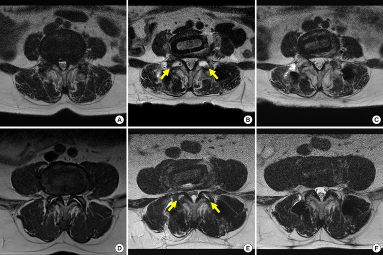
(A, D) Two cases of L4–5 oblique lumbar interbody fusion. Panels A–C are the same patients, panels D–F are the same patient. Central spinal stenosis in preoperative axial magnetic resonance images (B, E), early (postoperative 2 days), late (postoperative 12 months) axial images (C, F). In panels B and E, the area of the foramen and spinal canal were enlarged by facet joint release (arrows) and ligament flavum and disc annulus stretching (early effects). In panels C and F, the area of the foramen and spinal canal were widened by atrophy of the ligament flavum and disc annulus (late effects).
