



Abstract
Background
Prone positioning is a well‐known supportive approach for increasing oxygenation and reducing mortality in non‐COVID‐19 patients with moderate to severe acute respiratory distress syndrome. However, studies highlighting the effects of proning in patients with COVID‐19 are limited.
Aim
To investigate the effects of awake‐prone positioning (APP) on oxygenation and physiological outcomes in non‐intubated patients with COVID‐19.
Study Design
A randomized controlled trial was carried out with two parallel groups at 1:1 ratio.
Adult awake non‐intubated patients with confirmed COVID‐19, non‐rebreathing face mask or continuous positive airway pressure, PaO2/FiO2 ratio ≤150 mmHg were randomly assigned to the APP group or control group. The control group was subjected to conventional positioning interventions. Outcome measures were PaO2/FiO2 ratio, ROX index, PaO2, PaCO2, SaO2, respiratory rate, blood pressure, and shock index. These parameters were recorded immediately before positioning, 10 min after patient positioning, and 1 h after patient positioning.
Results
Of 115 patients assessed for eligibility, 82 were randomized to the APP group or control group (41 patients in each group). The use of APP for non‐intubated patients with COVID‐19 resulted in statistically significant improvements in oxygenation parameters, that is, SpO2, PaO2/FiO2, ROX index, PaO2, and SaO2, at the three study time points (p = .000, .007, .000, .011, and .000 respectively). The SpO2 was increased to 92.15 ± 2.735 mmHg for the APP group versus 88.17 ± 4.847 for the control group after 1 h of patients' positioning. The PaO2/FiO2 ratio increased in the APP group before proning compared with 1 h after proning (79.95 ± 22.508 vs. 98.91 ± 34.44) respectively. APP improved the SpO2, PaO2/FiO2, ROX index, PaO2, and SaO2 values for the APP group, representing an increase of 5.85%, 23.71%, 30.79%, 22.59%, and 5.26%, respectively.
Conclusion
Awake proning in non‐intubated patients with COVID‐19 showed marked improvement in oxygenation and physiological parameters.
Relevance to Clinical Practice
This study provides evidence for critical care nurses to implement APP in non‐intubated patients with COVID‐19 to improve oxygenation and physiological parameters, as it was tolerated by most of the patients without serious adverse events.
Keywords: acute respiratory distress syndrome (ARDS), awake‐prone positioning, coronavirus disease 2019 (COVID‐19), non‐intubated, oxygenation
What is known about this topic
The burden of coronavirus disease 2019 (COVID‐19) pandemic remains on a rising course putting great stress on medical resources worldwide.
Many retrospective cohort studies have examined the effectiveness of awake‐prone positioning (APP) in non‐intubated patients with COVID‐19. Despite a lack of evidence from randomised clinical studies, current guidelines recommend APP for COVID‐19 patients as a safe and beneficial technique.
What this paper adds
This study showed that exposing non‐intubated patients with COVID‐19 to APP resulted in improvements in oxygenation parameters and RR. Also, APP was tolerable by most patients without serious adverse events and detectable hemodynamic differences during proning.
1. INTRODUCTION
The burden of the coronavirus disease 2019 (COVID‐19) pandemic continues to rise, putting a strain on medical resources worldwide. Although most patients with COVID‐19 require non‐invasive ventilation (NIV), 61%–81% of them might have hypoxemic respiratory failure and ultimately acute respiratory distress syndrome (ARDS) faster. 1 , 2 , 3 , 4 As the COVID‐19 pandemic evolves throughout the world, resources, such as mechanical ventilators and critical care beds, will be the rate‐limiting step in treating affected patients. 5 In Egypt, the progression of the COVID‐19 pandemic has pushed resources, such as intensive care beds and mechanical ventilators, to or beyond their limits, which is a major obstacle in treating patients with COVID‐19. 6
Hence, strategies for avoiding intubation and mechanical ventilation are urgently needed. Prone positioning (PP) is a simple supportive ventilation strategy for improving oxygenation in patients with ARDS that critical care nurses (CCNs) may use. 2 , 7 , 8 In non‐COVID‐19‐associated ARDS, PP has been previously shown to enhance oxygenation and reduce mortality from 32.8% to 16%. 7 , 9 Recent studies have emphasized the potential benefits of using this technique for non‐intubated awake hypoxic patients with COVID‐19. It has been proposed that PP leads to the production of more uniform lung perfusion by relieving compression of the abdominal organs and the heart and lungs and decreasing ventilation/perfusion (V/Q) mismatch. 10 , 11
Moreover, it has been reported that awake PP (APP) can improve dorsal lung region recruitment, facilitate drainage of secretions, enhance gas exchange, and decrease the work of breathing and the risk of intubation. In addition, pronation is associated with a higher functional residual capacity and better venous return, which can interpret the relationship between PP and the improvement of physiological parameters in patients with COVID‐19. 11 , 12 For these reasons, the Surviving Sepsis Campaign COVID‐19 guidelines recommend PP as a treatment choice for COVID‐19‐related ARDS. 13
APP has been extensively adopted by clinicians worldwide 14 ; however, there is little evidence to support its effectiveness. 5 , 15 The guideline recommendations for PP in mechanically ventilated patients with ARDS are well established. 7 , 9 However, the guidelines for APP in patients with COVID‐19 remain unclear 16 although various retrospective cohort studies, case series, and case reports have been conducted to determine the effects of APP on non‐intubated patients with COVID‐19. To date, few experimental studies have been conducted. 17 , 18 , 19 Well‐structured experimental trials are necessary to identify which patients with COVID‐19 will benefit the most from APP.
2. AIM OF THE STUDY
This study was designed to investigate the effects of APP on oxygenation and physiological outcomes in non‐intubated patients with COVID‐19.
2.1. Research hypothesis
Awake non‐intubated patients with COVID‐19, who are subjected to PP, will have higher oxygenation indices and more stable physiological parameters than patients in the control group for whom the PP is not applied.
3. MATERIAL AND METHODS
3.1. Research design
A randomized controlled trial was conducted with two parallel groups at 1:1 ratio. The Consolidated Standards of Reporting Trials (CONSORT) criteria were fulfilled within the scope of this study. 20
3.2. Setting
This study was conducted in the General Intensive Care Unit (GICU) of the Chest Hospital in Damanhour City, El Beheira Governorate, Egypt. The Chest Hospital became a quarantine hospital in Egypt and provides service for patients with COVID‐19 in El Beheira Governorate. The GICU has 17 beds distributed into three rooms well‐suited to patients with COVID‐19.
3.3. Recruitment and eligibility
From 20 February 2021 to 20 April 2021, newly admitted patients were assessed daily for 2 months. Patients who met the following inclusion criteria were eligible to participate in this study: aged ≥18 years; awake non‐intubated spontaneously breathing patients; verified COVID‐19 diagnosis; presenting with dyspnea, respiratory rate (RR) of ≥30 breaths/min, oxygen saturation of ≤93%, or partial pressure of arterial oxygen (PaO2) to fraction of inspired oxygen (FiO2) ratio of ≤150 mmHg, needing supplementary oxygen (non‐rebreathing face mask or non‐invasive continuous positive airway pressure [CPAP]); and capable of performing PP on their own. A positive real‐time polymerase chain reaction (RT‐PCR) for severe acute respiratory syndrome coronavirus 2 was required, as well as a chest X‐ray revealing bilateral infiltrations or CT images indicating exudation or consolidation. The exclusion criteria were as follows: life‐threatening arrhythmias, hemodynamic instability, intracranial hypertension, recent abdominal surgery, and pregnancy.
3.4. Sample size
According to the findings of Gad 21 in which the mean difference in PaO2/FiO2 between before and after PP was 14 mmHg and assuming an alpha coefficient of 5% and two‐sided confidence interval at 95% with a test power of 80%, the sample size was estimated to be 77 patients. However, due to the possibility of dropouts, we expanded the sample size to 82 patients (41 in each group). The sample size was calculated according to Rosner. 22
3.5. Randomization
After admission, all eligible patients were randomly allocated to one of the two following groups—the intervention (PP) or the control group (conventional positioning)—after being enrolled by the research assistant (a person who was independent of the researchers) using envelope‐based randomization into two parallel groups at 1:1 ratio. The patients were provided a sequential number, and the research assistant placed it in an opaque envelope. Then, numbers were assigned as follows: odd numbers were assigned to the control group, whereas even numbers were assigned to the intervention group. When it was time to place the patients, the data collector opened the envelope and then applied the positioning. Because of the nature of the intervention, the allocation was known to the patients, treating physicians, nurses, and data collectors. However, principal investigators and the data analyst were blinded to the allocation.
3.6. Research instruments
A patients' position assessment record was developed by the researchers to record the patients' oxygenation and physiological parameters before and after positioning. It included the following three parts:
Part one included patients' socio‐demographics and baseline characteristics (i.e., age, gender, diagnosis, history, smoking history, and comorbidities). It also included the mode of oxygen delivery, baseline arterial blood gas (ABG) measurements (i.e., pH, PaO2 mmHg, PaCO2 mmHg, and SaO2), and laboratory biomarkers (i.e., d‐dimer and haemoglobin level and chest X‐ray findings). Furthermore, it included oxygenation parameters such as the oxygenation index (arterial oxygen pressure/fractional inspired oxygen PaO2/FiO2 ratio mmHg) and the respiratory rate oxygenation (ROX) index (combination of peripheral oxygen saturation to the fraction of inspired oxygen and RR [SpO2/FiO2]/RR).
Part two was designed by the researchers to record the oxygenation and physiological responses to positioning. The parameters of this part were organized in a flow sheet to record patients' data before and after positioning (i.e., immediately before positioning, 10 min after patient positioning, and 1 h after patient positioning). It included the oxygenation index (PaO2/FiO2 ratio), ROX index, peripheral oxygen saturation (SpO2), and ABG parameters. Moreover, it included vital signs (i.e., temperature, RR, heart rate [HR], systolic blood pressure [SBP], and diastolic blood pressure [DBP]), as well as the shock index.
Part three is a tolerability and adverse event observational checklist. This part was used by the researchers to record the patients' tolerability to APP in the intervention group. It involved the tolerability of three parameters in the APP session: comfort, acceptability, and PP lasting for ≥60 min. Moreover; it included seven adverse event parameters, that is, dyspnea, oxygen desaturation SpO2 ≤70%, hypotension SBP ≤90 mmHg, aspiration, vomiting, musculoskeletal pain, and general discomfort. Each item was either present or absent.
3.7. Data collection procedure
Both groups received standard COVID‐19 care according to the recommendations of the Egyptian Ministry of Health. 23 In the study setting, patients were routinely positioned every 2 h in the right lateral position, semi‐recumbent position with 45° elevation and left lateral position. Patients in the control group were managed by the previously mentioned unit routine of patients' positioning. Additionally, those patients were permitted to change positions as needed for their comfort (supine, semi‐recumbent, sitting or lateral). In this group, nurses and the treatment team did not actively encourage PP. The oxygenation indexes and physiological parameters of the control group were collected at three‐time points (i.e., baseline just before position change, 10 min after position change, and 1 h after position change) for whatever reason, including unit routine or patient comfort.
Patients in the intervention group received APP (APP group) and standard COVID‐19 care. Each patient in the APP group was assisted into the PP and instructed to remain in that posture for as long as it was tolerated (at least 1 h). In the PP, the patients were placed on their stomachs with the judicious use of pillows under the pelvis and head on the side. The patients' arms were placed at the sides or abducted to less than 90° at the shoulder and flexed. To avoid gastrointestinal adverse effects, APP was conducted 45 min to 1 h after meals. During PP, every effort was taken to keep the oxygenation adjuncts from being displaced. The researchers trained nurses (local study coordinators) about study procedures and data extraction. The oxygenation indexes and physiological parameters of the APP group were collected at three‐time points (i.e., immediately before positioning, 10 min after patient positioning, and 1 h after patient positioning).
3.8. Outcome parameters
The primary outcomes included the oxygenation indexes (i.e., PaO2/FiO2 ratio, ROX index, PaO2, PaCO2, and SaO2) and physiological parameters (i.e., RR, blood pressure, and shock index). The vital signs and oxygenation indexes (i.e., ROX index and SpO2) were recorded at three study time points (i.e., immediately before positioning, 10 min after patient positioning, and 1 h after patient positioning), while ABG parameters and the PaO2/FiO2 ratio were recorded at two‐time points (i.e., immediately before patient positioning and 1 h after patient positioning). Data on secondary outcomes of APP (patients' tolerability to APP session and appearance of adverse events) were also recorded using part three of the study tool.
3.9. Data analysis
Data were analysed using Statistical Package for the Social Sciences (SPSS), version 20. The Shapiro–Wilk test was used to determine the normality of the data. Shapiro–Wilk test indicated that the data were normally distributed. Categorical variables were described as numbers and percentages. The chi‐square test was used to investigate the relationship between categorical variables between the study and control groups. Student's t test was used for normally distributed data. F‐test (one‐way ANOVA) was conducted to compare groups over three‐time points. Differences with p‐values of less than .05 (two‐sided) were considered statistically significant, whereas differences with p‐values of less than .001 were regarded as highly statistically significant.
3.10. Ethical approval
The Ethics Committee of the Faculty of Medicine, Alexandria University, approved the study on January 21, 2021 (No. 0304974; IRB No: 00012098), and the study was registered at ClinicalTrials.gov (ID: NCT04760561). Informed consent was obtained from the patients. The patients' privacy and the confidentiality of the obtained data were protected. Moreover, the patients had the choice to withdraw from the research at any time.
4. RESULTS
A total of 115 patients were screened for eligibility. Among them, 20 patients were excluded from the study because they did not meet the inclusion criteria, and 11 declined to participate in the trial. Therefore, 84 patients were finally enrolled in this study and randomly allocated to either the APP group or the semi‐recumbent posture group (control group) (42 patients in each group). However, due to hemodynamic instability, one patient did not perform APP. Also, the procedure was discontinued for one patient in the control group due to endotracheal intubation and assisted ventilation. Ultimately, 82 patients in the control group and the APP group participated in this study (41 patients in each group) (Figure 1).
FIGURE 1.
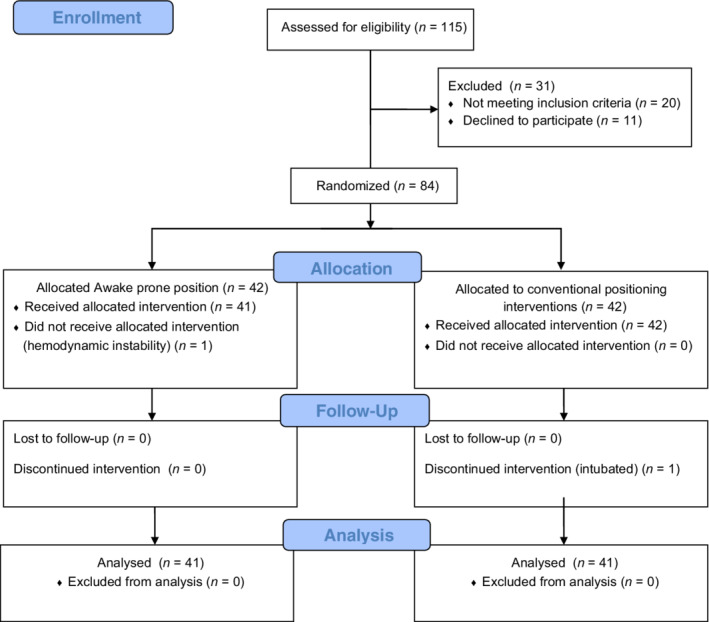
CONSORT flow diagram of participants through the trial (figure can be viewed at wileyonlinelibrary.com). CONSORT, Consolidated Standards of Reporting Trials
The results presented in Table 1 show that the groups under study were comparable; no differences in demographic or baseline characteristics were observed between the two groups. 70.7% of APP patients were male, compared with 56.1% in the control group. The mean age was 51.20 ± 13.10 and 51.95 ± 15.95 years for patients in the APP and control groups respectively. The most common comorbidities were hypertension and diabetes among both groups. According to chest X‐ray findings, the majority of patients in both groups had bilateral lower and upper lobe airspace it accounts for 92.7% of the PP group and 80.5% of the control group. Oxygen was delivered through mask reservoir in 63.4% among patients in both groups.
TABLE 1.
Patients' baseline demographic and clinical characteristics
| Characteristic | APP group (n = 41) | Control group (n = 41) | t or χ 2 | p |
|---|---|---|---|---|
| Age (years) (mean ± SD) | 51.20 ± 13.10 | 51.95 ± 15.95 | 0.05 | .81 |
| Sex n (%) | 1.89 | .16 | ||
| Male | 29 (70.7) | 23 (56.1) | ||
| Female | 12 (29.3) | 18 (43.9) | ||
| Smoking status n (%) | 4.25 | .11 | ||
| Non smoker | 21 (51.2) | 24 (58.5) | ||
| Former smoker | 0 (0.0) | 3 (7.3) | ||
| Active smoker | 20 (48.8) | 14 (34.1) | ||
| Comorbidities a n (%) | 3.03 | .55 | ||
| Hypertension | 15 (53.6) | 14 (34.1) | ||
| Diabetes | 14 (34.1) | 14 (34.1) | ||
| CHF | 4 (9.8) | 4 (9.8) | ||
| Kidney diseases | 1 (2.4) | 0 (0.0) | ||
| Cancer | 0 (0.0) | 2 (4.9) | ||
| Chest X‐ray n (%) | 2.62 | .10 | ||
| Bilateral lower lobe airspace | 3 (7.3) | 8 (19.5) | ||
| Bilateral lower and upper lobe airspace | 38 (92.7) | 33 (80.5) | ||
| Oxygen delivery interface n (%) | 0.00 | 1.00 | ||
| Mask reservoir | 26 (63.4) | 26 (63.4) | ||
| NCPAP | 15 (36.6) | 15 (36.6) | ||
| FiO2 at baseline (%) (mean ± SD) | 87.32 ± 9.75 | 87.56 ± 9.69 | −0.11 | .91 |
| SpO2/FiO2 ratio (mean ± SD) | 73.07 ± 27.28 | 80.95 ± 40.61 | −1.35 | .08 |
| Haemoglobin g/L (mean ± SD) | 12.07 ± 1.46 | 11.75 ± 1.62 | 0.93 | .35 |
| d‐Dimer mg/L (mean ± SD) | 0. 99 ± 0. 58 | 1.12 ± 0.78 | −0.87 | .38 |
Note: Data are presented as n (%), mean ± SD; χ 2, chi‐square test; t, Student's t test.
Abbreviations: CHF, congestive heart failure; FiO2, fraction of inspired oxygen; NCPAP, noninvasive continuous positive airway pressure; PP, prone position; SpO2, peripheral oxygen saturation.
Multiple answers were allowed.
4.1. Primary outcome measures
APP resulted in statistically significant improvements in oxygenation parameters, that is, SpO2, PaO2/FiO2, ROX index, PaO2, and SaO2, at the three study time points (p < .001, 0.007, p < .001, .011, and p < .001respectively) (Table 2). Furthermore, it can be noted that APP improved the SpO2, PaO2/FiO2, ROX index, PaO2, and SaO2 values for the APP group, representing an increase of 5.85%, 23.71%, 30.79%, 22.59%, and 5.26%, respectively. No statistically significant differences in pH and PaCO2 values were observed between the two groups (Table 2). However, PaCO2 values were decreased by 3.42% after APP. Similarly, the RR and HR were significantly decreased in the APP group by 18.35% and 1.8%, respectively (p < .001 and .030, respectively). The shock index revealed no discernible hemodynamic changes between the two groups during the three study time periods (Table 2).
TABLE 2.
Comparison between the APP and control groups according to oxygenation, arterial blood gases, and vital signs parameters at different time points
| Parameters | Timing of monitoring | APP group (n = 41) | Control group (n = 41) | % Change for APP | F‐test significance p‐value |
|---|---|---|---|---|---|
| (Mean ± SD) | (Mean ± SD) | ||||
| Oxygenation parameters | |||||
| SpO2 | Before | 87.05 ± 3.68 | 87.61 ± 5.03 | 5.85 |
8.15 .000** |
| After 10 min | 89.54 ± 2.77 | 88.12 ± 5.04 | |||
| After 1 h | 92.15 ± 2.73 | 88.17 ± 4.84 | |||
| PaO2/FiO2 ratio (mmHg) | Before | 79.95 ± 22.50 | 88.64 ± 34.28 | 23.71 |
4.162 .007* |
| After 1 h | 98.91 ± 34.44 | 77.61 ± 28.77 | |||
| ROX index | Before | 3.15 ± 0.41 | 3.20 ± 0.51 | 30.79 |
19.640 .000** |
| After 10 min | 3.62 ± 0.50 | 3.30 ± 0.57 | |||
| After 1 h | 4.12 ± 0.67 | 3.20 ± 0.56 | |||
| Arterial blood gases | |||||
| pH | Before | 7.40 ± 0.07 | 7.39 ± 0.09 | 0.27 |
0.944 0.421 |
| After 1 h | 7.42 ± 0.08 | 7.40 ± 0.10 | |||
| PaCO2 (mmHg) | Before | 43.27 ± 13.98 | 39.10 ± 9.04 | −3.42 |
0.830 0.479 |
| After 1 h | 41.79 ± 14.19 | 40.98 ± 10.77 | |||
| PaO2 (mmHg) | Before | 69.12 ± 17.31 | 77.39 ± 29.78 | 22.59 |
3.867 0.011* |
| After 1 h | 84.74 ± 26.00 | 68.38 ± 26.17 | |||
| SaO2 | Before | 87.51 ± 3.45 | 88.17 ± 6.53 | 5.26 |
8.267 0.000** |
| After 1 h | 92.12 ± 3.08 | 88.43 ± 4.81 | |||
| Vital signs | |||||
| Heart rate (b/m) | Before | 79.59 ± 10.83 | 83.78 ± 12.02 | −1.84 |
2.530 0.030* |
| After 10 min | 78.76 ± 7.78 | 81.83 ± 10.92 | |||
| After 1 h | 78.12 ± 8.17 | 83.83 ± 10.43 | |||
| Respiratory rate (b/m) | Before | 32.20 ± 1.83 | 31.80 ± 1.70 | −18.35 |
51.23 0.000** |
| After 10 min | 28.80 ± 2.25 | 31.05 ± 2.14 | |||
| After 1 h | 26.29 ± 2.29 | 32.05 ± 2.33 | |||
| SBP (mmHg) | Before | 111.22 ± 10.53 | 114.15 ± 15.16 | −0.65 |
0.946 0.452 |
| After 10 min | 111.22 ± 9.27 | 113.17 ± 12.33 | |||
| After 1 h | 110.49 ± 9.20 | 114.88 ± 13.25 | |||
| DBP (mmHg) | Before | 71.22 ± 5.99 | 72.93 ± 8.13 | 1.02 |
0.699 0.625 |
| After 10 min | 70.93 ± 12.05 | 73.66 ± 8.59 | |||
| After 1 h | 71.95 ± 6.00 | 73.17 ± 8.78 | |||
| Shock index | Before | 0.71 ± 0.12 | 0.74 ± 0.14 | −1.40 |
0.575 0.719 |
| After 10 min | 0.70 ± 0.09 | 0.72 ± 0.12 | |||
| After 1 h | 0.70 ± 0.08 | 0.73 ± 0.12 | |||
| Temperature (°C) | Before | 37.24 ± 0.33 | 37.24 ± 0.30 | 0.00 |
0.130 0.986 |
| After 10 min | 37.21 ± 0.33 | 37.22 ± 0.26 | |||
| After 1 h | 37.24 ± 0.34 | 37.21 ± 0.25 | |||
Abbreviations: APP, awake prone position; DBP, diastolic blood pressure; shock index, ratio of heart rate to systolic arterial pressure; F, F‐test (one‐way ANOVA); ROX index, ratio of SpO2/FiO2 ratio to respiratory rate; SBP, systolic blood pressure; SpO2, peripheral oxygen saturation.
Significant p at ≤.05.
Highly significant; % change, percent difference between before and after prone positioning.
4.2. Secondary outcome measures
Among the patients in the APP group, 51.2% tolerated PP for more than 3 h (Table 3). According to our findings, 65.8% of the patients in the APP group highly accepted this manoeuvre, whereas only 7.4% indicated a low degree of acceptance. Moreover, no adverse events related to the APP procedure were recorded among 48.7% of the patients in the APP group. Meanwhile, 29.3% of APP patients had general discomfort, 12.2% had nausea and vomiting, and 9.8% had musculoskeletal pain.
TABLE 3.
Secondary outcomes of prone positioning among the patients in the APP group
| Secondary outcomes | No. | % |
|---|---|---|
| Tolerability (session duration in hours) | ||
| 1 to <2 | 7 | 17.1 |
| 2 to <3 | 13 | 31.7 |
| ≥3 | 21 | 51.2 |
| Acceptability | ||
| High | 27 | 65.8 |
| Moderate | 11 | 26.8 |
| Low | 3 | 7.4 |
| Adverse events | ||
| No | 20 | 48.7 |
| General discomfort | 12 | 29.3 |
| Musculoskeletal pain | 4 | 9.8 |
| Nausea and vomiting | 5 | 12.2 |
5. DISCUSSION
This study is one of the first studies in Egypt that assessed the effects of APP on oxygenation and physiological outcomes in non‐intubated patients with COVID‐19 admitted to the ICU. The results of this study indicated that APP significantly improved oxygenation parameters, that is, SpO2, PaO2/FiO2 ratio, ROX index, and SaO2. One of the reasons for this outcome is the availability of a higher proportion of potentially recruitable alveoli in the early stages of respiratory distress, as we started the APP sessions in this study early. Other physiological parameters supporting this improvement are the reduction of shunting and V/Q mismatch caused by APP. 10 , 11
The findings in this study agree with a study conducted in São Paulo, Brazil by Sztajnbok et al., 10 who evaluated the effects of APP on oxygenation among non‐intubated patients with COVID‐19, suggesting that APP was associated with a significant improvement in SpO2 and the PaO2/FiO2 ratio. Similarly, a Spanish study directed by Taboada et al. 24 found that SpO2 and PaO2/FiO2 significantly increased after PP compared with those before PP implementation. Moreover, Sanz‐Moncusi et al. 25 investigated the use of NIV with APP. They recorded an improvement in the oxygenation parameters after placing the patient prone. Sartini et al. 26 used sessions of PP with CPAP in Italian patients with mild‐to‐moderate ARDS. During and after PP, all patients exhibited a substantial reduction in RR and a significant improvement in SpO2 and the PaO2/FiO2 ratio. Only 13% had no improvement. Contrary to the findings of this study, a study conducted in Aix‐en‐Provence Hospital (France) by Elharrar et al. 27 reported that only 25% of patients showed oxygenation improvement during PP.
In relation to the ROX index, the results of this study indicated that the ROX index values significantly improved in the APP group compared with those in the control group. This improvement may be explained by the decrease in the work of breathing, which in turn helped avoid disease progression. The ROX index, which presents a composite of both oxygenation and work of breathing, has previously been approved in patients with ARDS treated with high‐flow nasal cannula therapy. 28 In this study, the improvement in the ROX index achieved using APP on CPAP or a non‐rebreather mask implies that using this technique prevents disease progression by lowering the work of breathing. Many studies have addressed improvement in the ROX index for patients with COVID‐19 achieved by PP on NIV. A study carried out in England by Winearls et al. 29 reported that the use of PP with CPAP significantly improved the ROX index in patients with COVID‐19. Similarly, Sryma et al. 30 found significant improvement in the ROX index values in the study group compared with that in the control group 30 min after PP initiation.
Regarding the effects of APP on ABG parameters, our findings did not demonstrate a significant improvement in PaCO2 after APP implementation. The findings of this study agree with those of Clarke et al., 31 Coppo et al., 32 and Elharra et al. 27 APP is thought to minimize regions of over‐distension in the lung, resulting in less physiological dead space and perhaps lower PaCO2. 16 Because of variations in RR, tidal volume, and minute ventilation, PaCO2 outcomes in awake spontaneously breathing patients in this study may not be reliable. In contrast, it was reported that in mechanically ventilated patients, controlled ventilation with PP reduced the shunt fraction and improved CO2 removal. 33 Furthermore, the findings of this study showed no significant differences in the mean values of pH among the groups under study; however, we found a significant improvement in gas exchange parameters among patients receiving PP, as assessed by better before and after PaO2 and SaO2 values. The findings of this study agree with those of Liu et al., 34 who found that PP did not significantly affect the mean values of pH and PaCO2 among the groups under study. In contrast, Jouffroy et al. 35 have reported that the mean values of pH and PaCO2 significantly improved after PP implementation, whereas the mean values of PaO2 and SaO2 showed no significant improvement with PP.
Regarding the effects of APP on patients' vital signs, the results of this study suggested that PP significantly improved the mean values of HR and RR. No detectable significant hemodynamic differences were observed in the mean values of SBP, DBP, temperature, and shock index. The findings of this study agree with those of the studies by Padrao et al., 36 Jouffroy et al., 35 Liu et al., 34 and Sztajnbok et al. 10 Moreover, Damarla et al. 37 compared the improvement in the RR before using PP and approximately 1 h after initial PP. They found that all patients showed a significant reduction in the RR 1 h after PP implementation.
The findings of this study illustrated that most patients tolerated PP for more than 3 h. These findings agree with the results of Padrao et al., 36 who reported that more than half of the patients tolerated PP sessions for more than 4 h. Furthermore, our findings are consistent with those of Coppo et al., 32 who indicated that PP was feasible (i.e., sustained for at least 3 h) in most patients investigated.
Our findings showed that most patients in the APP group had positive responses to PP, as evidenced by a 23.71% rise in the PaO2/FiO2 ratio and an 18.35% decline in the RR. Similarly, Sartini et al. 26 reported that most patients who underwent PP responded positively to the procedure. Elharrar et al. 27 found that only 25% of the patients responded to therapy. The findings of this study showed that nearly half of the patients in the APP group had no adverse events related to APP. This may be attributed to conscious patients being able to adjust their posture when they were prone, which likely contributed to the lower complication rate. Moreover, it is thought that APP was associated with fewer adverse effects in this study because the duration of PP was decided by the patients as tolerated. Similarly, Coppo et al. 32 found no adverse events related to the APP procedure. In contrast, Shoults et al. in United States 38 discovered that all patients rated discomfort as the primary factor for being unable to resume a PP session. Since the beginning of the pandemic, there have been few case reports of patients with COVID‐19 with respiratory difficulties after self‐proning. 10 , 39 , 40
Overall, APP improved oxygenation and physiological indices in our study, and it was well tolerated by most patients without serious adverse events. However, it should be emphasized that the measurements in our study were performed immediately before patient positioning, 10 min after patient positioning, and 1 h after patient positioning, and were not repeated in the same patient at different time periods. So, it's important for the CCNs to keep in mind that these improvements after an hour may vary at a later period. A recent meta‐trial of six randomized clinical trials involving hospitals from six countries: Canada, France, Ireland, Mexico, USA, Spain 41 conducted to assess both short‐term and long‐term outcomes of APP revealed that RR, and ROX index were all significantly improved during the first session of APP, which lasted a median of 3 h, while at day 28 of the meta‐trial decrease in intubation and mortality rates were similar between the intervention and the control group. Moreover, in a more recent non‐randomized controlled trial conducted in USA 42 to identify whether the recommendation of APP is associated with better outcomes among awake non‐intubated patients with COVID‐19. They found significant evidence of worsening clinical outcomes among patients allocated for the APP intervention on study day 5, indicating potential harm.
5.1. Limitations
This study must be interpreted within the light of its limitations. First, this study was conducted in a single location; the results of this study may not be generalizable. Second, both groups were not blinded to the researchers due to the nature of the intervention, increasing the risk of bias. Another limitation in our study is that the clinical outcomes were only followed for a short period of time (1 h). As a result, establishing causality from observable changes is challenging. Future randomized control studies with long‐term follow‐up are needed to confirm these findings. If a new research is done, a multicenter study with a larger sample size is recommended. Further studies on the effects of APP on intubation delay and avoidance, duration of weaning from oxygen support, length of ICU stay, and mortality is needed.
5.2. Implications and recommendations for practice
The evidence gained from our study will add knowledge to critical care nursing practices regarding APP as a rescue therapy for COVID‐19 patients especially with limited advanced ICU resources during the pandemic. In this study APP safely improve oxygenation and physiological parameters. Additionally, it is simple, easy to apply and does not increase workload on nurses or need an additional staff. So, CCNs should use specific protocols when applying APP to support patient decisions and limit the occurrence of complications.
6. CONCLUSION
The findings of this study showed that exposing non‐intubated patients with COVID‐19 to APP resulted in a considerable improvement in oxygenation parameters and RR. Furthermore, APP was tolerable by most patients without serious adverse events and no detectable hemodynamic differences during proning.
Othman SY, El‐Menshawy AM, Mohamed AM. Effects of awake‐prone positioning on oxygenation and physiological outcomes in non‐intubated patients with COVID‐19: A randomized controlled trial. Nurs Crit Care. 2022;1‐9. doi: 10.1111/nicc.12833
REFERENCES
- 1. Cotton S, Zawaydeh Q, LeBlanc S, Husain A, Malhotra A. Proning during covid‐19: challenges and solutions. Heart Lung. 2020;49(6):686‐687. [DOI] [PMC free article] [PubMed] [Google Scholar]
- 2. Binda F, Marelli F, Galazzi A, Pascuzzo R, Adamini I, Laquintana D. Nursing management of prone positioning in patients with COVID‐19. Crit Care Nurse. 2020;41:e1‐e9. [DOI] [PubMed] [Google Scholar]
- 3. Pattison N. End‐of‐life decisions and care in the midst of a global coronavirus (COVID‐19) pandemic. Intensive Crit Care Nurs. 2020;58:102862. [DOI] [PMC free article] [PubMed] [Google Scholar]
- 4. Binda F, Galazzi A, Marelli F, et al. Complications of prone positioning in patients with COVID‐19: a cross‐sectional study. Intensive Crit Care Nurs. 2021;67:10308. [DOI] [PMC free article] [PubMed] [Google Scholar]
- 5. Anand S, Baishya M, Singh A, Khanna P. Effect of awake prone positioning in COVID‐19 patients—a systematic review. Trends Anaesth Crit Care. 2021;36:17‐22. [DOI] [PMC free article] [PubMed] [Google Scholar]
- 6. Ramadan HK, Mahmoud MA, Aburahma MZ, et al. Predictors of severity and co‐infection resistance profile in COVID‐19 patients: first report from upper Egypt. Infect Drug Resist. 2020;13:3409‐3422. [DOI] [PMC free article] [PubMed] [Google Scholar]
- 7. Schittek GA, Bornemann‐Cimenti H, Sandner‐Kiesling A. Wellbeing of ICU patients with COVID‐19. Intensive Crit Care Nurs. 2021;65:103050. [DOI] [PMC free article] [PubMed] [Google Scholar]
- 8. Pickler RH, Abshire DA, Chao AM, et al. Nursing science and COVID‐19. Nurs Outlook. 2020;68(5):685‐688. [DOI] [PMC free article] [PubMed] [Google Scholar]
- 9. Guerin C, Reignier J, Richard JC, et al. Prone positioning in severe acute respiratory distress syndrome. N Engl J Med. 2013;368(23):2159‐2168. [DOI] [PubMed] [Google Scholar]
- 10. Sztajnbok J, Maselli‐Schoueri JH, de Resende C, et al. Prone positioning to improve oxygenation and relieve respiratory symptoms in awake, spontaneously breathing non‐intubated patients with COVID‐19 pneumonia. Respir Med Case Rep. 2020;30:101096. [DOI] [PMC free article] [PubMed] [Google Scholar]
- 11. Flynn Makic MB. Prone position of patients with COVID‐19 and acute respiratory distress syndrome. J Perianesth Nurs. 2020;35(4):437‐438. [DOI] [PMC free article] [PubMed] [Google Scholar]
- 12. Weatherald J, Solverson K, Zuege DJ, Loroff N, Fiest KM, Parhar KKS. Awake prone positioning for COVID‐19 hypoxemic respiratory failure: a rapid review. J Crit Care. 2021;61:63‐70. [DOI] [PMC free article] [PubMed] [Google Scholar]
- 13. Alhazzani W, Moller MH, Arabi YM, et al. Surviving sepsis campaign: guidelines on the management of critically ill adults with coronavirus disease 2019 (COVID‐19). Intensive Care Med. 2020;46(5):854‐887. [DOI] [PMC free article] [PubMed] [Google Scholar]
- 14. Caputo ND, Strayer RJ, Levitan R. Early self‐proning in awake, non‐intubated patients in the emergency department: a single ED's experience during the COVID‐19 pandemic. Acad Emerg Med. 2020;27(5):375‐378. [DOI] [PMC free article] [PubMed] [Google Scholar]
- 15. Bamford P, Bentley A, Dean J, Whitmore D, Wilson‐Baig N. Intensive care society guidance for prone positioning of the conscious COVID patient 2020. https://emcrit.org/wp-content/uploads/2020/04/2020-04-12-Guidance-for-conscious-proning.pdf
- 16. Chua EX, Zahir S, Ng KT, et al. Effect of prone versus supine position in COVID‐19 patients: a systematic review and meta‐analysis. J Clin Anesth. 2021;74:110406. [DOI] [PMC free article] [PubMed] [Google Scholar]
- 17. Thompson AE, Ranard BL, Wei Y, Jelic S. Prone positioning in awake, nonintubated patients with COVID‐19 hypoxemic respiratory failure. JAMA Intern Med. 2020;180:1537. [DOI] [PMC free article] [PubMed] [Google Scholar]
- 18. Dubosh NM, Wong ML, Grossestreuer AV, et al. Early, awake proning in emergency department patients with COVID‐19. Am J Emerg Med. 2020;46:640‐645. [DOI] [PMC free article] [PubMed] [Google Scholar]
- 19. Solverson K, Weatherald J, Parhar KKS. Tolerability and safety of awake prone positioning COVID‐19 patients with severe hypoxemic respiratory failure. Can J Anaesth. 2021;68(1):64‐70. [DOI] [PMC free article] [PubMed] [Google Scholar]
- 20. Schulz KF, Altman DG, Moher D. CONSORT 2010 statement: updated guidelines for reporting parallel group randomised trials. BMC Med. 2010;8:18. [DOI] [PMC free article] [PubMed] [Google Scholar]
- 21. Gad S. Awake prone positioning versus non invasive ventilation for COVID‐19 patients with acute hypoxemic respiratory failure. Egypt J Anaesth. 2021;37(1):85‐90. [Google Scholar]
- 22. Rosner B. Fundamentals of Biostatistics 5th Edition (without Data Disk). Duxbury Thomson Learning; 1999. [Google Scholar]
- 23. Masoud HH, Elassal G, Zaky S, Kamal EK. Management protocol for COVID‐19 patients version 1.4/30th May 2020 Ministry of Health and Population (MOHP), Egypt. Coronavirus Disease 2019 (COVID‐19); SARS COV2 Management Guide Line. Ministry of Health and Population; 2020. Accessed May 27, 2021. http://www.mohp.gov.eg/JobsDetails.aspx?job_id=3061 [Google Scholar]
- 24. Taboada M, Rodriguez N, Riveiro V, Baluja A, Atanassoff PG. Prone positioning in awake non‐ICU patients with ARDS caused by COVID‐19. Anaesth Crit Care Pain Med. 2020;39(5):581‐583. [DOI] [PMC free article] [PubMed] [Google Scholar]
- 25. Sanz‐Moncusi M, Rossello‐Sancho J, Garcia‐Alamino JM. Use of high‐flow nasal cannula in COVID‐19 has improved effectiveness, safety and tolerability when applied in lateral position compared with prone positioning. Intensive Crit Care Nurs. 2021;66:103061. [DOI] [PMC free article] [PubMed] [Google Scholar]
- 26. Sartini C, Tresoldi M, Scarpellini P, et al. Respiratory parameters in patients with COVID‐19 after using noninvasive ventilation in the prone position outside the intensive care unit. JAMA. 2020;323(22):2338‐2340. [DOI] [PMC free article] [PubMed] [Google Scholar]
- 27. Elharrar X, Trigui Y, Dols AM, et al. Use of prone positioning in nonintubated patients with COVID‐19 and hypoxemic acute respiratory failure. JAMA. 2020;323(22):2336‐2338. [DOI] [PMC free article] [PubMed] [Google Scholar]
- 28. Roca O, Messika J, Caralt B, et al. Predicting success of high‐flow nasal cannula in pneumonia patients with hypoxemic respiratory failure: the utility of the ROX index. J Crit Care. 2016;35:200‐205. [DOI] [PubMed] [Google Scholar]
- 29. Winearls S, Swingwood EL, Hardaker CL, et al. Early conscious prone positioning in patients with COVID‐19 receiving continuous positive airway pressure: a retrospective analysis. BMJ Open Respir Res. 2020;7(1):000711. [DOI] [PMC free article] [PubMed] [Google Scholar]
- 30. Sryma PB, Mittal S, Mohan A, et al. Effect of proning in patients with COVID‐19 acute hypoxemic respiratory failure receiving noninvasive oxygen therapy. Lung India. 2021;38(Supplement):S6‐S10. [DOI] [PMC free article] [PubMed] [Google Scholar]
- 31. Clarke J, Geoghegan P, McEvoy N, et al. Prone positioning improves oxygenation and lung recruitment in patients with SARS‐CoV‐2 acute respiratory distress syndrome; a single centre cohort study of 20 consecutive patients. BMC Res Notes. 2021;14(1):20. [DOI] [PMC free article] [PubMed] [Google Scholar]
- 32. Coppo A, Bellani G, Winterton D, et al. Feasibility and physiological effects of prone positioning in non‐intubated patients with acute respiratory failure due to COVID‐19 (PRON‐COVID): a prospective cohort study. Lancet Respir Med. 2020;8(8):765‐774. [DOI] [PMC free article] [PubMed] [Google Scholar]
- 33. Gattinoni L, Vagginelli F, Carlesso E, et al. Decrease in PaCO2 with prone position is predictive of improved outcome in acute respiratory distress syndrome. Crit Care Med. 2003;31(12):2727‐2733. [DOI] [PubMed] [Google Scholar]
- 34. Liu X, Liu H, Lan Q, Zheng X, Duan J, Zeng F. Early prone positioning therapy for patients with mild COVID‐19 disease. Med Clin. 2021;156(8):386‐389. [DOI] [PMC free article] [PubMed] [Google Scholar]
- 35. Jouffroy R, Darmon M, Isnard F, et al. Impact of prone position in non‐intubated spontaneously breathing patients admitted to the ICU for severe acute respiratory failure due to COVID‐19. J Crit Care. 2021;64:199‐204. [DOI] [PMC free article] [PubMed] [Google Scholar]
- 36. Padrao EMH, Valente FS, Besen B, et al. Awake prone positioning in COVID‐19 hypoxemic respiratory failure: exploratory findings in a single‐center retrospective cohort study. Acad Emerg Med. 2020;27(12):1249‐1259. [DOI] [PubMed] [Google Scholar]
- 37. Damarla M, Zaeh S, Niedermeyer S, et al. Prone positioning of nonintubated patients with COVID‐19. Am J Respir Crit Care Med. 2020;202(4):604‐606. [DOI] [PMC free article] [PubMed] [Google Scholar]
- 38. Shoults B, Barber M, Millham L, et al. Feasibility and limitations of proning protocol for nonintubated patients with COVID‐19. J Patient Exp. 2021;8:2374373520981486. [DOI] [PMC free article] [PubMed] [Google Scholar]
- 39. Slessarev M, Cheng J, Ondrejicka M, Arntfield R. Patient self‐proning with high‐flow nasal cannula improves oxygenation in COVID‐19 pneumonia. Can J Anaesth. 2020;67(9):1288‐1290. [DOI] [PMC free article] [PubMed] [Google Scholar]
- 40. Elkattawy S, Noori M. A case of improved oxygenation in SARS‐CoV‐2 positive patient on nasal cannula undergoing prone positioning. Respir Med Case Rep. 2020;30:101070. [DOI] [PMC free article] [PubMed] [Google Scholar]
- 41. Ehrmann S, Li J, Ibarra‐Estrada M, et al. Awake prone positioning for COVID‐19 acute hypoxaemic respiratory failure: a randomised, controlled, multinational, open‐label meta‐trial. Lancet Respir Med. 2021;9(12):1387‐1395. [DOI] [PMC free article] [PubMed] [Google Scholar]
- 42. Qian ET, Gatto CL, Amusina O, et al. Assessment of awake prone positioning in hospitalized adults with COVID‐19: a nonrandomized controlled trial. JAMA Intern Med. 2022;182(6):612‐621. [DOI] [PMC free article] [PubMed] [Google Scholar]