Abstract
The toxin and antitoxin modules in bacteria consist of a toxin molecule that has activity to inhibit various cellular processes and its cognate antitoxin that neutralizes the toxin. This system is considered taking part in the formation of persister cells, which are a subpopulation of recalcitrant cells able to survive antimicrobial treatment without any resistance mechanisms. Importantly, persisters have been associated with long-term infections and treatment failures in healthcare settings. It is a public health concern since persisters can be involved in the evolution and dissemination of antimicrobial resistance amidst the aggravating spread of multidrug-resistant bacteria and insufficient novel antimicrobial therapy to tackle this issue. Salmonella enterica serovar Typhimurium is one of the most prevalent Salmonella serotypes in the world and is a leading cause of food-borne salmonellosis. S. Typhimurium has been known to cause persistent infection and a wealth of investigations on Salmonella persisters indicates that toxin and antitoxin modules play a role in mediating the phenotypic switch of persisters, rendering its survival ability in the presence of antimicrobial agents. In this review, we discuss findings regarding mechanisms that underly persistence in S. Typhimurium, especially the involvement of toxin and antitoxin modules.
Keywords: toxin, antitoxin, antimicrobial resistance, persisters, Salmonella
Introduction
Salmonella enterica is a complex and the largest species in the genus of Salmonella, comprising six subspecies which are further classified into serovars according to the White-Kauffman-Le Minor scheme.1,2 These Gram-negative and facultative intracellular enteropathogenic bacteria can colonize different types of hosts, causing diseases with various degree of severity. The majority of the serovars that are associated with humans and animals belong to subspecies I (subsp. enterica) and they are generally classified into typhoidal (TS) and non-typhoidal Salmonella (NTS) based on the disease and host specificity. The former includes serovars that cause invasive diseases and can only infect humans or higher primates, whereas the latter has a broader host range with the clinical manifestations depending on the host susceptibility and responses.3,4 The Salmonella enterica subsp. enterica serovar Typhimurium (S. Typhimurium) is one of the most prevalent NTS responsible for self-limiting inflammation in human intestines, yet has also been reported to emerge as the causative agent of invasive NTS (iNTS) infection.3,5,6
The resolution of disease following Salmonella infection can be achieved even when the pathogen has not completely cleared from the host. In this state, which is referred to as persistence of infection, the bacteria can survive for a prolonged period beyond the intestinal mucosa and evade the host immune response without necessarily establishing clinical manifestations.7 Persistence is typically associated with TS serovars that can remain in the carrier host between months to years. However, it has also been documented to a lesser extent in NTS infection and generally occurs for not more than 12 months.3,8,9 The sustained colonization can lead to relapse as it induces organ lesions at localization sites or re-seeding of the surviving bacteria to intestinal mucosa. In this context, the reappearance of symptoms is caused by the recrudescence of initial infection.3,7 Previous reports showed that recrudescence predominates the relapse cases in S. Typhimurium infection since the isolates from initial infection and during relapse were clonally related.10,11 Those findings present the challenge for bacterial eradication and are persuasive of the importance to better understand the Salmonella pathogenesis during long-term infection.
Intermittent shedding of Salmonella in asymptomatic carriers and acute relapse of NTS infection are often associated with antibiotic treatment.11–13 The majority of the relapse cases in some regions were also reported to involve multidrug-resistant (MDR) strains.4 The intracellular localization of bacteria was presumed to provide a protective niche from antibiotic killing effect.13 However, the existence of recalcitrant cells seems more plausible to render Salmonella persistence based on experimental findings.7 These subpopulations of bacterial cells are termed “persisters” and have the ability to survive antibiotic treatment by reducing cell growth through a transient phenotypic switch. Strikingly, they can resume growth under normal conditions and produce antibiotic-sensitive progeny.14 Antibiotic persisters have been observed in invasive S. Typhimurium infection through a murine typhoid model.15–17 The reduced growth rate and physiological properties of the surviving cells following antibiotic treatment have implicated its significance and further provide insights about persistence of Salmonella infection. However, it is noteworthy that defining a persister is not trivial as it represents a tiny proportion of the clonal bacterial population. Other survival phenomena with very similar characteristics, such as tolerant cells and viable but nonculturable (VBNC) state, may consequently confound the study of persisters as well.
Several mechanisms that underly the occurrence of persisters have been proposed through experimental studies.18–21 It is mostly associated with bacterial strategy to survive environmental stressors or antimicrobials, which results in a state of dormancy and temporal tolerant phenotype. In S. Typhimurium, several toxin-antitoxin (TA) modules have been reported to increase the proportion of persisters under host microenvironment conditions and therefore suggested to contribute to the formation of Salmonella persisters.16,22–24 The toxins from TA modules can interfere with various vital processes which are linked to cell growth (i.e. DNA replication, transcription, or translation).25–27 This implicates an interesting point regarding the role of TA module in switching the phenotype of antibiotic-recalcitrant Salmonella and influencing the physiological dynamics of bacterial cell growth. Moreover, understanding the complexity of persister formation is essential to address its relevance in long-term infection, as well as devise a strategy for pathogen eradication. The occurrence of persisters has also been proposed to promote emergence of antimicrobial resistance through horizontal gene transfer.28 It is indeed becoming another public health concern, particularly in the efforts to control the emergence of resistant pathogens. This review therefore focuses on the significance of S. Typhimurium persisters with particular emphasis in the involvement of TA modules. We first elaborate the clinical relevance of Salmonella persisters and highlight the prominent features that distinguish them from other survival phenomena. Then, we discuss both corroborating and contradictory findings regarding the role of TA modules in Salmonella persisters.
Persistence of S. Typhimurium Infection and Its Implications
S. Typhimurium belongs to the NTS group that can infect a broad range of hosts with different clinical manifestations. In humans, NTS infection normally causes self-limiting acute gastroenteritis and is rarely associated with invasive disease. The NTS infections are considerably less studied than those caused by TS serovars, which perhaps is due to the lower fatality in the general population. Infection of S. Typhimurium in susceptible mice, on the other hand, notably causes typhoid-like disease with similar clinical manifestations to those observed in TS infection in humans. This serovar was therefore used to develop a murine typhoid infection model due to the restricted host of TS serovars.29,30 It has then become an important experimental model to study Salmonella pathogenesis and the host response during systemic infection and persistence.
NTS infection in humans has emerged as one of the leading causes of foodborne illness globally. The prevalence is estimated to be 1.4 million cases per year in the US with S. Typhimurium being the most frequently identified serovar.31 It is also regarded as the most common cause of diarrhea in Asia and the emergence of its invasive form is rising in some regions, despite limited documented data.32–34 According to several clinical reports, systemic infections of S. Typhimurium, such as bacteremia, are prevalent in the population with immunocompromised conditions.35–37 Approximately 5% of NTS cases in high-income countries will develop to systemic infection with relatively low mortality. In contrast, iNTS is more problematic in Sub-Saharan Africa where the fatality rate is high (20–45%), especially in the population with high incidence of HIV, malaria, and malnutrition.5,6,10,38
One of the key features in Salmonella pathogenesis is the ability of this pathogen to invade extraintestinal tissues. The host immune response additionally restricts intracellular replication and thus allows bacteria to reside within the preferred host for prolonged periods without causing any symptoms.3,4 The compounding effect of survivors in the colonized tissues and its growth resumption is able to cause the symptoms of acute relapse during persistence. Despite the scarcity of studies on relapse episodes, a report showed a 2.2% prevalence of persistent NTS infection among the total Salmonella infection in Israel. Around 65% of those cases were associated with relapsing diarrhea, which is notably different from the majority of asymptomatic carriers in TS infection. The study also indicated that young age and antibiotic treatment are likely to contribute in the development of symptomatic relapse.11 Similarly, a randomized controlled trial suggested that antibiotic use during initial infection is a risk factor for acute relapse. The symptom resolution and early negative stool cultures was achieved within the first week of antibiotic treatment in patients with acute NTS infection, yet positive stool cultures will likely occur in the third week followed by relapse episode.39 Inadequate antibiotic treatment in NTS infection was also found to cause relapse among cancer patients.36
S. Typhimurium entry into host systemic circulation occurs mainly through invasion of phagocytic microfold (M) cells that cover the Peyer’s patches of intestinal lumen. The destruction of M cells is particularly mediated by expression of type III secretion system (TTSS-1) effectors encoded in Salmonella Pathogenicity Island-1 (SPI-1). As a result, bacteria can penetrate further into deeper enterocytes and reach the lamina propria where they are taken up by phagocytes and transported to mesenteric lymph nodes (MLNs). The alternative bacterial uptake pathway mediated by dendritic cells in the lamina propria has also been reported which does not involve M cell invasion, but rather dendritic cells expansion to the intestinal mucosa that actively captures the bacteria.40,41 Salmonella-infected phagocytes are vital for systemic infection since they provide access for the bacteria to reach the lymphatics and bloodstream, in which they can eventually colonize many organs, such as liver, spleen, gall bladder, and bone marrow.7,40
Tissue-associated S. Typhimurium has been shown to contain a small amount of persisters. This is because an analysis of surviving colonies in infected mice after antibiotic treatment revealed that the recalcitrant cell can resume growth, as well as remain antibiotic-sensitive and fully virulent when reinfected to the naive host.15,16 In addition, a distinct level of antibiotic tolerance due to phenotypic variation has been described in S. Typhimurium. A single-cell analysis using fluorescence reporter revealed the heterogenous growth rate of S. Typhimurium in the murine typhoid model. Slower growth rates are likely to display better tolerance since slow growing/non-dividing cells had the highest survival in the study, yet their rarity is deemed negligible to impact the outcome of antibiotic treatment. The moderately growing cells, on the other hand, had a greater proportion among survivors and were thus suggested to be responsible for delayed eradication.17 Proteomic analysis of those different subsets showed that ribosomal proteins are more abundant in the fast-growing population, whereas the proteome content of the slow growing/ non-dividing subset is more related to nutrient limitation response.17 Other studies have also demonstrated that S. Typhimurium persisters inside the host cell maintain their metabolic status, although being in non-growing state.16,42
The low metabolic activity in persisters is maintained through inhibition of protein and DNA synthesis, which suggests that the availability of antibiotic targets is reduced. Since the antibiotics mostly target essential components of a bacterial cell (i.e., peptidoglycan in the cell wall and enzymes involved in DNA synthesis), the transiently non-growing or slowly growing persister become temporarily non-viable for antibiotic killing mechanisms.21,43–45 Consequently, the antibiotic persister can be difficult to eradicate despite extended and aggressive antibiotic treatment, especially when they reside in the protective niche that can promote persister phenotype (i.e., biofilm, host microenvironment). Delayed eradication of S. Typhimurium can become the cause of treatment failure and further develop into relapse episodes when persisters reseed at the infection site in the gastrointestinal tract. In the clinical context, it will require multiple courses of antibiotics or adjustment of treatment duration which can possibly promote the selection of antimicrobial resistance traits.
There has also been evidence indicating the contribution of persisters in the evolution of antimicrobial resistance. A study using S. Typhimurium persisters demonstrated their potential role as both recipient and donor of transferable plasmids containing antimicrobial resistance determinants in murine gut.46,47 Interestingly, a case control study has described the occurrence of plasmid acquisition conferring extended-spectrum beta-lactamase activity in S. Typhimurium serial isolates from a NTS patient.11 Beside the acquired resistance involving a mobile genetic element, prolonged and repetitive antibiotic exposure may also potentially drive genetic alterations that eventually result in tolerance or resistance. Some laboratory experiments have shown that cyclical exposure to antibiotics in persister-derived culture leads to tolerance mutations which further can facilitate in the selection of stable antibiotic-resistant phenotype.45,48,49 However, it remains inconclusive if the bacterial persister in a clinical setting will encounter a similar fate as shown in the laboratory experimental model. The whole genome sequence analysis of S. Typhimurium isolates from infected individuals associated with relapse revealed that mutation in the coding regions had no correlation with tolerance,49 but may affect the virulence traits (motility, capsule production, and biofilm formation).50 In contrast, a study involving patients infected with methicillin-resistant Staphylococcus aureus (MRSA) showed the emergence of tolerance mutation leading to the rapid evolution of resistant strains.51 The disparity across species shown in those studies may be due to their differing mutation rates, as well as characteristics of host-pathogen interactions that can possibly give additional factors to promote genetic alteration during persistence.
Apart from relapsing infection and delayed bacterial eradication, persistence of Salmonella infection also implicates the potential risk for person-to-person transmission since the carrier is able to excrete viable bacteria through feces and urine. The age-dependent host susceptibility has been suggested to influence the fecal-oral route of NTS transmission from infected family members to neonates or young infants.52 However, it may not be considered as the main route of transmission in the general population due to the low prevalence of persistent NTS infection and relatively short fecal shedding period.8 According to several studies, community outbreaks typically involve contamination of such serovar in food products.53–55 Dissemination of S. Typhimurium through the food chain may be attributed to its vast animal host range that serves as an environmental reservoir (i.e., pigs, cattle, and poultry). Moreover, this serovar is known to produce a subset of super-shedder phenotype in asymptomatic animals8 which may become the main source of transmission between livestock animals.
Importance of Defining Persistence in the Study of Persisters
The term “persister” was first introduced in the 1940s to refer to a bacterial cell that can escape the antibiotic killing after it was observed that penicillin could not completely sterilize the Staphylococci culture. The descendant cells derived from surviving colonies were particularly able to regrow in the absence of antibiotics and remain susceptible to penicillin. One of the hypotheses from that study was that persisters are present in a dormant and non-dividing state which allows them to withstand the lethal effect of penicillin since only the actively dividing cells are viable for antibiotic action.56 Nearly four decades later, cells with high-frequency persistence (Hip) phenotype were isolated from Escherichia coli K-12 which supports the previous evidence regarding the occurrence of persisters in a bacterial population.57 By using the microfluidic device and time-lapse microscopy, another study further attempted to observe bacterial growth dynamics at single-cell level and revealed the phenotypic switch of persisters in E. coli K-12 as a result of heterogeneous growth rates within a clonal population. Three subsets of cell population were proposed to exist in the wildtype culture based on their growth dynamics, namely the normal cell, slow growing persister, and growth-arrested persister. The persisters were shown to display a distinct growth characteristic compared with its normal counterpart prior to antibiotic treatment.58 To date, persisters have been described in several medically important bacteria, such as Staphylococcus aureus, Mycobacterium tuberculosis, Pseudomonas aeruginosa, uropathogenic E. coli, Candida albicans, and S. Typhimurium.20,59–62
Regarding the nature of persisters in bacterial populations, there have been considerable efforts to understand their formation and survival mechanisms. Their presence as a small subset of cells in a clonal population of bacteria was one of the considerations to develop an appropriate experimental model that can explain the biological pathways involved in persister formation. Moreover, the transient antibiotic tolerance phenotype displayed by persisters is often confused with the genetic tolerance phenomenon that presents more homogeneously in the whole population. Limitations in experimental procedures to define bacterial survival strategy can confound and complicate the investigation. One important aspect in this regard is the variability of growth rates within a clonal bacterial population, from which the different fractions of cells may arise with distinct or even very similar phenotypic traits. As discussed in several previous reviews,14,63–65 it is therefore crucial to pinpoint a reliable definition and assess experimental models that are suitable to study persisters. The definitions and practical guidelines to study antibiotic persistence in vitro have been proposed by a group of investigators working in this field.14 Notably, the three terms to describe survival mechanism of bacteria (i.e., resistance, persistence, and tolerance), as well as their characteristics, were emphasized to further allow the discrimination between persistence and other survival phenomena.
Resistance is defined as the ability to replicate and survive in the presence of certain antimicrobial agents within the minimum inhibitory concentration (MIC). Resistance is a heritable trait since it involves genetic alterations of the bacterial genome which are responsible for survival and a higher concentration of antibiotic is required to achieve a similar bactericidal effect as in susceptible bacteria. The elevation of MIC is a prominent feature of drug-resistant bacteria that can be observed through standard antimicrobial susceptibility testing. Meanwhile, tolerance is defined as the ability to survive the duration of a transient antibiotic treatment several times above the MIC without resistance mechanism.14 Both tolerance and persistence are survival phenomena that are associated with reduced growth rate, display lower cellular activity, and are not characterized by the change of MIC. However, tolerance occurs at the whole population level and is governed by either genetic mutation or growth condition resulting in slower killing of the bacteria when antibiotic is given at the MIC level. It becomes a point to consider that the duration of antibiotic exposure is crucial in killing tolerant cell populations and thus minimum duration for killing (MDK) was proposed as a parameter to predict the tolerance level of bacterial populations.14,60,64
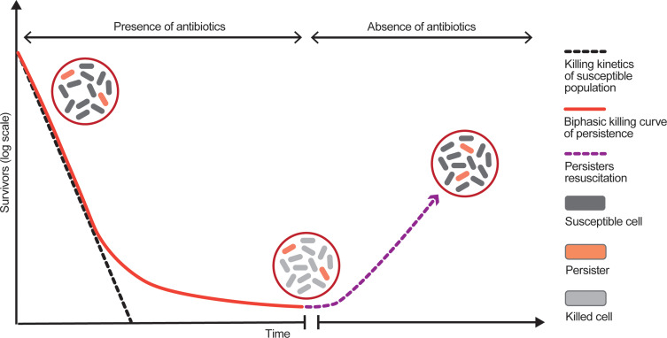
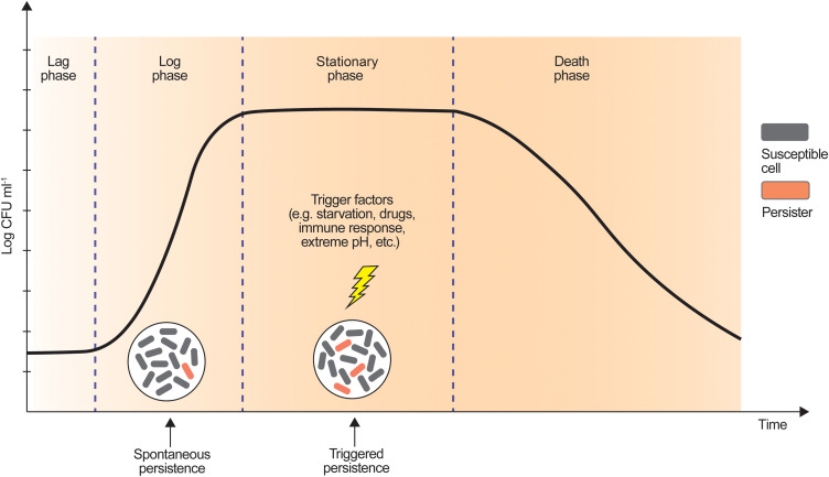
Although persistence may seem similar to tolerance, it particularly involves persisters that undergo a transient phenotypic switch to survive exposure to an antibiotic. It is important to note that persisters appear only in a small fraction of the clonal population and consequently cannot be explained by MIC and MDK which are more representative for the whole population.14,64,66 The presence of persisters can be revealed by biphasic killing curves (Figure 1) which reflect a killing pattern of antibiotics and the survival of tolerant persisters when antibiotic concentration exceeded certain thresholds. In the presence of antibiotic concentration several times higher than MIC, the subpopulation of recalcitrant cells also sustains the tolerance phenotype due to their non-viability for antibiotic action. The genetically tolerant population, on the other hand, can be efficiently killed with the kinetics as in susceptible population.14,66 Persisters are divided into two types based on the presence of trigger factors and which growth phase they occur in, spontaneous persisters and triggered persisters (Figure 2). Spontaneous persisters may be observed in the steady-state exponential growth without involving any trigger factors. Their occurrence is based on the hypothesis that a fraction of cells is stochastically converted into persister phenotype at a constant rate during growth. However, this type of persister is less common than the other one. The triggered persisters constitute the majority of recalcitrant cells in a clonal population and they are generated in the stationary phase due to the presence of triggering factors, such as stress signal as a response of cell starvation. When the resuscitation occurs in the absence of stressors, spontaneous persister can swiftly resume growth in a few hours while a subset of triggered persisters population can display an extended lag time before they eventually regrow.14,58
Figure 1.
Illustration of biphasic killing curves of bacterial growth during antibiotic treatment. The biphasic killing curves indicate the presence of two subpopulations, consisting susceptible cells that are eliminated rapidly by antibiotic treatment according to the killing kinetics (dashed black curve) and tolerant persister cells that may survive (red curve). Termination of antibiotic treatment allows persisters to resume growth, displaying similar phenotype to the parental population (dashed purple curve).
Figure 2.
Difference of triggered and spontaneous persisters based on the presence of trigger factors generating formation of persisters in a culture.
In addition to the survival strategies mentioned above, bacteria can also establish a viable but non-culturable (VBNC) state to elude bactericidal activity of antibiotics. This phenomenon exhibits similar characteristics to persisters and they are both related to cell dormancy, but not exactly identical at some points. However, it seems that VBNC state is rarely addressed together in the investigations related to persisters. Previous studies have hypothesized that VBNC and persisters coexist in the clonal population and become part of the dormancy continuum.67,68 Through population dynamics modeling, it is further described that generation of persisters leads to a VBNC state as prolonged or sustained exposure to stress induces a deeper dormancy state. Accordingly, their occurrence is thought to include some similar mechanisms that orchestrate the conversion of persisters into VBNC state consecutively.69 The overlapping phenotype between these two survival strategies was corroborated by the evidence that they share similar morphology and metabolic activity prior to ampicillin treatment, yet subsequently differed with the presence of antibiotics.67 Furthermore, the conditions required for resuscitation and lag time period upon stress removal appear to be two major points that can be used to discriminate VBNC state and persisters. The cells in the VBNC state are presumed to require a long period to restore the metabolic activity until they are eventually able to resume growth, which is indicated by the significantly prolonged lag.69 However, there might be confusion in defining this survival strategy with triggered persisters since they are also characterized by an extended lag time period.14,58 In this regard, it is worth noting that the VBNC state has been described as an extensive level of dormancy and consequently reflects a considerably longer period for the resuscitation when compared with persisters and necessitate a broader stress factor to select it. For instance, the isolation of cells in VBNC state is often involved exposure to further stressors after antibiotic treatment, such as osmotic pressure and temperature stress.68,70 The lower metabolic activity in VBNC state also implies the need for a more complex and specific condition to exit the dormancy state, hence common growth media are typically insufficient to recover the cells.69,71
Pathogenesis of S. Typhimurium Associated with Persister Formation
As it has been mentioned earlier, the spontaneous persisters are stochastically generated in a tiny proportion during logarithmic phase due to heterogeneity in the growth rate between cells in a clonal population. Subsequently, the proportion of persisters will significantly increase during the stationary phase due to the presence of environmental stimuli related to stress conditions, such as nutrient limitation, diauxic shift, ATP depletion, extreme pH, and DNA damage. In the context of persistent infection, the tissue localization where persisters survive determines the physiological niche with varied stressors that can influence the proportion of recalcitrant cells per se.60,72 In this part, we discuss the evidence regarding progression of S. Typhimurium infection that led to persistence and the factors that have been reported to influence the proportion of persister cell.
Enteric invasion of S. Typhimurium is initiated by the pathogen binding to the host cell surface and followed by expression of the SPI-1 to generate the needle-like complex and inject the TTSS-1 effectors into M cells and enterocytes. Secretion of these proteins induces cytoskeleton rearrangement that is characterized by membrane ruffling, as well as manipulation of the host signaling pathway.40 S. Typhimurium then penetrates the intestinal epithelium and migrates from the apical to basolateral side via transcytosis mediated by expression of type III secretion system encoded in SPI-2 (TTSS-2). Finally, the bacterial cells are released into lamina propria through an exocytosis mechanism.73 The active uptake by certain resident dendritic cells in the lamina propria can also provide direct transepithelial migration without requiring TTSS-1 expression.40,41
Interestingly, TSS-1 expression has been recently considered to have a fitness cost and drives within-host evolution. Cells expressing this virulence factor were demonstrated to penetrate epithelial tissues yet grow with a slower rate than the avirulent cells lacking TTSS-1 expression (this phenotype was shown to only colonize the gut lumen and not involved in transepithelial penetration). Accordingly, the heterogeneous phenotypes of S. Typhimurium during intestinal infection facilitate the establishment of cooperative virulence to outcompete intestinal microbiota and efficiently penetrate the epithelial tissue.74–76 Based on a study in a mouse model for Salmonella diarrhea, the concept of cooperative virulence is suggested to render different spatial localization of S. Typhimurium which further influences the antibiotic tolerance and promotes tissue persistence.75 However, the antibiotic perfusion in the tissue was mentioned as one of the factors that should be evaluated in order to confirm that the difference of antibiotic sensitivity is due to phenotypic variation between tissue-associated cells and those in gut lumen. Another study that investigates the tissue localization of S. Typhimurium persisters indicated that ciprofloxacin, a bactericidal antibiotic often used to treat invasive Salmonella infection, has a high bioavailability in the tissues where persisters were identified.15 In regard to persister formation, those findings indicate that phenotypic heterogeneity of S. Typhimurium during the initial stage of infection may predispose the factors to select or activate a transient antibiotic-tolerance phenotype that can eventually colonize the host cell for a prolonged period. The impact of within-host adaptation to metabolic activity and genetic alterations at single-cell level also appears to remain unexplored. This insight may lead to a better understanding of the differential tolerance phenotype generated in order to achieve cooperative virulence, as well as its association with antibiotic persister.
Upon reaching the lamina propria, S. Typhimurium is carried into MLN by mononuclear phagocytes and uses the lymphatic circulation as an entrance site for systemic dissemination. A more recent study revealed that the delivery of S. Typhimurium into lamina propria occurs through two scenarios: cell-mediated transport (uptake by dendritic cells, not macrophages) and autonomous migration in a cell-free form. In the latter mechanism, the pathogens are captured by the macrophages and B cells in the MLN.77 According to several studies in a murine typhoid model, the cells that enter MLN were shown to develop the characteristics of persisters, such as non-replicating, slower growth rate, temporary tolerance to high concentration of antibiotic (indicated with biphasic killing curve), and notably recover as the wildtype antibiotic sensitive cells after resuscitation. The S. Typhimurium persisters were identified in MLN, caecum lymph node (cLN), spleen, gall bladder, and liver.16,17,30,78,79 However, the persister fraction was predominantly found in MLN and thus considered as the primary site of persistence responsible for relapsing infection.30,78 Another report showed the liver as the main localization site of Salmonella persisters, contradicting the previous findings with the difference in experimental design assumed to underly the discrepancy.79
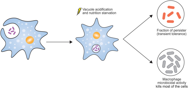
As part of the host defense mechanism, macrophages exert a harsh environment in order to eliminate the pathogen, such as vacuole acidification, production of free radicals, vacuolar ATPase, lysosomal digestive enzymes, and antimicrobial peptides. In S. Typhimurium-infected macrophages, the pathogen survives within a membrane-bound vacuole called a Salmonella-containing vacuole (SCV), evades host immunity, and undergoes intramacrophage adaptation through regulation of several virulence factors. In particular, the expression of TTSS-1 effectors involves the initial step of SCV biogenesis, followed by secretion of TTSS-2 effectors for SCV maturation and intracellular survival.42,80–82 The acidification of SCV has been demonstrated to promote survival and manipulate the replication of S. Typhimurium.83 Since the Salmonella persisters were notably found to reside within SCV, the physiological environment inside macrophage was further suggested to play a crucial role in Salmonella in persister formation and dormancy state. The in vitro infection of S. Typhimurium to bone-marrow derived-macrophages (BMM) showed that the phagocytosis of bacteria by macrophages serves as a cardinal process to induce persister formation. In particular, the vacuolar acidification (pH 4.5) and nutrient starvation were proposed to influence the persister fraction based on the exposure of corresponding conditions in laboratory media and infection to BMM (Figure 3).16 In addition, infection of clinically invasive S. Typhimurium isolates into human primary macrophages supported the idea that internalization to macrophages could significantly increase the proportion of persister bacteria.84
Figure 3.
Phagocytosis by macrophage is an important step to initiate Salmonella persisters. Vacuole acidification and the lack of nutrients for cell growth induce the activation of stress response and TA modules, leading to persister formation.
The evidence mentioned above was contradicted by a recent finding which shows that both vacuolar acidification and nutrient starvation do not influence persister formation, despite a similar experimental design being applied.85 However, the experiment was conducted in Luria-Bertani (LB)-treated media and did not include a study of macrophage infection. One possible factor that can underly the contradictory results between those studies is the use of LB with uncontrolled concentration of sugar and divalent cations since it may cause growth variations, as previously discussed.86 In another study that used a different medium, investigation of persister fraction was achieved at pH 3.524 which suggests a more specific pH value may be required to induce persister formation upon phagocytosis by macrophages. It is also important to note that the mechanism of pathogen internalization into macrophages was found to correlate with the vacuole acidification process87 and therefore can possibly affect the differential pH across vacuoles.
Despite conflicting findings regarding the vacuole acidification, localization of Salmonella persisters within macrophages indicates the importance of the host-specific environment as one of the key aspects that should not be overlooked when investigating S. Typhimurium persisters. Additionally, the interplay between macrophage defense mechanisms and persisters may be much more complex and involves various virulence factors. It includes the consequences of vacuole acidification or other environmental stimuli to the action of molecular effectors that have been suggested to take part in persister formation. For example, an assessment of 32 reported SPI-2 T3SS effectors revealed that some of those effectors influence the intracellular replication within macrophages.88 Another investigation also showed that translocation of SPI-2 T3SS effectors to the host cells are maintained within SCV, although persisters are in a nongrowing state.42 However, it seems that the contribution of vacuole acidification in the expression of SPI-2 T3SS effectors remains unclear due to contradictory results between several studies.87
Toxin-Antitoxin Modules That Affect the Proportion of Salmonella Persisters
Manipulation of several major metabolic activities, including inhibition of DNA and protein syntheses, has been typically associated with persister cells as part of a survival strategy from environmental stressors. There are several types of stress response in bacteria that have been described to mediate persistence, such as general stress response induced by environmental stress condition, DNA damage-induced SOS response, as well as stringent response mediated by the alarmones (p)ppGpp (guanosine tetraphosphate and guanosine pentaphosphate).25,44,63,89 Accordingly, the role of those stress responses in reprogramming bacterial growth and metabolic activity during stress conditions has become one of particular interest to elucidate the mechanism of persister formation.
As briefly mentioned in the introduction, several studies have indicated the link between stress responses and TA modules in S. Typhimurium. This genetic element contains the toxin which has the ability to inhibit cellular function and its cognate antitoxin which neutralizes the action of the toxin. Initially identified in F plasmid, the TA operon is now known to be widely distributed in various bacteria and archaea. Based on its genetic structure and regulation, there are six classes (I–VI) of TA modules. They have a wide range of cellular targets and can interfere with biological processes within the bacterial cells, such as protein translation, cell wall synthesis, and DNA replication.26,27,90 The toxin activities of TA modules can affect various biological functions, yet the detailed molecular mechanisms on how they are being activated remain enigmatic. Several biological functions of TA modules have been associated with the regulation of cell growth and cellular adaptation under stress conditions, including post-segregational killing, abortive infection, and persister formation/antibiotic tolerance.25
Among six classes of TA modules, the type II is considered the most abundant and well-studied in many bacteria. Both toxin and antitoxin molecules in this type are proteins. The latter one generally has two domains: the N-terminal domain that binds inverted repeats in the operon promoter for regulatory purposes and C-terminal domain that binds the cognate toxin to neutralize its toxic activity.25,90,91 One of the early investigations regarding the role of TA modules in persistence identified the bicistronic hipBA operon that is responsible for high persistence levels in E. coli K-12. The operon was further characterized and known to comprise the high persistence gene A (hipA) and its counterpart hipB gene, producing a toxin-antitoxin complex.92,93 Mutations in the toxin hipA gene were shown to impair the complex formation, thus allowing HipA protein to exert its toxic activity through phosphorylation of glutamate-tRNA ligase (GltX) leading to inhibition of protein translation and cell growth.94
In S. Typhimurium, bioinformatic analysis in the genome of a virulent strain revealed 27 TA loci (four loci located in plasmids); seven of them belong to type I while the rest are type II.95 In addition, the type II toxin-antitoxin database (TADB 2.0) has listed at least 18 type II TA loci from the genome of S. Typhimurium strain LT2.96 The regulation and role of TA modules in intracellular survival was shown to vary between host cell types, indicating the specificity of physiological conditions inside the host that promote the biological function of each TA module.24,95 Remarkably, S. Typhimurium 12023 mutants carrying a single deletion of 14 from those identified putative type II TA loci have been shown to exhibit around 10–30% reduction in the proportion of macrophage-induced persisters relative to its wildtype strain.16
The potential role of each type II TA operon in Salmonella persistence has also been demonstrated in several reports (Table 1). For example, a study used transposon mutagenesis to generate highly persistent mutants of S. Typhimurium LT2 which led to the identification of shpAB TA operon and its contribution to a persistent phenotype. This type II TA module encodes ShpA toxin with RNase sequence signature and its cognate ShpB antitoxin. Based on the experimental results, it was proposed that the shpAB regulation depends on the Lon protease activity which degrades the antitoxin but is not mediated by (p)ppGpp stringent response.22 The vapBC of TA locus identified in S. Typhimurium LT2 also indicates the activity that may promote persister formation. This TA module encodes the PiIN (PilT N-terminal) domain toxin and its cognate antitoxin. It was shown that amino acid starvation and antibiotic chloramphenicol induce the ectopically-expressed Salmonella VapC toxin to mediate bacteriostatic condition which is responsible for the inhibition of cell growth.97 Knocking out the shpAB or vapBC TA loci of S. Typhimurium 12023 was also shown to decrease the proportion of macrophage-induced persisters. Additionally, protein sequence homology analysis of the three previously uncharacterized TA modules exhibiting association with persister formation in S. Typhimurium has unraveled a novel indicator of toxin biological activity. In specific, the toxins from those TA modules share similarities with Gcn5 N-acetyltransferases (GNAT) based on the presence of a conserved N-acetyltransferase superfamily domain and further were named as TacT, TacT2, and TacT3.23,84
Table 1.
Several Characterized Type II TA Modules Involve in Salmonella Persistence
| TA Module | Predicted Mechanism of Toxin Activity | Molecular Class of Toxin | Cellular Target | References |
|---|---|---|---|---|
| ShpAB | Protein translation inhibition: cleave mRNA | RelE-like RNases | mRNA | [22] |
| VapBC | Protein translation inhibition: cleave initiator tRNA | PIN-domain ribonucleases | tRNAfMet | [97,101,103] |
| TacAT | Protein translation inhibition: acetylating aminoacyl-tRNA | GNAT | tRNA(gly) | [23,84,102] |
| TacAT2 | Protein translation inhibition: acetylating aminoacyl-tRNA | GNAT | tRNA | [23,84] |
| TacAT3 | Protein translation inhibition: acetylating aminoacyl-tRNA | GNAT | tRNA | [23,84] |
Overall, the studies mentioned above provide a constructive indication that TA modules may take part in promoting the persistent phenotype of intracellular Salmonella persisters. Further assessment of these TA modules and verification of their significance in allowing Salmonella to survive within its host through persistence is essential. Understanding the target specificity of TA modules as well as the regulation of TA operons, which are discussed in the next section, are some of the rising concerns due to the increased abundance of TA modules that may be found within Salmonella genomes and considering the complexity of the molecular basis that may underly the formation of persisters.
Mechanisms and Regulation of Toxin and Antitoxin Modules in Promoting Salmonella Persisters Formation
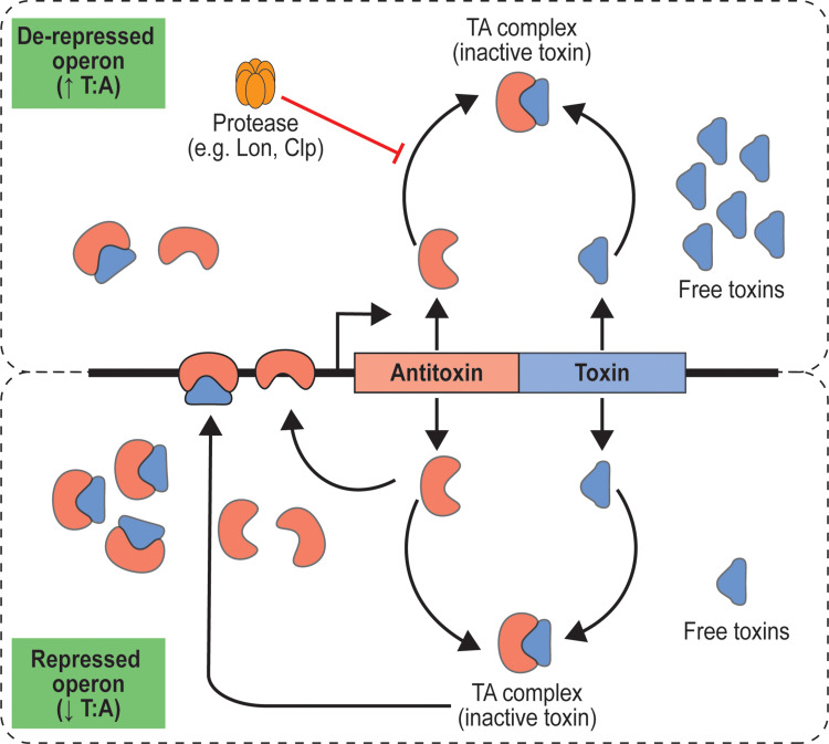
Several models have been proposed to describe the regulation of TA modules in which it may occur at transcriptional or translational level. In type II TA modules, the gene expression is often autoregulated at transcriptional level through the principle of coordination cooperativity (Figure 4), which apparently is the most well-studied model. Explicitly, the repression and de-repression are influenced by the ratio between toxin and antitoxin (T:A) in TA complexes which implicate the bifunctional role of toxin as either co-repressor or de-repressors depending on the toxin level.25 The N-terminal DNA-binding domain of the antitoxin binds to the promoter resulting in low to moderate repression. On the other hand, formation of TA complexes mostly contributes to an increased binding affinity of antitoxin in the promoter region. Based on this knowledge, strong repression will be achieved when the T:A is low owing to the steady-state stoichiometry of TA complexes formation and the reduced availability of free toxins. Conversely, the excess level of toxin increases the T:A ratio, leading to a de-repression state and eventually activation of TA module.25,90,98 This type of TA operon autoregulation has been observed in ShpAB and VapBC of S. Typhimurium.22,97
Figure 4.
Illustration of type II TA system autoregulation through coordination cooperativity. The ratio of toxin and antitoxin influence the switch between repressed and de-repressed state of operon. Degradation of antitoxin by cellular protease prevent the formation of TA complex.
The stress response has been proposed to mediate TA module activation that led to persister formation. It appears that the different stability between toxin and antitoxin underlies this hypothesis. The free antitoxins are generally unstable proteins and prone to proteolytic degradation while the free toxins are relatively more stable. Meanwhile, the stress-dependent activation of cellular proteases; such as Lon, ClpXP, and ClpAP, contributes to the degradation of antitoxins and consequently impairs the stoichiometry of TA complex formation. As a result, free toxins can bind to their targets and exert biological activities.25,27,98 Some investigations suggest that the stringent response may involve the activation of type II TA module. The alarmones (p)ppGpp is a secondary messenger known for its important role in mediating stringent response as the bacteria attempts to regulate its cellular physiology under stress conditions.99 Notably, it was proposed that (p)ppGpp is involved in the activation of Lon protease through the accumulation of polyphosphate (polyP) which subsequently causes antitoxin degradation of TA module.100 The regulation of cellular (p)ppGpp is carried out by RelA-SpoT Homologue (RSH) family proteins which have a bifunctional role in both synthesizing and degrading the (p)ppGpp.99
In the study of macrophage-induced Salmonella persister, assessment of mutants carrying deletion of relA and spoT or lon showed a significant decrease in persister fraction. Moreover, the lon mutant and wildtype strains displayed no significant difference in the proportion of persister when grown in laboratory media which indicates the critical influence of intracellular environments. Accordingly, it was inferred that this evidence may be linked to the activation of TA module.16 However, this model may not be generally applied in all Salmonella TA modules and depends on the organization or regulation of individual operon. In ShpAB TA module, the persistence was found to be independent of (p)ppGpp level although the Lon protease was shown to take part in the process.22 The toxin-degrading activity of Lon protease was also observed in VapBC TA module, but the association with stringent response remains elusive.101
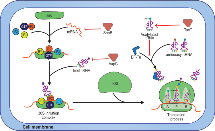
As mentioned above, liberation of the toxin through antitoxin degradation by Lon protease allows it to generate biological activities. Consequently, investigating the cellular process that becomes the target of toxin activity is essential in order to elucidate its mechanism of action in promoting persistence. Among the characterized toxins from Salmonella TA modules, their target has been proposed based on structural analysis and some laboratory experiments (Figure 5). In TacT, the radiolabeled pulse-chase assays indicated that the toxin inhibits protein translation while the DNA replication and transcription were likely not influenced.23 Protein structure analysis and cell-free translation assay revealed that the substrate of TacT is charged aminoacyl-tRNA in which the acetyl moiety is transferred from Ac-CoA to the alpha amine group of amino acids.23 Similar to TacT, the TacT2 and TacT3 also showed tRNA-acetylating activity, but TacT3 exhibited the highest acetylating potency. The in vitro experiments showed that those toxins can transfer the acetyl moiety to a different range of aminoacyl-tRNAs, indicating their target specificity.84 Another in vitro study suggested that the glycyl-tRNA is the most efficient substrate of TacT although it can acetylate other aminoacyl-tRNAs. For confirmation, in vivo study was performed, and it showed glycyl-tRNA as the only aminoacyl-tRNA species acetylated by TacT. Moreover, it was predicted that TacT2 share similar specificity with TacT based on their structural comparison analysis.102 The activity of toxin from VapBC TA module was shown to inhibit the translation process as well, but with different targets. Characterization of VapC toxin revealed that it cleaves the initiator tRNA between anticodon stem and loop, thus may interrupt the initiation step of mRNA translation.103 On the other hand, the RelE-like RNases signature shown in the sequence of ShpB toxin22 indicates that it may share a similar activity with the well-characterized RelE toxin in E. coli in mediating growth arrest. RelE toxin is part of RelBE TA module in E. coli which was known to exhibit mRNA interferase activity by cleaving the mRNA at the ribosomal A site.104,105 However, the activity of ShpB seems to be still unclear and has not yet been investigated in S. Typhimurium.
Figure 5.
Proposed target of the toxins within bacterial translational machinery. The ShpB, VapC, and TacTs toxins target the mRNA, initiator tRNA, and aminoacyl-tRNAs respectively.
The protein translation is likely the primary target of toxins from TA modules in S. Typhimurium. Although both acetylation of tRNAs and cleavage of initiator tRNA could potentially affect the dynamics of the translation process, further investigation within this context has yet to be performed to clarify the details of the toxin mechanism in interfering protein translation. Initiator tRNA is one of the key players in the formation of 30S initiation complex in which it interacts with initiation factor 2 (IF2) to enable specific recognition by IF1 and IF3 in the peptidyl (P) site of the ribosome.106 On the other hand, the aminoacyl-tRNAs are involved in the elongation process and binding with elongation factor thermal unstable (EF-Tu) is essential to be recognized in the aminoacyl (A) site of the ribosomal complex during translation. Therefore, it is speculated that the tRNA-acetylating toxins may prevent aminoacyl-tRNA to bind with EF-Tu which eventually disrupts the elongation of polypeptide chains. Also, it appears that VapC toxin acts in a more upstream step by inhibiting the complex formation between initiator tRNA and IF2 in the initiation step which probably generates a more global impact in the protein translation of bacterial cells.
However, the activation and regulation of TA modules in S. Typhimurium seem to remain elusive and some conflicting results between one study and the others are generating more questions to be investigated. Most importantly, the inconsistency of the TA activation model in E. coli persisters has raised debates concerning the role of (p)ppGpp, Lon protease, and TA modules in promoting bacterial persister formation, including in S. Typhimurium. Evaluation in E. coli found that infection of cryptic prophages in the E. coli strains used to study the TA modules was responsible for the artifact results and corresponds to the interlaboratory variation.11 Further reassessment of the E. coli strains confirmed that activation of TA modules does not correlate with persister formation.107 In S. Typhimurium, a study reported that (p)ppGpp and TA modules are dispensable for persistence and also highlighted the negligible impact of acidic environment85 as discussed above. The latter study has provided a significant contradictory result from the previous findings about the role of (p)ppGpp and TA modules in Salmonella persister formation. However, the influence of physiological properties encountered by S. Typhimurium during intracellular persistence was likely not addressed. Regarding the link between stringent response and TA module, a recent study reported that the RelA-SpoT Homolog (RSH) protein family can inhibit protein biosynthesis which is responsible for growth arrest.108 This finding may indicate the independent role of (p)ppGpp in promoting persisters and could be another caveat to consider when clarifying the role of TA module in persister formation. On top of that, it is also crucial to do thorough assessments with a clear experimental procedure that can be widely accepted, produce less variability, and considers the factors from the perspective of bacterial pathogenesis and host response that may promote persister phenotype in S. Typhimurium.
Concluding Remarks
In summary, there have been pieces of evidence indicating the involvement of the toxin-antitoxin system in the persister formation of S. Typhimurium. These include the role of TA modules in inhibiting protein biosynthesis with indications of the alteration of bacterial physiology, such as metabolic activity and cell growth. As an outcome, the bacterial cell will undergo a transient phenotypic switch that enables survival through a persistence phenomenon in the presence of antimicrobial agents due to reduced availability or access to the cellular target. The conflicting results between studies suggest the need to clarify the molecular mechanism of persister formation in S. Typhimurium using appropriate and comparable experimental models, considering the nature and characteristic of persisters in a clonal bacterial population. It is also important to note that the host microenvironments should not be neglected since there have been evidence suggesting its influence to offer a more suitable intracellular condition for persister formation in S. Typhimurium. For instance, the role of within-host adaptation and the macrophage microenvironment that can influence the proportion and tissue localization of persisters. Furthermore, the valuable knowledge regarding Salmonella persisters can be used as future directions to address treatment issues in Salmonella persistent infections, as well as growing concern that persisters may take part in the evolution of antimicrobial resistance.
Acknowledgments
This work was partially supported by Scholarship for Ph.D. Student from Mahidol University. The authors wish to thank staff in the Department of Microbiology, Faculty of Pharmacy, Mahidol University, for suggestions on this work. Their support and collaboration are gratefully acknowledged.
Disclosure
The authors declare that they have no competing interests.
References
- 1.Issenhuth-Jeanjean S, Roggentin P, Mikoleit M, et al. Supplement 2008–2010 (no. 48) to the White-Kauffmann-Le Minor scheme. Res Microbiol. 2014;165(7):526–530. doi: 10.1016/j.resmic.2014.07.004 [DOI] [PubMed] [Google Scholar]
- 2.Grimont PAD, Weill FX. Antigenic Formulae of the Salmonella Serovars. World Health Organization Collaborating Center for Reference and Research on Salmonella. 9th ed. Paris: Pasteur Institute; 2007. Available from: https://www.pasteur.fr/sites/default/files/veng_0.pdf. [Google Scholar]
- 3.Gal-Mor O. Persistent infection and long-term carriage of typhoidal and nontyphoidal salmonellae. Clin Microbiol Rev. 2019;32(1). doi: 10.1128/CMR.00088-18 [DOI] [PMC free article] [PubMed] [Google Scholar]
- 4.Monack DM. Salmonella persistence and transmission strategies. Curr Opin Microbiol. 2012;15(1):100–107. doi: 10.1016/j.mib.2011.10.013 [DOI] [PubMed] [Google Scholar]
- 5.Feasey NA, Dougan G, Kingsley RA, et al. Invasive non-typhoidal Salmonella disease: an emerging and neglected tropical disease in Africa. Lancet. 2012;379(9835):2489–2499. doi: 10.1016/S0140-6736(11)61752-2 [DOI] [PMC free article] [PubMed] [Google Scholar]
- 6.GBD 2017 Non-Typhoidal Salmonella Invasive Disease Collaborators.The global burden of non-typhoidal Salmonella invasive disease: a systematic analysis for the Global Burden of Disease Study 2017. Lancet Infect Dis. 2019;19(12):1312–1324. doi: 10.1016/S1473-3099(19)30418-9 [DOI] [PMC free article] [PubMed] [Google Scholar]
- 7.Hill PSW, Helaine S. Antibiotic persisters and relapsing Salmonella enterica infections. In: Lewis K, editor. Persister Cells and Infectious Disease. 1st ed. Switzerland: Springer Cham; 2019:19–38. [Google Scholar]
- 8.Gopinath S, Carden S, Monack D. Shedding light on Salmonella carriers. Trends Microbiol. 2012;20(7):320–327. doi: 10.1016/j.tim.2012.04.004 [DOI] [PubMed] [Google Scholar]
- 9.Gunn JS, Marshall JM, Baker S, et al. Salmonella chronic carriage: epidemiology, diagnosis, and gallbladder persistence. Trends Microbiol. 2014;22(11):648–655. doi: 10.1016/j.tim.2014.06.007 [DOI] [PMC free article] [PubMed] [Google Scholar]
- 10.Okoro CK, Kingsley RA, Connor TR, et al. Intracontinental spread of human invasive Salmonella Typhimurium pathovariants in sub-Saharan Africa. Nat Genet. 2012;44(11):1215–1221. doi: 10.1038/ng.2423 [DOI] [PMC free article] [PubMed] [Google Scholar]
- 11.Marzel A, Desai PT, Goren A, et al. Persistent infections by nontyphoidal Salmonella in humans: epidemiology and genetics. Clin Infect Dis. 2016;62(7):879–886. doi: 10.1093/cid/civ1221 [DOI] [PMC free article] [PubMed] [Google Scholar]
- 12.Aserkoff B, Bennett JV. Effect of antibiotic therapy in acute salmonellosis on the fecal excretion of salmonellae. N Engl J Med. 1969;281(12):636–640. doi: 10.1056/NEJM196909182811202 [DOI] [PubMed] [Google Scholar]
- 13.Murase T, Yamada M, Muto T, et al. Fecal excretion of Salmonella enterica serovar Typhimurium following a food-borne outbreak. J Clin Microbiol. 2000;38(9):3495–3497. doi: 10.1128/JCM.38.9.3495-3497.2000 [DOI] [PMC free article] [PubMed] [Google Scholar]
- 14.Balaban NQ, Helaine S, Lewis K, et al. Definitions and guidelines for research on antibiotic persistence. Nat Rev Microbiol. 2019;17(7):441–448. doi: 10.1038/s41579-019-0196-3 [DOI] [PMC free article] [PubMed] [Google Scholar]
- 15.Kaiser P, Regoes RR, Dolowschiak T, et al. Cecum lymph node dendritic cells harbor slow-growing bacteria phenotypically tolerant to antibiotic treatment. PLoS Biol. 2014;12(2):e1001793. doi: 10.1371/journal.pbio.1001793 [DOI] [PMC free article] [PubMed] [Google Scholar]
- 16.Helaine S, Cheverton AM, Watson KG, et al. Internalization of Salmonella by macrophages induces formation of nonreplicating persisters. Science. 2014;343(6167):204–208. doi: 10.1126/science.1244705 [DOI] [PMC free article] [PubMed] [Google Scholar]
- 17.Claudi B, Sprote P, Chirkova A, et al. Phenotypic variation of Salmonella in host tissues delays eradication by antimicrobial chemotherapy. Cell. 2014;158(4):722–733. doi: 10.1016/j.cell.2014.06.045 [DOI] [PubMed] [Google Scholar]
- 18.Kaldalu N, Hauryliuk V, Turnbull KJ, et al. In vitro studies of persister cells. Microbiol Mol Biol Rev. 2020;84(4). doi: 10.1128/MMBR.00070-20 [DOI] [PMC free article] [PubMed] [Google Scholar]
- 19.Wainwright J, Hobbs G, Nakouti I. Persister cells: formation, resuscitation and combative therapies. Arch Microbiol. 2021;203(10):5899–5906. doi: 10.1007/s00203-021-02585-z [DOI] [PMC free article] [PubMed] [Google Scholar]
- 20.Wilmaerts D, Windels EM, Verstraeten N, et al. General mechanisms leading to persister formation and awakening. Trends Genet. 2019;35(6):401–411. doi: 10.1016/j.tig.2019.03.007 [DOI] [PubMed] [Google Scholar]
- 21.Van den Bergh B, Fauvart M, Michiels J. Formation, physiology, ecology, evolution and clinical importance of bacterial persisters. FEMS Microbiol Rev. 2017;41(3):219–251. doi: 10.1093/femsre/fux001 [DOI] [PubMed] [Google Scholar]
- 22.Slattery A, Victorsen AH, Brown A, et al. Isolation of highly persistent mutants of Salmonella enterica serovar Typhimurium reveals a new toxin-antitoxin module. J Bacteriol. 2013;195(4):647–657. doi: 10.1128/JB.01397-12 [DOI] [PMC free article] [PubMed] [Google Scholar]
- 23.Cheverton AM, Gollan B, Przydacz M, et al. A Salmonella toxin promotes persister formation through acetylation of tRNA. Mol Cell. 2016;63(1):86–96. doi: 10.1016/j.molcel.2016.05.002 [DOI] [PMC free article] [PubMed] [Google Scholar]
- 24.Silva-Herzog E, McDonald EM, Crooks AL, et al. Physiologic stresses reveal a Salmonella persister state and TA family toxins modulate tolerance to these stresses. PLoS One. 2015;10(12):e0141343. doi: 10.1371/journal.pone.0141343 [DOI] [PMC free article] [PubMed] [Google Scholar]
- 25.Harms A, Brodersen DE, Mitarai N, et al. Toxins, targets, and triggers: an overview of toxin-antitoxin biology. Mol Cell. 2018;70(5):768–784. doi: 10.1016/j.molcel.2018.01.003 [DOI] [PubMed] [Google Scholar]
- 26.Unterholzner SJ, Poppenberger B, Rozhon W. Toxin-antitoxin systems: biology, identification, and application. Mob Genet Elements. 2013;3(5):e26219. doi: 10.4161/mge.26219 [DOI] [PMC free article] [PubMed] [Google Scholar]
- 27.Yamaguchi Y, Park JH, Inouye M. Toxin-antitoxin systems in bacteria and archaea. Ann Rev Genet. 2011;45:61–79. doi: 10.1146/annurev-genet-110410-132412 [DOI] [PubMed] [Google Scholar]
- 28.Bakkeren E, Diard M, Hardt WD. Evolutionary causes and consequences of bacterial antibiotic persistence. Nat Rev Microbiol. 2020;18(9):479–490. doi: 10.1038/s41579-020-0378-z [DOI] [PubMed] [Google Scholar]
- 29.Zhang S, Kingsley RA, Santos RL, et al. Molecular pathogenesis of Salmonella enterica serotype Typhimurium-induced diarrhea. Infect Immun. 2003;71(1):1–12. doi: 10.1128/IAI.71.1.1-12.2003 [DOI] [PMC free article] [PubMed] [Google Scholar]
- 30.Monack DM, Bouley DM, Falkow S. Salmonella Typhimurium persists within macrophages in the mesenteric lymph nodes of chronically infected Nramp1+/+ mice and can be reactivated by IFNgamma neutralization. J Exp Med. 2004;199(2):231–241. doi: 10.1084/jem.20031319 [DOI] [PMC free article] [PubMed] [Google Scholar]
- 31.Crum-Cianflone NF. Salmonellosis and the gastrointestinal tract: more than just peanut butter. Curr Gastroenterol Rep. 2008;10(4):424–431. doi: 10.1007/s11894-008-0079-7 [DOI] [PMC free article] [PubMed] [Google Scholar]
- 32.Akhtar S, Sarker MR, Jabeen K, et al. Antimicrobial resistance in Salmonella enterica serovar Typhi and Paratyphi in South Asia-current status, issues and prospects. Crit Rev Microbiol. 2015;41(4):536–545. doi: 10.3109/1040841X.2014.880662 [DOI] [PubMed] [Google Scholar]
- 33.Whistler T, Sapchookul P, McCormick DW, et al. Epidemiology and antimicrobial resistance of invasive non-typhoidal Salmonellosis in rural Thailand from 2006–2014. PLoS Negl Trop Dis. 2018;12(8):e0006718. doi: 10.1371/journal.pntd.0006718 [DOI] [PMC free article] [PubMed] [Google Scholar]
- 34.Phu Huong Lan N, Le Thi Phuong T, Nguyen Huu H, et al. Invasive non-typhoidal Salmonella infections in Asia: clinical observations, disease outcome and dominant serovars from an infectious disease hospital in Vietnam. PLoS Negl Trop Dis. 2016;10(8):e0004857. doi: 10.1371/journal.pntd.0004857 [DOI] [PMC free article] [PubMed] [Google Scholar]
- 35.Gordon MA. Salmonella infections in immunocompromised adults. J Infect. 2008;56(6):413–422. doi: 10.1016/j.jinf.2008.03.012 [DOI] [PubMed] [Google Scholar]
- 36.Mori N, Szvalb AD, Adachi JA, et al. Clinical presentation and outcomes of non-typhoidal Salmonella infections in patients with cancer. BMC Infect Dis. 2021;21(1):1021. doi: 10.1186/s12879-021-06710-7 [DOI] [PMC free article] [PubMed] [Google Scholar]
- 37.Yen YF, Wang FD, Chiou CS, et al. Prognostic factors and clinical features of non-typhoid Salmonella bacteremia in adults. J Chin Med Assoc. 2009;72(8):408–413. doi: 10.1016/S1726-4901(09)70397-1 [DOI] [PubMed] [Google Scholar]
- 38.Kingsley RA, Msefula CL, Thomson NR, et al. Epidemic multiple drug resistant Salmonella Typhimurium causing invasive disease in sub-Saharan Africa have a distinct genotype. Genome Res. 2009;19(12):2279–2287. doi: 10.1101/gr.091017.109 [DOI] [PMC free article] [PubMed] [Google Scholar]
- 39.Onwuezobe IA, Oshun PO, Odigwe CC. Antimicrobials for treating symptomatic non-typhoidal Salmonella infection. Cochrane Database Syst Rev. 2012;11:CD001167. doi: 10.1002/14651858.CD001167.pub2 [DOI] [PMC free article] [PubMed] [Google Scholar]
- 40.Griffin AJ, McSorley SJ. Development of protective immunity to Salmonella, a mucosal pathogen with a systemic agenda. Mucosal Immunol. 2011;4(4):371–382. doi: 10.1038/mi.2011.2 [DOI] [PMC free article] [PubMed] [Google Scholar]
- 41.Martinoli C, Chiavelli A, Rescigno M. Entry route of Salmonella Typhimurium directs the type of induced immune response. Immunity. 2007;27(6):975–984. doi: 10.1016/j.immuni.2007.10.011 [DOI] [PubMed] [Google Scholar]
- 42.Stapels DAC, Hill PWS, Westermann AJ, et al. Salmonella persisters undermine host immune defenses during antibiotic treatment. Science. 2018;362(6419):1156–1160. doi: 10.1126/science.aat7148 [DOI] [PubMed] [Google Scholar]
- 43.Lewis K. Persister cells. Annu Rev Microbiol. 2010;64:357–372. doi: 10.1146/annurev.micro.112408.134306 [DOI] [PubMed] [Google Scholar]
- 44.Cohen NR, Lobritz MA, Collins JJ. Microbial persistence and the road to drug resistance. Cell Host Microbe. 2013;13(6):632–642. doi: 10.1016/j.chom.2013.05.009 [DOI] [PMC free article] [PubMed] [Google Scholar]
- 45.Barrett TC, Mok WWK, Murawski AM, et al. Enhanced antibiotic resistance development from fluoroquinolone persisters after a single exposure to antibiotic. Nat Commun. 2019;10(1):1177. doi: 10.1038/s41467-019-09058-4 [DOI] [PMC free article] [PubMed] [Google Scholar]
- 46.Bakkeren E, Huisman JS, Fattinger SA, et al. Salmonella persisters promote the spread of antibiotic resistance plasmids in the gut. Nature. 2019;573(7773):276–280. doi: 10.1038/s41586-019-1521-8 [DOI] [PMC free article] [PubMed] [Google Scholar]
- 47.Bakkeren E, Herter JA, Huisman JS, et al. Pathogen invasion-dependent tissue reservoirs and plasmid-encoded antibiotic degradation boost plasmid spread in the gut. Elife. 2021;10. doi: 10.7554/eLife.69744 [DOI] [PMC free article] [PubMed] [Google Scholar]
- 48.Windels EM, Michiels JE, Fauvart M, et al. Bacterial persistence promotes the evolution of antibiotic resistance by increasing survival and mutation rates. ISME J. 2019;13(5):1239–1251. doi: 10.1038/s41396-019-0344-9 [DOI] [PMC free article] [PubMed] [Google Scholar]
- 49.Hill PWS, Moldoveanu AL, Sargen M, et al. The vulnerable versatility of Salmonella antibiotic persisters during infection. Cell Host Microb. 2021;29(12):1757–1773. doi: 10.1016/j.chom.2021.10.002 [DOI] [PubMed] [Google Scholar]
- 50.Octavia S, Wang Q, Tanaka MM, et al. Genomic variability of serial human isolates of Salmonella enterica serovar Typhimurium associated with prolonged carriage. J Clin Microbiol. 2015;53(11):3507–3514. doi: 10.1128/JCM.01733-15 [DOI] [PMC free article] [PubMed] [Google Scholar]
- 51.Liu J, Gefen O, Ronin I, et al. Effect of tolerance on the evolution of antibiotic resistance under drug combinations. Science. 2020;367(6474):200–204. doi: 10.1126/science.aay3041 [DOI] [PubMed] [Google Scholar]
- 52.Zhang K, Dupont A, Torow N, et al. Age-dependent enterocyte invasion and microcolony formation by Salmonella. PLoS Pathog. 2014;10(9):e1004385. doi: 10.1371/journal.ppat.1004385 [DOI] [PMC free article] [PubMed] [Google Scholar]
- 53.Xiang Y, Li F, Dong N, et al. Investigation of a salmonellosis outbreak caused by multidrug resistant Salmonella Typhimurium in China. Front Microbiol. 2020;11:801. doi: 10.3389/fmicb.2020.00801 [DOI] [PMC free article] [PubMed] [Google Scholar]
- 54.Moffatt CR, Musto J, Pingault N, et al. Salmonella Typhimurium and outbreaks of egg-associated disease in Australia, 2001 to 2011. Foodborne Pathog Dis. 2016;13(7):379–385. doi: 10.1089/fpd.2015.2110 [DOI] [PubMed] [Google Scholar]
- 55.Bruun T, Sorensen G, Forshell LP, et al. An outbreak of Salmonella Typhimurium infections in Denmark, Norway and Sweden, 2008. Euro Surveill. 2009;14(10):19147. [PubMed] [Google Scholar]
- 56.Bigger JW. Treatment of Staphylococcal infections with penicillin by intermittent sterilisation. Lancet. 1944;244(6320):497–500. doi: 10.1016/S0140-6736(00)74210-3 [DOI] [Google Scholar]
- 57.Moyed HS, Bertrand KP. hipA, a newly recognized gene of Escherichia coli K-12 that affects frequency of persistence after inhibition of murein synthesis. J Bacteriol. 1983;155(2):768–775. doi: 10.1128/jb.155.2.768-775.1983 [DOI] [PMC free article] [PubMed] [Google Scholar]
- 58.Balaban NQ, Merrin J, Chait R, et al. Bacterial persistence as a phenotypic switch. Science. 2004;305(5690):1622–1625. doi: 10.1126/science.1099390 [DOI] [PubMed] [Google Scholar]
- 59.Harms A, Maisonneuve E, Gerdes K. Mechanisms of bacterial persistence during stress and antibiotic exposure. Science. 2016;354:6318. doi: 10.1126/science.aaf4268 [DOI] [PubMed] [Google Scholar]
- 60.Fisher RA, Gollan B, Helaine S. Persistent bacterial infections and persister cells. Nat Rev Microbiol. 2017;15(8):453–464. doi: 10.1038/nrmicro.2017.42 [DOI] [PubMed] [Google Scholar]
- 61.Moldoveanu AL, Rycroft JA, Helaine S. Impact of bacterial persisters on their host. Curr Opin Microbiol. 2021;59:65–71. doi: 10.1016/j.mib.2020.07.006 [DOI] [PubMed] [Google Scholar]
- 62.Defraine V, Fauvart M, Michiels J. Fighting bacterial persistence: current and emerging anti-persister strategies and therapeutics. Drug Resist Updat. 2018;38:12–26. doi: 10.1016/j.drup.2018.03.002 [DOI] [PubMed] [Google Scholar]
- 63.Ronneau S, Helaine S. Clarifying the link between toxin-antitoxin modules and bacterial persistence. J Mol Biol. 2019;431(18):3462–3471. doi: 10.1016/j.jmb.2019.03.019 [DOI] [PubMed] [Google Scholar]
- 64.Ronneau S, Hill PW, Helaine S. Antibiotic persistence and tolerance: not just one and the same. Curr Opin Microbiol. 2021;64:76–81. doi: 10.1016/j.mib.2021.09.017 [DOI] [PubMed] [Google Scholar]
- 65.Gollan B, Grabe G, Michaux C, et al. Bacterial persisters and infection: past, present, and progressing. Annu Rev Microbiol. 2019;73:359–385. doi: 10.1146/annurev-micro-020518-115650 [DOI] [PubMed] [Google Scholar]
- 66.Brauner A, Fridman O, Gefen O, et al. Distinguishing between resistance, tolerance and persistence to antibiotic treatment. Nat Rev Microbiol. 2016;14(5):320–330. doi: 10.1038/nrmicro.2016.34 [DOI] [PubMed] [Google Scholar]
- 67.Bamford RA, Smith A, Metz J, et al. Investigating the physiology of viable but non-culturable bacteria by microfluidics and time-lapse microscopy. BMC Biol. 2017;15(1):121. doi: 10.1186/s12915-017-0465-4 [DOI] [PMC free article] [PubMed] [Google Scholar]
- 68.Ayrapetyan M, Williams TC, Baxter R, et al. Viable but nonculturable and persister cells coexist stochastically and are induced by human serum. Infect Immun. 2015;83(11):4194–4203. doi: 10.1128/IAI.00404-15 [DOI] [PMC free article] [PubMed] [Google Scholar]
- 69.Ayrapetyan M, Williams T, Oliver JD. Relationship between the viable but nonculturable state and antibiotic persister cells. J Bacteriol. 2018;200(20). doi: 10.1128/JB.00249-18 [DOI] [PMC free article] [PubMed] [Google Scholar]
- 70.Kusumoto A, Miyashita M, Kawamoto K. Deletion in the C-terminal domain of ClpX delayed entry of Salmonella enterica into a viable but non-culturable state. Res Microbiol. 2013;164(4):335–341. doi: 10.1016/j.resmic.2013.01.011 [DOI] [PubMed] [Google Scholar]
- 71.Li L, Mendis N, Trigui H, et al. The importance of the viable but non-culturable state in human bacterial pathogens. Front Microbiol. 2014;5:258. doi: 10.3389/fmicb.2014.00258 [DOI] [PMC free article] [PubMed] [Google Scholar]
- 72.Levin-Reisman I, Ronin I, Gefen O, et al. Antibiotic tolerance facilitates the evolution of resistance. Science. 2017;355(6327):826–830. doi: 10.1126/science.aaj2191 [DOI] [PubMed] [Google Scholar]
- 73.Muller AJ, Kaiser P, Dittmar KE, et al. Salmonella gut invasion involves TTSS-2-dependent epithelial traversal, basolateral exit, and uptake by epithelium-sampling lamina propria phagocytes. Cell Host Microb. 2012;11(1):19–32. doi: 10.1016/j.chom.2011.11.013 [DOI] [PubMed] [Google Scholar]
- 74.Sturm A, Heinemann M, Arnoldini M, et al. The cost of virulence: retarded growth of Salmonella Typhimurium cells expressing type III secretion system 1. PLoS Pathog. 2011;7(7):e1002143. doi: 10.1371/journal.ppat.1002143 [DOI] [PMC free article] [PubMed] [Google Scholar]
- 75.Diard M, Sellin ME, Dolowschiak T, et al. Antibiotic treatment selects for cooperative virulence of Salmonella Typhimurium. Curr Biol. 2014;24(17):2000–2005. doi: 10.1016/j.cub.2014.07.028 [DOI] [PubMed] [Google Scholar]
- 76.Diard M, Garcia V, Maier L, et al. Stabilization of cooperative virulence by the expression of an avirulent phenotype. Nature. 2013;494(7437):353–356. doi: 10.1038/nature11913 [DOI] [PubMed] [Google Scholar]
- 77.Bravo-Blas A, Utriainen L, Clay SL, et al. Salmonella enterica Serovar Typhimurium travels to mesenteric lymph nodes both with host cells and autonomously. J Immunol. 2019;202(1):260–267. doi: 10.4049/jimmunol.1701254 [DOI] [PMC free article] [PubMed] [Google Scholar]
- 78.Griffin AJ, Li LX, Voedisch S, et al. Dissemination of persistent intestinal bacteria via the mesenteric lymph nodes causes typhoid relapse. Infect Immun. 2011;79(4):1479–1488. doi: 10.1128/IAI.01033-10 [DOI] [PMC free article] [PubMed] [Google Scholar]
- 79.Kurtz JR, Nieves W, Bauer DL, et al. Salmonella persistence and host immunity are dictated by the anatomical microenvironment. Infect Immun. 2020;88(8). doi: 10.1128/IAI.00026-20 [DOI] [PMC free article] [PubMed] [Google Scholar]
- 80.Pucciarelli MG, Garcia-Del Portillo F. Salmonella intracellular lifestyles and their impact on host-to-host transmission. Microbiol Spectr. 2017;5(4). doi: 10.1128/microbiolspec.MTBP-0009-2016 [DOI] [PMC free article] [PubMed] [Google Scholar]
- 81.Steele-Mortimer O. The Salmonella-containing vacuole: moving with the times. Curr Opin Microbiol. 2008;11(1):38–45. doi: 10.1016/j.mib.2008.01.002 [DOI] [PMC free article] [PubMed] [Google Scholar]
- 82.Anderson CJ, Kendall MM. Salmonella enterica serovar Typhimurium strategies for host adaptation. Front Microbiol. 2017;8:1983. doi: 10.3389/fmicb.2017.01983 [DOI] [PMC free article] [PubMed] [Google Scholar]
- 83.Rathman M, Sjaastad MD, Falkow S. Acidification of phagosomes containing Salmonella typhimurium in murine macrophages. Infect Immun. 1996;64(7):2765–2773. doi: 10.1128/iai.64.7.2765-2773.1996 [DOI] [PMC free article] [PubMed] [Google Scholar]
- 84.Rycroft JA, Gollan B, Grabe GJ, et al. Activity of acetyltransferase toxins involved in Salmonella persister formation during macrophage infection. Nat Commun. 2018;9(1):1993. doi: 10.1038/s41467-018-04472-6 [DOI] [PMC free article] [PubMed] [Google Scholar]
- 85.Pontes MH, Groisman EA. Slow growth determines nonheritable antibiotic resistance in Salmonella enterica. Sci Signal. 2019;12(592). doi: 10.1126/scisignal.aax3938 [DOI] [PMC free article] [PubMed] [Google Scholar]
- 86.Harms A, Fino C, Sorensen MA, et al. Prophages and growth dynamics confound experimental results with antibiotic-tolerant persister cells. mBio. 2017;8(6). doi: 10.1128/mBio.01964-17 [DOI] [PMC free article] [PubMed] [Google Scholar]
- 87.Drecktrah D, Knodler LA, Ireland R, et al. The mechanism of Salmonella entry determines the vacuolar environment and intracellular gene expression. Traffic. 2006;7(1):39–51. doi: 10.1111/j.1600-0854.2005.00360.x [DOI] [PubMed] [Google Scholar]
- 88.Figueira R, Watson KG, Holden DW, et al. Identification of salmonella pathogenicity island-2 type III secretion system effectors involved in intramacrophage replication of S. enterica serovar Typhimurium: implications for rational vaccine design. mBio. 2013;4(2):e00065. doi: 10.1128/mBio.00065-13 [DOI] [PMC free article] [PubMed] [Google Scholar]
- 89.Eisenreich W, Rudel T, Heesemann J, et al. Persistence of intracellular bacterial pathogens-with a focus on the metabolic perspective. Front Cell Infect Microbiol. 2020;10:615450. doi: 10.3389/fcimb.2020.615450 [DOI] [PMC free article] [PubMed] [Google Scholar]
- 90.Page R, Peti W. Toxin-antitoxin systems in bacterial growth arrest and persistence. Nat Chem Biol. 2016;12(4):208–214. doi: 10.1038/nchembio.2044 [DOI] [PubMed] [Google Scholar]
- 91.Fraikin N, Goormaghtigh F, Van Melderen L. Type II toxin-antitoxin systems: evolution and revolutions. J Bacteriol. 2020;202(7). doi: 10.1128/JB.00763-19 [DOI] [PMC free article] [PubMed] [Google Scholar]
- 92.Black DS, Irwin B, Moyed HS. Autoregulation of Hip, an operon that affects lethality due to inhibition of peptidoglycan or DNA synthesis. J Bacteriol. 1994;176(13):4081–4091. doi: 10.1128/jb.176.13.4081-4091.1994 [DOI] [PMC free article] [PubMed] [Google Scholar]
- 93.Black DS, Kelly AJ, Mardis MJ, et al. Structure and organization of Hip, an operon that affects lethality due to inhibition of peptidoglycan or DNA synthesis. J Bacteriol. 1991;173(18):5732–5739. doi: 10.1128/jb.173.18.5732-5739.1991 [DOI] [PMC free article] [PubMed] [Google Scholar]
- 94.Schumacher MA, Piro KM, Xu W, et al. Molecular mechanisms of HipA-mediated multidrug tolerance and its neutralization by HipB. Science. 2009;323(5912):396–401. doi: 10.1126/science.1163806 [DOI] [PMC free article] [PubMed] [Google Scholar]
- 95.Lobato-Marquez D, Moreno-Cordoba I, Figueroa V, et al. Distinct type I and type II toxin-antitoxin modules control Salmonella lifestyle inside eukaryotic cells. Sci Rep. 2015;5:9374. doi: 10.1038/srep09374 [DOI] [PMC free article] [PubMed] [Google Scholar]
- 96.Xie Y, Wei Y, Shen Y, et al. TADB 2.0: an updated database of bacterial type II toxin-antitoxin loci. Nucleic Acids Res. 2018;46(D1):D749–D753. doi: 10.1093/nar/gkx1033 [DOI] [PMC free article] [PubMed] [Google Scholar]
- 97.Winther KS, Gerdes K. Ectopic production of VapCs from Enterobacteria inhibits translation and trans-activates YoeB mRNA interferase. Mol Microbiol. 2009;72(4):918–930. doi: 10.1111/j.1365-2958.2009.06694.x [DOI] [PubMed] [Google Scholar]
- 98.Jurenas D, Fraikin N, Goormaghtigh F, et al. Biology and evolution of bacterial toxin-antitoxin systems. Nat Rev Microbiol. 2022;20(6):335–350. doi: 10.1038/s41579-021-00661-1 [DOI] [PubMed] [Google Scholar]
- 99.Hauryliuk V, Atkinson GC, Murakami KS, et al. Recent functional insights into the role of (p)ppGpp in bacterial physiology. Nat Rev Microbiol. 2015;13(5):298–309. doi: 10.1038/nrmicro3448 [DOI] [PMC free article] [PubMed] [Google Scholar]
- 100.Maisonneuve E, Castro-Camargo M, Gerdes K. (p)ppGpp controls bacterial persistence by stochastic induction of toxin-antitoxin activity. Cell. 2013;154(5):1140–1150. doi: 10.1016/j.cell.2013.07.048 [DOI] [PubMed] [Google Scholar]
- 101.Winther KS, Gerdes K. Regulation of enteric vapBC transcription: induction by VapC toxin dimer-breaking. Nucleic Acids Res. 2012;40(10):4347–4357. doi: 10.1093/nar/gks029 [DOI] [PMC free article] [PubMed] [Google Scholar]
- 102.Yashiro Y, Zhang C, Sakaguchi Y, Suzuki T, Tomita K. Molecular basis of glycyl-tRNA(Gly) acetylation by TacT from Salmonella Typhimurium. Cell Rep. 2012;37(12):110130. doi: 10.1016/j.celrep.2021.110130 [DOI] [PubMed] [Google Scholar]
- 103.Winther KS, Gerdes K. Enteric virulence associated protein VapC inhibits translation by cleavage of initiator tRNA. Proc Natl Acad Sci U S A. 2011;108(18):7403–7407. doi: 10.1073/pnas.1019587108 [DOI] [PMC free article] [PubMed] [Google Scholar]
- 104.Christensen SK, Mikkelsen M, Pedersen K, et al. RelE, a global inhibitor of translation, is activated during nutritional stress. Proc Natl Acad Sci U S A. 2001;98(25):14328–14333. doi: 10.1073/pnas.251327898 [DOI] [PMC free article] [PubMed] [Google Scholar]
- 105.Pedersen K, Zavialov AV, Pavlov MY, et al. The bacterial toxin rele displays codon-specific cleavage of mRNAs in the ribosomal A site. Cell. 2003;112(1):131–140. doi: 10.1016/S0092-8674(02)01248-5 [DOI] [PubMed] [Google Scholar]
- 106.Gualerzi CO, Pon CL. Initiation of mRNA translation in bacteria: structural and dynamic aspects. Cell Mol Life Sci. 2015;72(22):4341–4367. doi: 10.1007/s00018-015-2010-3 [DOI] [PMC free article] [PubMed] [Google Scholar]
- 107.Goormaghtigh F, Fraikin N, Putrins M, et al. Reassessing the role of type II toxin-antitoxin systems in formation of Escherichia coli type II persister cells. mBio. 2018;9(3). doi: 10.1128/mBio.00640-18 [DOI] [PMC free article] [PubMed] [Google Scholar]
- 108.Kurata T, Brodiazhenko T, Alves Oliveira SR, et al. RelA-SpoT Homolog toxins pyrophosphorylate the CCA end of tRNA to inhibit protein synthesis. Mol Cell. 2021;81(15):3160–3170 e3169. doi: 10.1016/j.molcel.2021.06.005 [DOI] [PubMed] [Google Scholar]


