Figure 3.

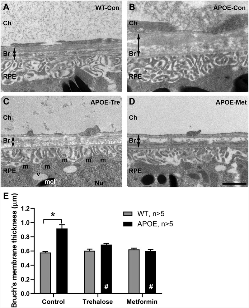
APOE-mice show thickening of Bruch’s membrane at 13 months that is ameliorated by treatment with trehalose or metformin. Bruch’s membrane thickness was investigated using transmission electron microscopy. (A-D) Representative images of Bruch’s membrane are presented for (A) WT-control, (B) APOE-control, and APOE-mice treated with (C) trehalose and (D) metformin. (E) Bruch’s membrane thickness was assessed using segmentation analysis and was found to be significantly thicker in 13-month-old control APOE-mice (black bars) than in age matched WT-mice (gray bars). Treatment of APOE-mice with either trehalose or metformin for 8 months resulted in Bruch’s membrane being similar in thickness to WT-control mice. For all groups n = >5; Two way ANOVA with post-hoc significance of p < 0.05 shown for genotype (*) and treatment (#). Scale: 1 µm. Ch, Choroid; Br, Bruch’s membrane; RPE, retinal pigment epithelium; m, mitochondria; v, vacuole; Nu, nucleus; mel, melanosomes.
