PLOS One logoLink to PLOS One
. 2022 Oct 7;17(10):e0272128. doi: 10.1371/journal.pone.0272128

Cervical length distribution among Brazilian pregnant population and risk factors for short cervix: A multicenter cross-sectional study

Kaline Gomes Ferrari Marquart 1, Thais Valeria Silva 1,2, Ben W Mol 3, José Guilherme Cecatti 1, Renato Passini Jr 1, Cynara M Pereira 1, Thaísa B Guedes 1, Tatiana F Fanton 1, Rodolfo C Pacagnella 1,*; The P5 working group
Editor: Federico Ferrari4
PMCID: PMC9544154  PMID: 36206265

Abstract

Objective

Since there are populational differences and risk factors that influence the cervical length, the aim of the study was to construct a populational curve with measurements of the uterine cervix of pregnant women in the second trimester of pregnancy and to evaluate which variables were related to cervical length (CL) ≤25 mm.

Materials and methods

This was a multicenter cross-sectional study performed at 17 hospitals in several regions of Brazil. From 2015 to 2019, transvaginal ultrasound scan was performed in women with singleton pregnancies at 18 0/7 to 22 6/7 weeks of gestation to measure the CL. We analyzed CL regarding its distribution and the risk factors for CL ≤25 mm using logistic regression.

Results

The percentage of CL ≤ 25mm was 6.67%. Shorter cervices, when measured using both straight and curve techniques, showed similar results: range 21.0–25.0 mm in straight versus 22.6–26.0 mm in curve measurement for the 5th percentile. However, the difference between the two techniques became more pronounced after the 75th percentile (range 41.0–42.0 mm straight x 43.6–45.0 mm in curve measurement). The risk factors identified for short cervix were low body mass index (BMI) (OR: 1.81 CI: 1.16–2.82), higher education (OR: 1.39 CI: 1.10–1.75) and personal history ([one prior miscarriage OR: 1.41 CI: 1.11–1.78 and ≥2 prior miscarriages OR: 1.67 CI: 1.24–2.25], preterm birth [OR: 1.70 CI: 1.12–2.59], previous low birth weight <2500 g [OR: 1.70 CI: 1.15–2.50], cervical surgery [OR: 4.33 CI: 2.58–7.27]). By contrast, obesity (OR: 0.64 CI: 0.51–0.82), living with a partner (OR: 0.76 CI: 0.61–0.95) and previous pregnancy (OR: 0.46 CI: 0.37–0.57) decreased the risk of short cervix.

Conclusions

The CL distribution showed a relatively low percentage of cervix ≤25 mm. There may be populational differences in the CL distribution and this as well as the risk factors for short CL need to be considered when adopting a screening strategy for short cervix.

Introduction

Transvaginal ultrasound (TVU) is the gold standard method of assessing cervical length in pregnant women, established by drawing a straight line between the internal and external orifice of the cervix; it provides objective and reproducible measurements [14]. TVU can also help to prevent prematurity because cervical length is one of the best predictors of preterm birth (PTB), and short cervical length may trigger interventions. Progesterone has a role in reducing spontaneous preterm in singleton pregnancies with cervical length (CL) ≤ 25 mm [5].

Although randomized studies have demonstrated benefits for the treatment of women with short cervix with progesterone in the reduction of PTB and consequently prevention of neonatal morbidity and mortality [6], the cutoff point defining short cervix that justifies interventions remains a matter of debate. The recommended cutoff point for intervention varied from 10 to 30 mm, with 25 mm being the most accepted cervical length that would trigger intervention [2, 79], including the recommendation of the American College of Obstetricians and Gynecologists (ACOG) [10].

The standard technique for measuring the cervix using TVU is to draw a straight line between the internal and external os [14]. Previous studies have already compared the straight technique with the curved technique, as well as the contribution of the volume of the uterine cervix for the diagnosis of short cervix. No technique showed better results compared to the standard technique [3, 4, 6].

There are populational differences in terms of the genesis of preterm labor and evidence associating cervical structure (length and dilation) with race and other social factors [11]. A retrospective cohort of singleton gestations without prior PTB undergoing universal second trimester ultrasound screening found that African-American women had a 2.8–fold increased risk of cervical length ≤25 mm compared to non-Hispanic white women in a low-risk population [12]. Another study of a prospective cohort of 5092 low risk women with singleton pregnancies who underwent TVU showed a relationship between mid-trimester cervical length and BMI, maternal age, maternal ethnicity, and parity [13]. These findings suggest that different groups of women may present specific characteristics and, therefore, it is necessary to identify conditions that may influence the cervical length and its ability to predict preterm labor.

The use of distribution curves of cervical length from generic population without consider populational differences can leading to unnecessary treatments or inaccurate risk estimation. Conversely, underestimating the risk for short cervix, may lead to failure to intervene. Nevertheless, few studies have been devoted to construction of specific population curves. Therefore, the aim of the present study was to describe curves for cervical length in singleton pregnant Brazilian women and to assess the risk factors associated with CL ≤25 mm.

Materials and methods

This is a cross-sectional study from women including in the screening phase of the P5 Trial (Pessary Plus Progesterone to Prevent Preterm Birth Study). The P5 Trial was a randomized controlled trial that compared the effectiveness of vaginal progesterone alone versus progesterone plus cervical pessary in women with short cervix, coordinated by the University of Campinas (Trial registration RBR-3t8prz) and approved by the Brazilian National Review Board (CONEP)—number 1.055.555 [14]. In July 2015, a TVU screening program was implemented in 17 institutions (nine states in three regions: South, Southeast and Northeast of Brazil) for 44 months period as the standard of care during routine second trimester ultrasonographic examination. In the current analysis, we studied 8167 singleton pregnant women using an online database from the screening phase of the P5 Trial.

All pregnant women attending the ultrasound department of these facilities at gestational age between 18 0/7 to 22 6/7 weeks of gestation were invited to participate. Before the exam, the women received information about the technique of ultrasound and about the P5 Study; all provided written informed consent. Women with painful contractions, vaginal bleeding, cerclage during current pregnancy before the screening, ruptured membranes diagnosed before screening, severe liver disease, cholestasis during this pregnancy, previous or current thromboembolism, placenta previa, cervical dilation greater than 1 cm, monoamniotic twin pregnancy, higher order multiple pregnancies (triplets or higher), and major fetal malformation or at least one fetus and stillbirth were not eligible for the study. All twin gestations were excluded from our current analysis.

The gestational age was calculated using the date of the last menstrual period (LMP) and was confirmed by a first trimester ultrasound. When there were discrepancies ≥ 7 days, the first trimester ultrasound was used to calculate gestational age. Sociodemographic information, personal and previous gestational history and information about the current pregnancy were collected. After data collection and second trimester gestational US, TVU was performed using a GE Logic C5® equipment or similar with a 5–9-MHz transvaginal probe.

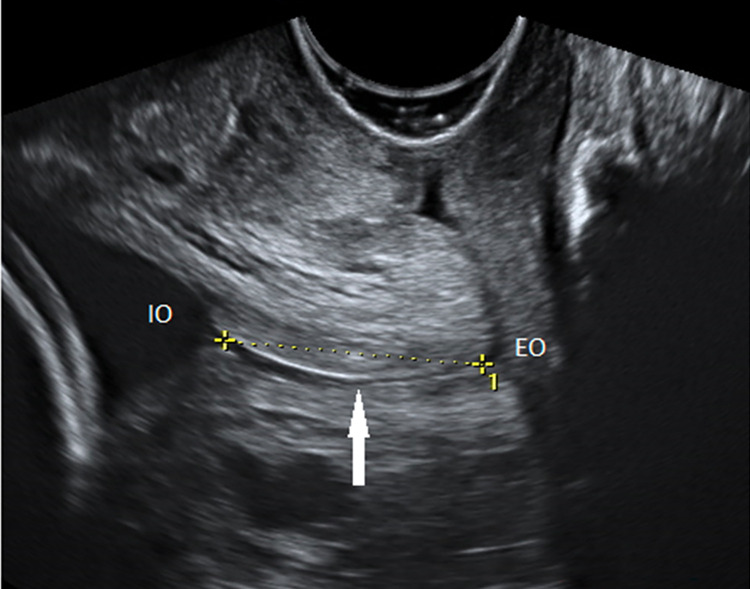
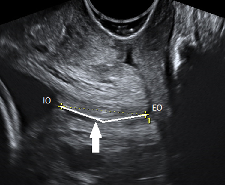
All sonographers were trained in cervical measurement according to the Fetal Medicine Foundation training program [15] and an additional training regarding the volume measurement. After emptying the bladder, the participant was placed in the dorsal lithotomy position. The transvaginal ultrasound probe was introduced and directed toward the anterior fornix, avoiding exerting undue pressure on the cervix, which may artificially increase the length. A sagittal view of the cervix was obtained and the endocervical mucosa was used as a guide to the proper position of the internal os. Four strategies of uterine cervical measurements were used in our study: straight line measurement (SL) between the internal to the external os, used for the primary outcome (distribution); curved measurement (CM) with two straight measurements respecting the endocervical canal pathway between the internal and external os (Figs 1 and 2); anteroposterior measurement near the insertion of the uterine arteries, in the middle third of the cervix; and transverse measurement rotating the transducer 90 degrees to allow transverse visualization of the cervix. The volume of the cervix was calculated using the formula for the volume of a cylinder, πR2h, where R is half the transverse diameter of the cervix, and h is the length. The curved measurement and the measurements for calculating the volume were used only for comparison purposes with the standard straight measurement. The presence or absence of sludge and funneling were also evaluated. Funneling was present when the internal os opening was in the form of “Y,” “U” or “V,” with a width greater than 5 mm. The time required to complete the exam was approximately 10 minutes.

Fig 1. CL measurement in straight line technique.

Fig 1

Transvaginal ultrasonography in sagittal section. The endocervical mucosa (arrow) is used as a guide to identify the internal (IO) and external (EO) os. The straight-line technique is presented (dashed line).

Fig 2. CL measurement curve line technique.

Fig 2

Transvaginal ultrasonography in sagittal section. The endocervical mucosa (arrow) is used as a guide to identify the internal (IO) and external (EO) os. The curve technique is presented (continue line): two lines are drawn respecting the curvature of the endocervical canal.

The calculation of the sample size considered a standard deviation (SD) of 4.0 mm, a type I error of 0.05 and type II error of 0.2. The number estimated to be necessary to adequately power the study was 1500 women for each gestational age between 18 to 22 weeks, totaling a minimum number of 7500 pregnant women.

For the descriptive analysis, mean and percentiles for each measurement were obtained. Distribution curves were presented in graphics. The odds ratio (OR) and 95% confidence intervals (95% CI) for CL≤25mm were calculated. A stepwise multiple logistic regression analysis was used to select the variables to identify risk factors for short cervix. The following variables were used to estimate the model: maternal age (≤ 19, 20 to 34 and ≥ 35 years), schooling (until middle school and beyond high school), body mass index (BMI: low weight <18.5, normal weight 18.6 to 24.9, overweight 25 to 29.9 and obese ≥30), history of PTB and PTB <28 weeks, previous low birth weight (< 2500 g), cerclage in previous pregnancy, previous cervix surgeries, Mullerian malformations, non-spontaneous conception, marital status, number of births and miscarriage and the region of Brazil. Statistical analysis was performed using R software from the R Project for Statistical Computing (version 4.1.2).

Results

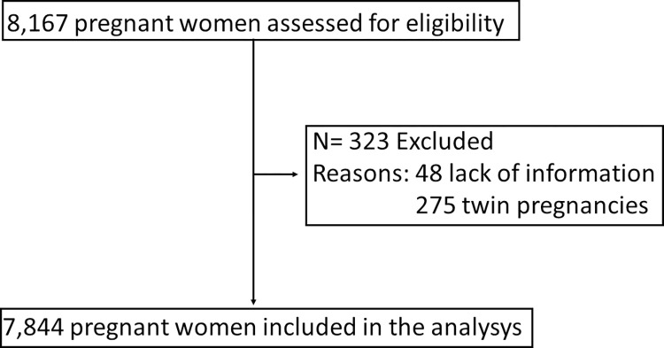
A total of 7,844 of the 8,167 eligible pregnant women were included in the analysis. We excluded 323 participants: 48 due to lack of information and 275 twin pregnancies (Fig 3).

Fig 3. Inclusion and exclusion flowchart.

Fig 3

Eligible pregnant women, excluded and included in the analysis.

In our sample, almost 70% of women were between 20 and 34 years old, a total of 61.8% were overweight or obese, a quarter studied until middle school, 62.5% were non-white and 82.8% lived with their partner predominantly in the south and southeastern region. About obstetric history, 63% had previous pregnancies and 55% had previous births, 10.7% had PTBs, 3.5% had previous PTB <28 weeks and 9.1% with birth weight <2500 g. Regarding delivery, 2801 (35.7%) women had ≥ 1 previous vaginal deliveries and 2020 (25.7%) had previous C-sections; 0.4% had non-spontaneous conception, 0.4% had a previous cerclage, 1.3% had previous cervix surgeries, 1.5% presented uterine malformations, 1.3% women reported active bleeding until the second trimester, and 4.6% and 3.0% presented sludge and funneling in TVU assessment, respectively (Table 1).

Table 1. Main sociodemographic characteristics of the sample population, mean, median of the ultrasonographic measurements in straight line of the uterine cervix in millimeters and percentage of cervical length ≤ 25 mm.

Characteristics n (%) Mean Median CL ≤ 25 mm (%) p- value
Maternal age       0.0019
≤ 19 924 (11.8) 34 .99 35.00 9.09  
20 to 34 5425 (69.2) 36.80 36.60 6.60  
≥ 35 1495 (19.0) 38.44 38.00 5.42  
Schooling       0.0043
Preschool, Elementary and Middle School 1889 (24.1) 37.33 37.00 5.24  
High School and Higher education 5955 (75.9) 36.77 36.60 7.12  
Marital status       0.0018
Without partner 1351 (17.2) 36.20 36.00 8.59  
With partner 6493 (82.8) 37.05 37.00 6.27  
Region       0.0285
Northeast 2900 (37.0) 37.03 37.00 5.86  
South, southeast 4944 (63.0) 36.82 36.90 7.14  
Race 0.7630
White 2943 (37.5) 37.10 37.00 6.56
Non-white 4901 (62.5) 36.78 36.50 6.73
BMI (kg/m2)         <0.0001
<18.5 178 (2.3) 33.97 33.50 15.17  
18.6–24.9 2814 (35.9) 35.84 35.70 8.17  
25–29.9 2630 (33.5) 37.12 37.00 5.93  
≥ 30 2222 (28.3) 38.22 38.00 4.95  
Numbers of pregnancies         0.0804
0 2900 (37.0) 35.89 35.70 7.31  
≥ 1 4944 (63.0) 37.49 37.20 6.29  
Numbers of births         <0.0001
0 3528 (45.0) 35.71 35.50 8.13  
≥ 1 4316 (55.0) 37.88 38.00 5.47  
Numbers of vaginal births         0.62
0 5043 (64.3) 36.64 36.20 6.56  
≥ 1 2801 (35.7) 37.38 37.00 6.85  
Number of C-section         <0.0001
0 5824 (74.3) 36.27 36.00 7.73  
≥ 1 2020 (25.7) 38.72 38.10 3.61  
Number of miscarriage       0.0025
0 5822 (74.2) 37.06 37.00 6.15  
1 1369 (17.5) 36.81 37.00 7.60  
≥ 2 653 (8.3) 35.69 36.00 9.34  
History of preterm birth <0.0001
Yes 840 (10.7) 35.41 36.00 13.10
No 7004 (89.3) 37.08 37.00 5.90
History of preterm birth < 28 weeks <0.0001
Yes 272 (3.5) 32.39 33.80 22.43
No 7572 (96.5) 37.06 37.00 6.10
History birth weight (< 2500g) <0.0001
Yes 717 (9.1) 35.28 35.70 13.95
No 7127 (90.9) 37.06 37.00 5.94
Cerclage in previous pregnancy 0.0073
Yes 35 (0.4) 32.09 33.00 20.00
No 7809 (99.6) 36.92 36.90 6.61
Previous cervix surgeries <0.0001
Yes 102 (1.3) 34.06 34.65 19.61
No 7742 (98.7) 36.94 37.00 6.50
Uterine malformations 0.941
Yes 117 (1.5) 37.51 38.00 6.84
No 7727 (98.5) 36.89 36.90 6.66
Non-spontaneous conception 0.2768
Yes 33 (0.4) 31.80 33.00 12.12
No 7811 (99.6) 36.92 37.00 6.64
Sludge <0.0001
Yes 347 (4.6) 29.00   30.50  30.55
No 7497 (95.4) 37.27 37.00 5.56
Cervical Funneling <0.0001
Yes 229 (3.0)  19.40 20.00 79.04
No 7615 (97.0) 37.48 37.00 4.49
n = 7844          

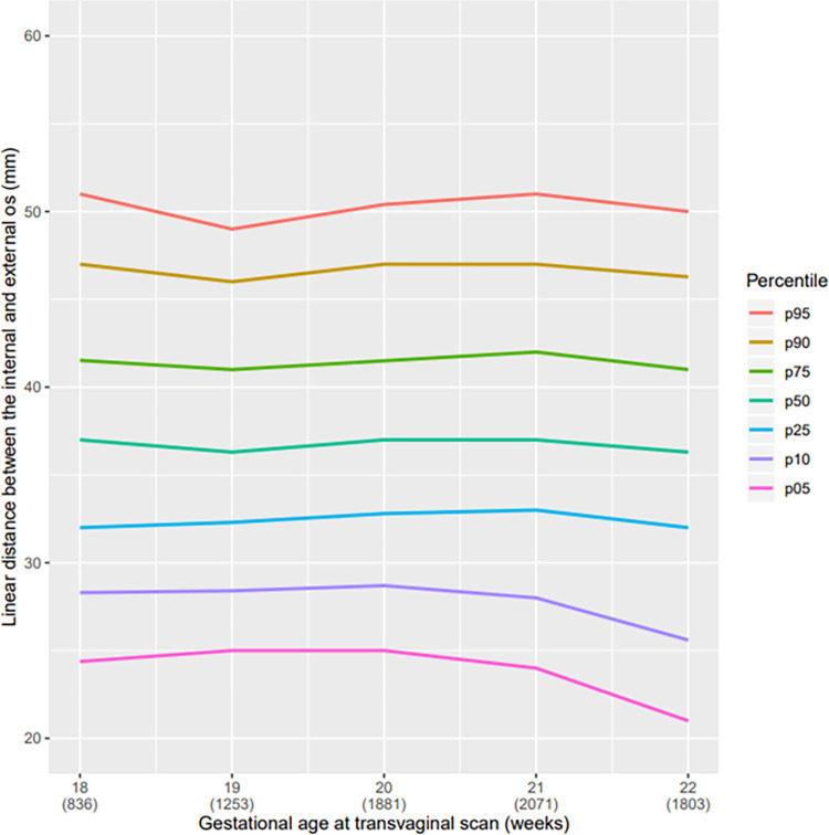
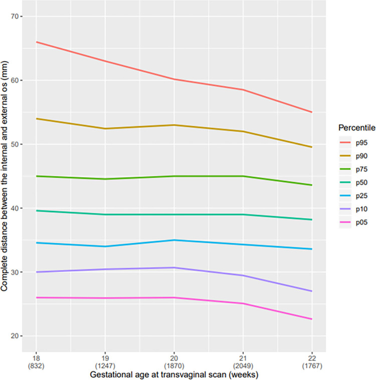
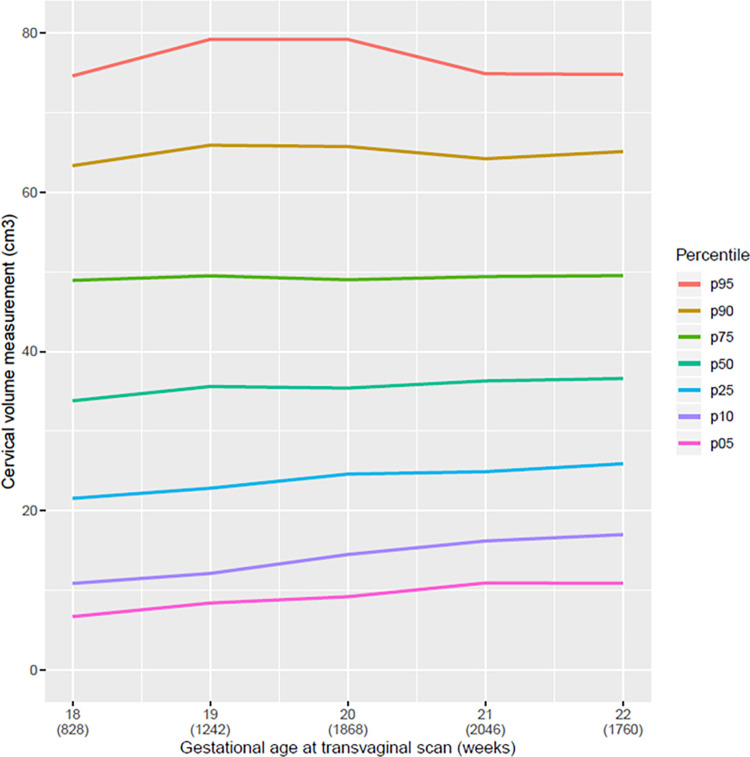
The mean cervical length in linear distance of our population was 36.9 mm, range 36.3 to 37.0 mm; in curve measurement, the mean was 40.1 mm range 38.2 to 39.6 mm (Table 2). In the descriptive analysis, a reduction in the CL from the twenty-first week of pregnancy, regardless of the technique used for measurement (straight or curve) can be observed (Figs 4 and 5). All pregnant women with SL CL measurement ≤25 mm was above the 5th percentile (Table 2 and Fig 4). Comparing the graphs for the straight and curve cervical length measurement grouped at the 5th percentile there was only a small amount of variation. However, in larger cervices, we observed a broader difference between the straight and curve measurement of the cervix. The median CL at 20 weeks of GA was 2 mm higher using the CM than using the SL; in the 95th percentile, this difference was almost 10 mm (Table 2). We observed that the cervical volume slightly increased with progression of gestational age (Fig 6).

Table 2. Values of percentile 5, 10, 25, 50, 75, 90 e 95 for the cervical length measurement in linear distance between internal and external os and in curve by ultrasonography according to gestational age.

Gestational age (weeks)  Cervical length in linear distance (mm)
Mean p 5 p 10 p 25 p 50 p 75 p 90 p 95 CL ≤ 25 mm (%)
18 37.0 24.4 28.3 32.0 37.0 41.5 47.0 51.0 5.7
19 36.9 25.0 28.4 32.3 36.3 41.0 46.0 49.0 5.3
20 37.2 25.0 28.7 32.8 37.0 41.5 47.0 50.4 5.4
21 37.2 24.0 28.0 33.0 37.0 42.0 47.0 51.0 6.4
22 36.2 21.0 25.6 32.0 36.3 41.0 46.3 50.0 9.6
Total 36.9 23.8 27.8 32.3 36.9 41.6 47.0 50.0 6.7
n = 7844
Gestational age (weeks)  Cervical length in curve (mm)
Mean p 5 p 10 p 25 p 50 p 75 p 90 p 95 CL ≤ 25 mm* (%)
18 41.8 26.0 30.0 34.6 39.6 45.0 54.0 66.0 5.1
19 40.3 25.9 30.4 34.0 39.0 44.5 52.4 63.0 4.9
20 40.5 26.0 30.7 35.0 39.0 45.0 53.0 60,2 4.5
21 40.1 25.1 29.5 34.3 39.0 45.0 52.0 58.5 5.6
22 38.5 22.6 27.0 33.6 38.2 43.6 49.5 55.0 8.3
Total 40.1 25.0 29.1 34.0 39.0 44.6 52.1 59.0 6.1
n = 7765 *Considering the curved technic

Fig 4. Curve of percentile values for the linear CL measurement.

Fig 4

Curve of percentile values for the linear distance between the internal and external os according to gestational age (weeks) at transvaginal scan.

Fig 5. Curve of percentile values for the curve CL measurement.

Fig 5

Curve of percentile values for the curve distance between the internal and external os according to gestational age (weeks) at transvaginal scan.

Fig 6. Curve of percentile values for the volume of the uterine cervix according to gestational age (weeks) at transvaginal scan.

Fig 6

Curve of percentile values for the volume of the uterine cervix according to gestational age (weeks) at transvaginal scan.

The lowest mean cervix lengths were observed in women with cervical funneling (19.40 mm) and sludge (29.0 mm) followed by those who had non-spontaneous conception (31.80 mm), previous history of cerclage (32.09 mm), preterm birth <28 weeks (32.39 mm) and in low-weight women (33.97 mm). Of 7844 women, 523 (6.67%) had CL ≤ 25 mm. The percentage of CL ≤25 mm was high among women with cervical funneling, sludge, and other clinical condition related to preterm birth were higher than in the total sample (Table 1).

Considering 25 mm as a cutoff point for risk of preterm birth, we sought to identify variables associated with it. The variables significantly associated with CL ≤ 25 mm were as follows: BMI ≤ 18.5 (aOR: 1.81 CI: 1.16–2.82), higher levels of education (aOR: 1.39 CI: 1.10–1.75), one or more miscarriages (respectively aOR: 1.41 CI: 1.11–1.78 and aOR: 1.67 CI: 1.24–2.25), previous history of preterm birth < 28 weeks (aOR: 2.72 CI: 1.79–4.15), preterm birth (aOR: 1.70 CI: 1.12–2.59), previous child with low birth weight < 2500 g (aOR: 1.70 CI: 1.15–2.50) and history of cervix surgery (aOR: 4.33 CI: 2.58–7.27). By contrast, characteristics inversely associated to CL ≤ 25 mm were living with a partner (aOR: 0.76 CI: 0.61–0.95), maternal overweight (aOR: 0.74 CI: 0.60–0.92), obesity (aOR: 0.64 CI: 0.51–0.82) and at least one previous delivery (aOR: 0.46 CI: 0.37–0.57) (Table 3).

Table 3. Multiple analysis for cervical length ≤ 25 mm.

Variables p-value aOR (95% CI)
Marital status (living with partner) 0,018 0.76 (0.61–0.95)
BMI (kg/m2)  
<18.5 0,009 1.81 (1.16–2.82)
25–29.9 0,007 0.74 (0.60–0.92)
≥ 30 0.000 0.64 (0.51–0.82)
Schooling (High School and Higher education) <0.0001 1.39 (1.10–1.75)
Numbers of previous births ≥ 1 <0.0001 0.46 (0.37–0.57)
Numbers of miscarriages    
1 0,005 1.41 (1.11–1.78)
≥ 2 0,001 1.67 (1.24–2.25)
Previous history of preterm birth < 28 weeks ≥ 1 0.000 2.72 (1.79–4.15)
Previous history of preterm birth 0,013 1.70 (1.12–2.59)
Previous birth weight (< 2500g) 0,008 1.70 (1.15–2.50)
Previous cervix surgeries <0.0001 4.33 (2.58–7.27)
n = 7844    

We also assessed women without previous pregnancies separately from those who had at least one previous pregnancy. Women with previous pregnancies and with previous deliveries had reduced risk of CL ≤ 25 mm (aOR: 0.30 CI: 0.22–0.41). Moreover, those who had a history of PTB birth < 28 weeks had 2.7-fold increased risk for CL ≤ 25 mm (aOR: 2.77 CI: 1.82–4.22) as well as women who had a previous child with low birthweight <2500 g (aOR: 1.74 CI: 1.17–2.57). In the group of women without previous pregnancies, those living with their partners had a lower frequency of CL ≤ 25 mm (aOR: 0.68 CI: 0.50–0.91) and living in the southeast and south regions were associated to a CL ≤ 25 mm (aOR: 1.41 CI: 1.04–1.90). In both groups, previous cervix surgery significantly increased the risk of CL ≤ 25 mm (multiparous: aOR: 4.54 CI: 2.43–8.47 and nulliparous: aOR: 3.77 CI: 1.48–9.60) (Tables 4 and 5).

Table 4. Multiple analysis for cervical length ≤ 25 mm in women with previous pregnancies.

Variables p-value aOR (95% CI)
BMI (kg/m2)    
<18.5 0.292 2.10 (1.08–4.10)
25–29.9 0.117 0.80 (0.61–1.06)
≥ 30 0.001 0.59 (0.43–0.81)
Schooling (High School and Higher education) 0.023 1.40 (1.05–1.87)
Numbers of previous births ≥ 1 <0.0001 0.30 (0.22–0.41)
Previous history of preterm birth < 28 weeks ≥ 1 <0.0001 2.77 (1.82–4.22)
Previous history of preterm birth 0,014 1.69 (1.11–2.58)
Previous birth weight (< 2500g) 0,006 1.74 (1.17–2.57)
Previous cervix surgeries <0.0001 4.54 (2.43–8.47)
n = 4944    

Table 5. Multiple analysis for cervical length ≤ 25 mm in women without previous pregnancies.

Variables p-value aOR (95% CI)
Marital status (living with partner) 0,011 0.68 (0.50–0.91)
BMI (kg/m2)    
<18.5 0,130 1.59 (0.87–2.89)
25–29.9 0,013 0.63 (0.44–0.91)
≥ 30 0,132 0.75 (0.51–1.09)
Region (South, southeast) 0,027 1.41 (1.04–1.90)
Previous cervix surgeries 0,005 3.77 (1.48–9.60)
N = 2900    

Discussion

We determined the CL distribution among second trimester Brazilian pregnant women. The distribution showed a low percentage of CL ≤25 mm. The risk factors associated with increased risk for CL ≤25 mm were as follows: low BMI, high levels of education, previous miscarriage, prior PTB (especially if <28 weeks), previous low birthweight <2500 g and prior cervical surgery.

Iams et al. were among the pioneers in proposing reference values for CL. For women at 22-week’s gestation, we found very similar measurements for the 5th, 10th and 25th percentiles [2]. Our 5th and 10th percentiles, however, were similar to those previously proposed. Nevertheless, even considering women with a CL ≤25 mm as having an increased risk for preterm birth, our data corroborates the fact that there may be populational differences in the CL distribution and its relation to preterm birth risk compared to the literature [2, 16, 19].

Studies have proposed cervical distributions curves for the Brazilian population considering population characteristics. In general, the 50th and 95th percentiles are similar to those of our study; however, for the lower percentiles, we obtained slightly different values than others for the lower percentiles [16, 17].

Even within a single country, it is also necessary to be aware of the importance of intra-population differences. A prospective cohort found that Afro-Caribbean women had a shorter cervix than did Caucasian women [18]. Similar findings were identified in a retrospective cohort conducted in the US involving 16,598 women in the second trimester of pregnancy, suggesting that a short cervix definition should differ between ethnic groups within the same population [19].

In 2020, a prospective Asian cohort study involving 1013 women found significant difference between the mean cervical measurement by population group (Chinese 32.2 ± 0.77 mm, Malay 31.3 ± 0.69 mm, Indian 29.7 ± 0.70, Others 33.3 ± 0.82 mm) [20]. Our study, thus, reinforce that a single distribution curve for cervical measurement, without considering the different population characteristics, may not represent all women equally and could inefficiently guide preventive measures for prematurity.

With respect to different techniques to measure the cervix, shorter cervixes when measured by both straight and curve techniques, do not differ substantially. In both techniques, there is a reduction of CL as the pregnancy advances [21, 22]. We observed that CL reduces more significantly after 21 weeks, regardless of the technique used for assessment and this pattern is reported in literature. However, in longer cervix, we observed that the difference between the two techniques becomes more pronounced.

This underestimation in the values of the last quartile changes the design of the distribution curve. Considering the importance of building reference curves that respect the cervical anatomy, we suggest that the most adequate method to measure the cervix is respecting the curvature, however, for short cervix, which is the main factor related to spontaneous PTB, the straight-line measurement for cervical length may be the best strategy. Previous studies have already compared the straight technique with the curved technique [23], as well as the contribution of the volume of the uterine cervix for the diagnosis of short cervix. No technique showed better results compared to the standard technique [2429].

We also observed that the volume of the cervix increased slightly over the course of gestation despite the progressive shortening of the longitudinal measurement of the cervix. In other words, the cervix becomes shorter but wider [23]. Although many studies have shown a correlation between cervical volume and the ability of this measure to contribute to the prediction of the risk of prematurity, none has demonstrated additional benefits in relation to the longitudinal cervical measurement technique [2428].

Regarding the risk factors for short cervix, level of schooling is a social aspect that is related to health improvement; however, it was found that extremely high maternal education did not confer more protection against PTB [29]. In high-income countries, a higher level of education is also associated with increasing working day for women. High-level education provokes an overloaded of responsibilities and stress, including employment relationships, excessive time into the traffic, less time to the physiological needs (like time to sleep, rest, and healthy nutrition), and less time for family care and domestic tasks [28].

We found that CL was shorter in pregnant women ≤19 years old than those >20 years old. In the literature, young pregnant women are at increased risk for spontaneous PTB [30], which may be due to biological immaturity of the female genital tract [31, 32], social and behavioral factors [18], and intra-amniotic infections as a consequence of genital tract infections [31].

We as well found that low BMI was associated with CL≤ 25 mm. This result confirms the findings from other studies showing the same relationship between shorter CL and lower BMI [13, 18, 33]. There appears to be a correlation between low pre-pregnancy BMI and low weight gain during pregnancy with spontaneous preterm birth [34, 35]. On the other hand, we found a lower frequency of CL ≤25 mm in obese than in underweight women. A systematic review showed that, compared to normal weight women, pre-obese women and those with grade I obesity had a 15% reduction in their risk of spontaneous PTB and the prevalence of short cervix was significantly lower in obese compared to normal or underweight women [34]. By contrast, other studies showed relationships between obesity and prematurity [3338], mostly related to therapeutic PTB [39]. A theory to explain this cervical behavior on pregnancies with low BMI is related to acquired collagen deficiency.

Among the strongest risk factors for PTB we found the previous history of spontaneous preterm delivery, particularly if it occurred early in pregnancy [40]. Our study reinforces this argument and shows an association between short cervix and history of PTB [37]. Nevertheless, in literature, nulliparity appears to increase the risk for PTB [30]. The mechanisms by which nulliparity can lead to PTB remain poorly understood. We noted a smaller mean and median of CL as well as a larger percentage of CL ≤25 mm in nulliparous as compared to multiparous women.

The strength of this study is that we included a large sample of cervical measurements from Brazilian pregnant women with singletons in the second trimester, establishing reference values; therefore, external validation is possible. Limitations of our study include the cross-sectional design that prevented establishment of correlations between the two techniques for measuring the cervix (straight and curve) and the outcome (PTB). In addition, we analyzed data from 836 and 1253 pregnant women with gestational ages of 18 and 19 weeks, respectively, thereby failing to reach the calculated sample size at these gestational ages. The measurements were performed by different, albeit trained, professionals from different facilities, which might have included a sort of bias in measurements. We also do not have information regarding the outcomes for most women, which would have added information.

Considering the results of the multiple analysis, because the universal screening of pregnant women in the second trimester remains controversial and is not recommended by the main gynecology and obstetrics societies [4143], as well as the fact that we recognize that women with CL ≤25 mm due to population differences are at different risks for PTB, we can propose that risk factors for CL ≤25mm in mid-trimester for Brazilian singleton pregnant women as follows: low BMI, high levels of education, previous miscarriage, prior PTB (especially if <28 weeks), previous low birth weight <2500 g and prior cervical surgery. However, as the prevalence of PTB in Brazil is high, in places where financial resources are available and easy access to transvaginal ultrasound, we recommend that universal screening in the second trimester of pregnancy should be implemented.

Conclusion

The reference CL distribution curves should consider populational characteristics since physicians may use it as a strategy to prevent preterm birth in clinical practice. Doing so, it will enable a more efficient diagnosis of short cervix and its association with prematurity, allowing assertive medical decisions. Moreover, we suggest that subsequent studies should consider these populational characteristics to build new distribution curves and define specific screening strategies for different populations to prevent premature delivery.

Supporting information

S1 Appendix. P5 trial description.

(DOCX)

S2 Appendix. STROBE statement checklist.

(DOCX)

S3 Appendix. The P5 working group.

(DOCX)

Acknowledgments

*P5 working group

Amanda Dantas; Anderson Borovac-Pinheiro; Antonio Fernandes Moron; Carlos Augusto Santos Menezes; Cláudio Sérgio Medeiros Paiva; Cristhiane B Marques; Cynara Maria Pereira; Djacyr Magna Cabral Paiva; Elaine Christine Dantas Moisés; Enoch Quinderé Sá Barreto; Felipe Soares; Fernando Maia Peixoto-Filho; Francisco Edson de Lucena Feitosa; Francisco Herlanio Costa Carvalho; Jessica Scremin Boechem; João Renato Benini-Jr.; Karayna Gil Fernandes; Kleber Cursino Andrade; Leila Katz; Maíra Rossmann Machado; Marcelo L Nomura; Marcelo Marques Souza Lima; Marcelo Santucci Franca; Marcos Nakamura-Pereira; Maria Julia Miele; Maria Laura Costa; Mário Dias Correia Jr; Nelson Sass; Renato T Souza; Rodrigo Pauperio Soares Camargo; Samira Maerrawi Haddad; Sérgio Martins-Costa; Silvana F Bento; Silvana Maria Quintana; Stéphanno Gomes Pereira Sarmento;

Data Availability

The database is available at https://doi.org/10.25824/redu/P8PUWR.

Funding Statement

This work was supported by the Bill & Melinda Gates Foundation [OPP1107597]. Under the grant conditions of the Foundation, a Creative Commons Attribution 4.0 Generic License has already been assigned to the Author Accepted Manuscript version that might arise from this submission. This work was supported by The Brazilian Ministry of Health, and the Brazilian National Council for Scientific and Technological Development (CNPq) [401615/20138]. The funders had no role in the design, development of the study, analysis, interpretation of data, writing the manuscript and in the decision to submit the article for publication.

References

  • 1.Andersen HF, Nugent CE, Wanty SD, Hayashi RH. Prediction of risk for preterm delivery by ultrasonographic measurement of cervical length. Am J Obstet Gynecol. 1990;163(3):859–67. doi: 10.1016/0002-9378(90)91084-p [DOI] [PubMed] [Google Scholar]
  • 2.Iams JD, Goldenberg RL, Meis PJ, Mercer BM, Moawad A, Das A, et al. The length of the cervix and the risk of spontaneous premature delivery. National Institute of Child Health and Human Development Maternal Fetal Medicine Unit Network. N Engl J Med. 1996;334(9):567–72. doi: 10.1056/NEJM199602293340904 [DOI] [PubMed] [Google Scholar]
  • 3.Berghella V, Baxter JK, Hendrix NW. Cervical assessment by ultrasound for preventing preterm delivery. Cochrane Database Syst Rev. 2013(1):Cd007235. doi: 10.1002/14651858.CD007235.pub3 [DOI] [PMC free article] [PubMed] [Google Scholar]
  • 4.AIUM Practice Parameter for the Performance of Limited Obstetric Ultrasound Examinations by Advanced Clinical Providers. J Ultrasound Med. 2018;37(7):1587–96. doi: 10.1002/jum.14677 [DOI] [PubMed] [Google Scholar]
  • 5.Romero R, Nicolaides KH, Conde-Agudelo A, O’Brien JM, Cetingoz E, Da Fonseca E, et al. Vaginal progesterone decreases preterm birth </ = 34 weeks of gestation in women with a singleton pregnancy and a short cervix: an updated meta-analysis including data from the OPPTIMUM study. Ultrasound Obstet Gynecol. 2016;48(3):308–17. doi: 10.1002/uog.15953 [DOI] [PMC free article] [PubMed] [Google Scholar]
  • 6.Parry S, Elovitz MA. Pros and cons of maternal cervical length screening to identify women at risk of spontaneous preterm delivery. Clin Obstet Gynecol. 2014;57(3):537–46. doi: 10.1097/GRF.0000000000000051 [DOI] [PubMed] [Google Scholar]
  • 7.Romero R, Conde-Agudelo A, Da Fonseca E, O’Brien JM, Cetingoz E, Creasy GW, et al. Vaginal progesterone for preventing preterm birth and adverse perinatal outcomes in singleton gestations with a short cervix: a meta-analysis of individual patient data. Am J Obstet Gynecol. 2018;218(2):161–80. doi: 10.1016/j.ajog.2017.11.576 [DOI] [PMC free article] [PubMed] [Google Scholar]
  • 8.Berghella V, Rafael TJ, Szychowski JM, Rust OA, Owen J. Cerclage for short cervix on ultrasonography in women with singleton gestations and previous preterm birth: a meta-analysis. Obstet Gynecol. 2011;117(3):663–71. doi: 10.1097/AOG.0b013e31820ca847 [DOI] [PubMed] [Google Scholar]
  • 9.Pedretti MK, Kazemier BM, Dickinson JE, Mol BW. Implementing universal cervical length screening in asymptomatic women with singleton pregnancies: challenges and opportunities. Aust N Z J Obstet Gynaecol. 2017;57(2):221–7. doi: 10.1111/ajo.12586 [DOI] [PubMed] [Google Scholar]
  • 10.Prediction and Prevention of Spontaneous Preterm Birth: ACOG Practice Bulletin, Number 234. Obstet Gynecol. 2021;138(2):e65–e90. doi: 10.1097/AOG.0000000000004479 [DOI] [PubMed] [Google Scholar]
  • 11.Harville EW, Knoepp LR, Wallace ME, Miller KS. Cervical pathways for racial disparities in preterm births: the Preterm Prediction Study. J Matern Fetal Neonatal Med. 2019;32(23):4022–8. doi: 10.1080/14767058.2018.1484091 [DOI] [PubMed] [Google Scholar]
  • 12.Buck JN, Orzechowski KM, Berghella V. Racial disparities in cervical length for prediction of preterm birth in a low risk population. J Matern Fetal Neonatal Med. 2017;30(15):1851–4. doi: 10.1080/14767058.2016.1228056 [DOI] [PubMed] [Google Scholar]
  • 13.Van der Ven AJ, van Os MA, Kleinrouweler CE, de Groot CJ, Haak MC, Mol BW, et al. Is cervical length associated with maternal characteristics? Eur J Obstet Gynecol Reprod Biol. 2015;188:12–6. doi: 10.1016/j.ejogrb.2015.02.032 [DOI] [PubMed] [Google Scholar]
  • 14.Pacagnella RC, Mol BW, Borovac-Pinheiro A, Passini R, Nomura ML, Andrade KC, et al. A randomized controlled trial on the use of pessary plus progesterone to prevent preterm birth in women with short cervical length (P5 trial). BMC Pregnancy Childbirth. 192019. doi: 10.1186/s12884-019-2513-2 [DOI] [PMC free article] [PubMed] [Google Scholar]
  • 15.The Fetal Medicine Foudation. The FMF certification cervical assessment. Available in: https://fetalmedicine.org/fmf-certification-2/cervical-assessment-1. Accessed on November 2, 2021.
  • 16.Silva SV, Damião R, Fonseca EB, Garcia S, Lippi UG. Reference ranges for cervical length by transvaginal scan in singleton pregnancies. J Matern Fetal Neonatal Med. 2010;23(5):379–82 doi: 10.3109/14767050903177169 [DOI] [PubMed] [Google Scholar]
  • 17.Andrade SGA, Andrade FM, Araujo Júnior E, Pires CR, Mattar R, Moron AF. Assessment of Length of Maternal Cervix between 18 and 24 weeks of Gestation in a Low-Risk Brazilian Population. Rev Bras Ginecol Obstet. 2017;39(12):647–52. [DOI] [PMC free article] [PubMed] [Google Scholar]
  • 18.Heath VC, Southall TR, Souka AP, Novakov A, Nicolaides KH. Cervical length at 23 weeks of gestation: relation to demographic characteristics and previous obstetric history. Ultrasound Obstet Gynecol. 1998;12(5):304–11. doi: 10.1046/j.1469-0705.1998.12050304.x [DOI] [PubMed] [Google Scholar]
  • 19.Bligard K TL, Stout MJ, Tuuli MG, Macones GA, Cahill AG. Performance of cervical length screening in african american women. American Journal of Obstetrics & Gynecology. 2020. [Google Scholar]
  • 20.Thain S, Yeo GSH, Kwek K, Chern B, Tan KH. Spontaneous preterm birth and cervical length in a pregnant Asian population. PLoS One. 2020;15(4):e0230125. doi: 10.1371/journal.pone.0230125 [DOI] [PMC free article] [PubMed] [Google Scholar]
  • 21.Papastefanou I, Pilalis A, Kappou D, Souka AP. Cervical length at 11–40 weeks: unconditional and conditional longitudinal reference ranges. Acta Obstet Gynecol Scand. 2016;95(12):1376–82. doi: 10.1111/aogs.13025 [DOI] [PubMed] [Google Scholar]
  • 22.van Zijl MD, Koullali B, Mol BWJ, Kazemier BM, Pajkrt E. How to measure the cervical length: A prospective cohort study in the Netherlands. American Journal of Obstetrics & Gynecology. 2020;218(1):S263–S4. [Google Scholar]
  • 23.Andrade KC, Bortoletto TG, Almeida CM, Daniel RA, Avo H, Pacagnella RC, et al. Reference Ranges for Ultrasonographic Measurements of the Uterine Cervix in Low-Risk Pregnant Women. Rev Bras Ginecol Obstet. 2017;39(9):443–52 [DOI] [PMC free article] [PubMed] [Google Scholar]
  • 24.Barber MA, Medina M, Cabrera F, Romero A, Valle L, Garcia-Hernández JA. Cervical length vs VOCAL cervical volume for predicting pre-term delivery in asymptomatic women at 20–22 weeks’ pregnancy. J Obstet Gynaecol. 2012;32(7):648–51. doi: 10.3109/01443615.2012.698330 [DOI] [PubMed] [Google Scholar]
  • 25.Dilek TU, Gurbuz A, Yazici G, Arslan M, Gulhan S, Pata O, et al. Comparison of cervical volume and cervical length to predict preterm delivery by transvaginal ultrasound. Am J Perinatol. 2006;23(3):167–72. doi: 10.1055/s-2006-934102 [DOI] [PubMed] [Google Scholar]
  • 26.Yilmaz NC, Yiğiter AB, Kavak ZN, Durukan B, Gokaslan H. Longitudinal examination of cervical volume and vascularization changes during the antepartum and postpartum period using three-dimensional and power Doppler ultrasound. J Perinat Med. 2010;38(5):461–5. doi: 10.1515/jpm.2010.087 [DOI] [PubMed] [Google Scholar]
  • 27.Park IY, Kwon JY, Hong SC, Choi HM, Kwon HS, Won HS, et al. Usefulness of cervical volume by three-dimensional ultrasound in identifying the risk for preterm birth. Ultrasound Med Biol. 2011;37(7):1039–45. doi: 10.1016/j.ultrasmedbio.2011.04.010 [DOI] [PubMed] [Google Scholar]
  • 28.Marinelli JVC, Filho AGA, de Barros MF, Rodrigues AS, Francisco RPV, Carvalho MHB. Women’s Obstetric History and Midtrimester Cervical Length Measurements by 2D/3D and Doppler Ultrasound. Rev Bras Ginecol Obstet. 2020. [DOI] [PMC free article] [PubMed] [Google Scholar]
  • 29.Auger N, Abrahamowicz M, Park AL, Wynant W. Extreme maternal education and preterm birth: time-to-event analysis of age and nativity-dependent risks. Ann Epidemiol. 2013;23(1):1–6. doi: 10.1016/j.annepidem.2012.10.007 [DOI] [PubMed] [Google Scholar]
  • 30.Mayo JA, Shachar BZ, Stevenson DK, Shaw GM. Nulliparous teenagers and preterm birth in California. J Perinat Med. 2017;45(8):959–67. doi: 10.1515/jpm-2016-0313 [DOI] [PubMed] [Google Scholar]
  • 31.D’Agostini C, de Oliveira M, D’Souza-Li L. Comparison of cervical length in adult and adolescent nulliparae at mid-gestation. J Pediatr Adolesc Gynecol. 2013;26(4):209–11. doi: 10.1016/j.jpag.2013.03.001 [DOI] [PubMed] [Google Scholar]
  • 32.Kaplanoglu M, Bulbul M, Konca C, Kaplanoglu D, Tabak MS, Ata B. Gynecologic age is an important risk factor for obstetric and perinatal outcomes in adolescent pregnancies. Women Birth. 2015;28(4):e119–23. doi: 10.1016/j.wombi.2015.07.002 [DOI] [PubMed] [Google Scholar]
  • 33.Kandil M, Sanad Z, Sayyed T, Ellakwa H. Body mass index is linked to cervical length and duration of pregnancy: An observational study in low risk pregnancy. J Obstet Gynaecol. 2017;37(1):33–7. doi: 10.1080/01443615.2016.1205555 [DOI] [PubMed] [Google Scholar]
  • 34.Torloni MR, Betran AP, Daher S, Widmer M, Dolan SM, Menon R, et al. Maternal BMI and preterm birth: a systematic review of the literature with meta-analysis. J Matern Fetal Neonatal Med. 2009;22(11):957–70. doi: 10.3109/14767050903042561 [DOI] [PubMed] [Google Scholar]
  • 35.Siega-Riz AM, Adair LS, Hobel CJ. Maternal underweight status and inadequate rate of weight gain during the third trimester of pregnancy increases the risk of preterm delivery. J Nutr. 1996;126(1):146–53. doi: 10.1093/jn/126.1.146 [DOI] [PubMed] [Google Scholar]
  • 36.Shaw GM, Wise PH, Mayo J, Carmichael SL, Ley C, Lyell DJ, et al. Maternal prepregnancy body mass index and risk of spontaneous preterm birth. Paediatr Perinat Epidemiol. 2014;28(4):302–11. doi: 10.1111/ppe.12125 [DOI] [PubMed] [Google Scholar]
  • 37.Salihu H, Mbah AK, Alio AP, Kornosky JL, Whiteman VE, Belogolovkin V, et al. Nulliparity and preterm birth in the era of obesity epidemic. J Matern Fetal Neonatal Med. 2010;23(12):1444–50. doi: 10.3109/14767051003678044 [DOI] [PubMed] [Google Scholar]
  • 38.Khatibi A, Brantsaeter AL, Sengpiel V, Kacerovsky M, Magnus P, Morken NH, et al. Prepregnancy maternal body mass index and preterm delivery. Am J Obstet Gynecol. 2012;207(3):212.e1–7. doi: 10.1016/j.ajog.2012.06.002 [DOI] [PubMed] [Google Scholar]
  • 39.Sharashova EE, Anda EE, Grjibovski AM. Early pregnancy body mass index and spontaneous preterm birth in Northwest Russia: a registry-based study. BMC Pregnancy Childbirth. 2014;14:303. doi: 10.1186/1471-2393-14-303 [DOI] [PMC free article] [PubMed] [Google Scholar]
  • 40.Mercer BM, Macpherson CA, Goldenberg RL, Goepfert AR, Hauguel-de Mouzon S, Varner MW, et al. Are women with recurrent spontaneous preterm births different from those without such history? Am J Obstet Gynecol. 2006;194(4):1176–84; discussion 84–5. doi: 10.1016/j.ajog.2006.01.069 [DOI] [PubMed] [Google Scholar]
  • 41.Lim K, Butt K, Crane JM. No. 257-Ultrasonographic Cervical Length Assessment in Predicting Preterm Birth in Singleton Pregnancies. J Obstet Gynaecol Can. 2018;40(2):e151–e64. doi: 10.1016/j.jogc.2017.11.016 [DOI] [PubMed] [Google Scholar]
  • 42.Practice bulletin no. 130: prediction and prevention of preterm birth. Obstet Gynecol. 2012;120(4):964–73. doi: 10.1097/AOG.0b013e3182723b1b [DOI] [PubMed] [Google Scholar]
  • 43.Progesterone and Preterm Birth Prevention: Translating Clinical Trials Data Into Clinical Practice. American journal of obstetrics and gynecology. 2012;206(5). doi: 10.1016/j.ajog.2012.03.010 [DOI] [PubMed] [Google Scholar]

Decision Letter 0

Federico Ferrari

6 Oct 2021

PONE-D-21-13533Cervical length distribution among Brazilian pregnant population and risk factors for short cervix: a multicenter cross-sectional studyPLOS ONE

Dear Dr. Pacagnella,

Thank you for submitting your manuscript to PLOS ONE. After careful consideration, we feel that it has merit but does not fully meet PLOS ONE’s publication criteria as it currently stands. Therefore, we invite you to submit a revised version of the manuscript that addresses the points raised during the review process.

Please submit your revised manuscript by Nov 20 2021 11:59PM. If you will need more time than this to complete your revisions, please reply to this message or contact the journal office at plosone@plos.org. When you're ready to submit your revision, log on to https://www.editorialmanager.com/pone/ and select the 'Submissions Needing Revision' folder to locate your manuscript file.

Please include the following items when submitting your revised manuscript:

  • A rebuttal letter that responds to each point raised by the academic editor and reviewer(s). You should upload this letter as a separate file labeled 'Response to Reviewers'.

  • A marked-up copy of your manuscript that highlights changes made to the original version. You should upload this as a separate file labeled 'Revised Manuscript with Track Changes'.

  • An unmarked version of your revised paper without tracked changes. You should upload this as a separate file labeled 'Manuscript'.

If you would like to make changes to your financial disclosure, please include your updated statement in your cover letter. Guidelines for resubmitting your figure files are available below the reviewer comments at the end of this letter.

If applicable, we recommend that you deposit your laboratory protocols in protocols.io to enhance the reproducibility of your results. Protocols.io assigns your protocol its own identifier (DOI) so that it can be cited independently in the future. For instructions see: https://journals.plos.org/plosone/s/submission-guidelines#loc-laboratory-protocols. Additionally, PLOS ONE offers an option for publishing peer-reviewed Lab Protocol articles, which describe protocols hosted on protocols.io. Read more information on sharing protocols at https://plos.org/protocols?utm_medium=editorial-email&utm_source=authorletters&utm_campaign=protocols.

We look forward to receiving your revised manuscript.

Kind regards,

Federico Ferrari

Academic Editor

PLOS ONE

Journal Requirements:

When submitting your revision, we need you to address these additional requirements.

1. Please ensure that your manuscript meets PLOS ONE's style requirements, including those for file naming. The PLOS ONE style templates can be found at 

https://journals.plos.org/plosone/s/file?id=wjVg/PLOSOne_formatting_sample_main_body.pdf and 

https://journals.plos.org/plosone/s/file?id=ba62/PLOSOne_formatting_sample_title_authors_affiliations.pdf

2. We note that the grant information you provided in the ‘Funding Information’ and ‘Financial Disclosure’ sections do not match. 

When you resubmit, please ensure that you provide the correct grant numbers for the awards you received for your study in the ‘Funding Information’ section.

3. We note that you have stated that you will provide repository information for your data at acceptance. Should your manuscript be accepted for publication, we will hold it until you provide the relevant accession numbers or DOIs necessary to access your data. If you wish to make changes to your Data Availability statement, please describe these changes in your cover letter and we will update your Data Availability statement to reflect the information you provide.

4. One of the noted authors is a group or consortium "The P5 working group". In addition to naming the author group, please list the individual authors and affiliations within this group in the acknowledgments section of your manuscript. Please also indicate clearly a lead author for this group along with a contact email address.

[Note: HTML markup is below. Please do not edit.]

Reviewers' comments:

Reviewer's Responses to Questions

Comments to the Author

1. Is the manuscript technically sound, and do the data support the conclusions?

The manuscript must describe a technically sound piece of scientific research with data that supports the conclusions. Experiments must have been conducted rigorously, with appropriate controls, replication, and sample sizes. The conclusions must be drawn appropriately based on the data presented.

Reviewer #1: Partly

Reviewer #2: Yes

**********

2. Has the statistical analysis been performed appropriately and rigorously?

Reviewer #1: Yes

Reviewer #2: Yes

**********

3. Have the authors made all data underlying the findings in their manuscript fully available?

The PLOS Data policy requires authors to make all data underlying the findings described in their manuscript fully available without restriction, with rare exception (please refer to the Data Availability Statement in the manuscript PDF file). The data should be provided as part of the manuscript or its supporting information, or deposited to a public repository. For example, in addition to summary statistics, the data points behind means, medians and variance measures should be available. If there are restrictions on publicly sharing data—e.g. participant privacy or use of data from a third party—those must be specified.

Reviewer #1: Yes

Reviewer #2: Yes

**********

4. Is the manuscript presented in an intelligible fashion and written in standard English?

PLOS ONE does not copyedit accepted manuscripts, so the language in submitted articles must be clear, correct, and unambiguous. Any typographical or grammatical errors should be corrected at revision, so please note any specific errors here.

Reviewer #1: Yes

Reviewer #2: No

**********

5. Review Comments to the Author

Please use the space provided to explain your answers to the questions above. You may also include additional comments for the author, including concerns about dual publication, research ethics, or publication ethics. (Please upload your review as an attachment if it exceeds 20,000 characters)

Reviewer #1: Abstract

A brief mention of why this topic is important would be helpful.

The mention of straight versus curved measurements does not appear to align with the rest of the abstract. This should be clearer.

Intro

Page 5 - Second paragraph - recommend mentioning ACOG's use of 25 mm cutoff

Page 5 - Paragraph 3 is well written but I would also include mention of the techniques for measuring CL. What does AIUM say? How did previous publications describe their approach to CL measurements?

Page 6 - First paragraph would use "distribution curves" or "population curves" as the wording is confusing

Materials/Methods

I believe this is a case-control study

Why were so many types of subjects excluded?

Page 7 Paragraph 3 - what is the reference for the training?

Results

Page 12 - The mention of cervical volume was not described in the intro and its utility/relevance was not described

Was an adjusted analysis done to assess for confounders? Were Tables 3/4/5 adjusted?

Table 5 isn't labeled in the text

Data points should be shown as bell curves as well and authors should consider overlapping these images

Some of the results are confusing regarding the difference in the two measurement techniques.

Discussion

Which values did the authors use the straight line or the curved for the discussion?

Comparison of previous studies evaluating cervical volume to their results would benefit this manuscript

Comparison of previous studies evaluating techniques for cervical length measurement would benefit this manuscript

Majority of discussion and conclusion are well written

Lastly, the authors should consider the write up of the methodology for cervical measurement (straight vs curved) as a separate paper and focus on use of cervical length and cervical volume, as the finer points of the paper get lost in the results and discussion due to the many analyses.

Reviewer #2: In this manuscript, Marquat and collaborators present results of the association between cervical length distribution and risk factors for short cervix. The study was conduct 8,167 pregnant women in Brazil. I can see interesting results in this study, this article has a major methodological flaw, mainly because it is limited and superficial. Thus, without some basic information, it is difficult to judge the quality of the study. In addition, the authors should clearly indicate several important limitations observed in this work and correct some errors before publication.

• Line 45-47: I don’t understand what are they saying, please work on it and more informative.

• Line 54-55: Didn’t show in paper those are not significant to your research.

• Line 76: Review these references to fit the journal guidelines and also Please check grammar.

• Line 78: Cannot use this type of word. Please review it.

• Line 109-186: In material and method section is not clear, rewrite again kindly, for data analysis which software are authors use?

• Line 159-167: Don’t use figure in methods and where is your statical analysis? and check grammar.

• Line 189-191: Please add these lines in “material and method” section

• Line 192: Don’t use figure, please rewrite this.

• Line 227-238: Didn’t need use any figure here.

• Line 245: Check Grammar

• Line 277: Check Grammar

• Line 296: Check the spelling

• Line 327 – 332: The first sentence is not clear. It seems incomplete. Rewrite the full lines.

• Line 378-383: Rewrite the conclusion again.

**********

6. PLOS authors have the option to publish the peer review history of their article (what does this mean?). If published, this will include your full peer review and any attached files.

If you choose “no”, your identity will remain anonymous but your review may still be made public.

Do you want your identity to be public for this peer review? For information about this choice, including consent withdrawal, please see our Privacy Policy.

Reviewer #1: Yes: Rachel Harrison

Reviewer #2: No

[NOTE: If reviewer comments were submitted as an attachment file, they will be attached to this email and accessible via the submission site. Please log into your account, locate the manuscript record, and check for the action link "View Attachments". If this link does not appear, there are no attachment files.]

While revising your submission, please upload your figure files to the Preflight Analysis and Conversion Engine (PACE) digital diagnostic tool, https://pacev2.apexcovantage.com/. PACE helps ensure that figures meet PLOS requirements. To use PACE, you must first register as a user. Registration is free. Then, login and navigate to the UPLOAD tab, where you will find detailed instructions on how to use the tool. If you encounter any issues or have any questions when using PACE, please email PLOS at figures@plos.org. Please note that Supporting Information files do not need this step.

Attachment

Submitted filename: Manuscript.docx

PLoS One. 2022 Oct 7;17(10):e0272128. doi: 10.1371/journal.pone.0272128.r002

Author response to Decision Letter 0


18 Nov 2021

Point-by-point responses

Reviewer 1

Abstract

A brief mention of why this topic is important would be helpful.

Thank you for the suggestion. We believe that the importance of this study is in contributing to screening strategies since there are populational differences and risk factors that influence the cervical length (line 69-72). We considered this

The mention of straight versus curved measurements does not appear to align with the rest of the abstract. This should be clearer.

As for the alignment of the straight and curved technique, we made changes to the text to make it clear that for statistical analysis (logistic regression), we used the straight-line technique, since it is the gold standard technique for performing measurements of the uterine cervix. We used the curved technique only to compare with the standard technique. We excluded this information from the abstract as this could bring confusion.

Intro

Page 5 - Second paragraph - recommend mentioning ACOG's use of 25 mm cutoff

Thank you very much. We acknowledge that ACOG’s uses 25 mm as the cutoff point for a short cervix and we included this reference: Prediction and Prevention of Spontaneous Preterm Birth: ACOG Practice Bulletin, Number 234. Obstet Gynecol. 2021;138(2):e65-e90.

Page 5 - Paragraph 3 is well written but I would also include mention of the techniques for measuring CL. What does AIUM say? How did previous publications describe their approach to CL measurements?

Thank you for the comment. We added a mention about the technique in the first paragraph to make it clearer that transvaginal ultrasound is the gold standard for measuring the cervix and that the straight-line technique is the established method.

The AIUM also advises that transvaginal ultrasound is the best method for evaluating the cervix and describes the technique for measuring the cervix in straight line. Thus, we add the following reference: AIUM Practice Parameter for the Performance of Limited Obstetric Ultrasound Examinations by Advanced Clinical Providers. J Ultrasound Med. 2018;37(7):1587-96.

As already established, the straight technique is the standard measure [2-4, 10, 15-17]. However, previous studies [18, 23] have already shown that the straight measurement can underestimate the measurement of the cervix, since longer cervix tend to adopt a more curvilinear shape. The curved measurement, as it follows the anatomy of the cervix, gives us a real measure of the cervical length. Our study confirmed these findings. Our objective was to construct distribution curves for the uterine cervix of Brazilian pregnant women in the second trimester, and a multicenter study had never been carried out in Brazil. Therefore, we think it would be important to compare the technique that respects the anatomy (curved technique) with the standard technique. However, as the short cevix tends to assume a straightened anatomical shape, we observe that the straight technique remains an effective method of diagnosis.

Page 6 - First paragraph would use "distribution curves" or "population curves" as the wording is confusing

We changed to distribution

Materials/Methods

I believe this is a case-control study

We understand the study design as a cross-sectional study. Although we identified short and no-short cervices within our population, we obtained data from all the participants (cervical measurement, sociodemographic data, and obstetric and gynecological history) regardless any characteristics of cervical length. We did not select our study population considering cervical length, we performed the group identification in the analysis only.

Why were so many types of subjects excluded?

Thank you for your question. We used data from a large randomized, multicenter study that aimed to compare the effectiveness of using progesterone versus progesterone and pessary in patients with a short cervix. Therefore, as our study is a secondary analysis of this large study, it was necessary to follow the exclusion criteria of the main study and, from this database, we only excluded twin pregnancies.

Page 7 Paragraph 3 - what is the reference for the training?

We appreciate your suggestion. We added the reference: The Fetal Medicine Foudation. The FMF certification cervical assessment. Available in: https://fetalmedicine.org/fmf-certification-2/cervical-assessment-1. Accessed on November 2, 2021.

Results

Page 12 - The mention of cervical volume was not described in the intro and its utility/relevance was not described

Thank you very much. We added one paragraph in the introduction: “The standard technique for measuring the cervix using TVU is to draw a straight line between the internal and external os [1-4]. Previous studies have already compared the straight technique with the curved technique [23], as well as the contribution of the volume of the uterine cervix for the diagnosis of short cervix. No technique showed better results compared to the standard technique [24-29]”.

We also described in the discussion that volume of the cervix was not relevant for diagnosis of short cervix, and we observed that short cervix tend to be rectified so curve straight line could be the best strategy.

Was an adjusted analysis done to assess for confounders? Were Tables 3/4/5 adjusted?

Yes, an adjusted analysis was performed to assess confounding factors. Tables 3 and 4 are analyzes of pre-determined subgroups in the study design and worked statistically with multiple logistic regression to reduce confounding factors.

Table 5 isn't labeled in the text

There was no table 5, however, I agree that the way table 4 was structured was confusing. In this way, we broke down table 4 and created table 5. Thanks for the suggestion.

Data points should be shown as bell curves as well and authors should consider overlapping these images

We understand that presenting data as we present in the text may be more useful for clinicians to use the curves as presenting in a bell shape.

Some of the results are confusing regarding the difference in the two measurement techniques.

We tried to reduce the confusion changing some wording and the data in tables.

Discussion

Which values did the authors use the straight line or the curved for the discussion?

Thank you very much for your observation, it was not clearly written. We used the straight technique for the analysis, which is the standardized technique for measuring the cervical length of the uterine cervix. We added this information in the first paragraph to make it clearer: “We determined the CL distribution (in straight line) among second trimester Brazilian pregnant women”.

Comparison of previous studies evaluating cervical volume to their results would benefit this manuscript.

We compared with other studies that also evaluated volume of the cervix, however, it has not been shown to be superior to diagnose short cervix compared to the standard method (straight technique): “We also observed that the volume of the cervix increased slightly over the course of gestation despite the progressive shortening of the longitudinal measurement of the cervix. In other words, the cervix becomes shorter but wider [23]. Although many studies have shown a correlation between cervical volume and the ability of this measure to contribute to the prediction of the risk of prematurity, none has demonstrated additional benefits in relation to the longitudinal cervical measurement technique [24-28].”

Comparison of previous studies evaluating techniques for cervical length measurement would benefit this manuscript

Thank you for your suggestion. We agreed and added comparison of previous study: “Similar results were found in a prospective Dutch cohort involving 508 women aged 18-22 weeks who identified greater differences between line and trace measurement techniques above P95 (line 51.1mm x trace 55mm, p <0.0001) [21]. This result is because the straight measurement underestimates the biometry of the cervix. The construction of the reference curve for the measurement of the cervix, considering the straight line as a reference, specially underestimates the values above 75th percentile when the difference between the straight and curve measurements assumes a tendency to be > 4mm, which according to the Fetal Medicine Foundation (FMF) protocol cannot be considered an operator-dependent difference [15].”

Majority of discussion and conclusion are well written

Thank you very much.

Reviewer 2

Line 45-47: I don’t understand what are they saying, please work on it and more informative.

Thank you for your suggestion. We tried to rewrite more clearly.

" Since there are populational differences and risk factors that influence the cervical length, the aim of the study was to construct a populational curve with measurements of the uterine cervix of pregnant women in the second trimester of pregnancy and to evaluate which variables was related to cervical length ≤25 mm".

Line 54-55: Didn’t show in paper those are not significant to your research.

Thank you very much for your observation.

"showed similar results: range 21.0–25.0 mm in straight versus 22.6– 26.0 mm in curve measurement for the 5th percentile".

Line 76: Review these references to fit the journal guidelines and also Please check grammar.

We adapted the references according to the journal guidelines.

"TVU can also help to prevent preterm birth (PTB) because cervical length is one of the best predictors of preterm birth, and short cervical length may trigger interventions"

Line 78: Cannot use this type of word. Please review it.

Thank you. We rephrased the sentence.

"The progesterone has a role in reducing spontaneous preterm in singleton pregnancies with cervical length (CL) ≤ 25 mm".

Line 109-186: In material and method section is not clear, rewrite again kindly, for data analysis which software are authors use?

We rephrased some sentences in order to make It clear. For data analysis we used the software “R” form R Foundation for Statistical Computing.

Line 159-167: Don’t use figure in methods and where is your statical analysis? and check grammar.

Thank you for your guidance. This study used ultrasonography to obtain measurements of the cervix, and in addition there are different techniques to measure the cervix. Therefore, we believe this is a clear way of exemplifying how we obtained the measurements. Other references used figure in methods:

Heath VC, Southall TR, Souka AP, Novakov A, Nicolaides KH. Cervical length at 23 weeks of gestation: relation to demographic characteristics and previous obstetric history. Ultrasound Obstet Gynecol. 1998;12(5):304-11.

The last two paragraphs of material and methods section describe statistical analyzes

Line 189-191: Please add these lines in “material and method” section

I appreciate your comment. In this study, we used the STROBE checklist (STrengthening the Reporting of Observational studies in Epidemiology). In the results, item number 13, the description of the participants is oriented: (a) Report numbers of individuals at each stage of study—eg numbers potentially eligible, examined for eligibility, confirmed eligible, included in the study, completing follow-up, and analyzed.

Line 192: Don’t use figure, please rewrite this.

The STROBE suggests considering the possibility of use a flow diagram. We chose to use the flowchart to better illustrate eligible and excluded participants.

Line 227-238: Didn’t need use any figure here.

I really consider your comments, however, as the main objective of this study was to construct curves with measurements of the cervix of pregnant women in the second trimester of pregnancy, we thought it would be interesting to present such curves.

Line 245: Check Grammar

Thank you for your comment. checked.

Line 277: Check Grammar

Thank you.

checked.

Line 296: Check the spelling

We corrected the spelling: “shorter cervix”.

Line 327 – 332: The first sentence is not clear. It seems incomplete. Rewrite the full lines.

We rewrote the sentence:

“We found that CL was shorter in pregnant women ≤19 years old than those >20 years old. In the literature, young pregnant women are at increased risk for spontaneous PTB [30], which may be due to biological immaturity of the female genital tract [31,32], social and behavioral factors [18], and intra-amniotic infections as a consequence of genital tract infections [31]”.

Line 378-383: Rewrite the conclusion again

Thank you for your suggestion. We rewrote the conclusion.

“The reference CL distribution curves should consider populational characteristics since physicians may use it as a strategy to prevent preterm birth in clinical practice. Doing so, it will enable a more efficient diagnosis of short cervix and its association with prematurity, allowing assertive medical decisions. Moreover, we suggest that subsequent studies should consider these populational characteristics to build new distribution curves and define specific screening strategies for different populations to prevent premature delivery”.

Attachment

Submitted filename: Response to Reviewers_rcp.docx

Decision Letter 1

Federico Ferrari

14 Jan 2022

PONE-D-21-13533R1Cervical length distribution among Brazilian pregnant population and risk factors for short cervix: a multicenter cross-sectional studyPLOS ONE

Dear Dr. Pacagnella,

Thank you for submitting your manuscript to PLOS ONE. After careful consideration, we feel that it has merit but does not fully meet PLOS ONE’s publication criteria as it currently stands. Therefore, we invite you to submit a revised version of the manuscript that addresses the points raised during the review process.

Please submit your revised manuscript by Feb 28 2022 11:59PM. If you will need more time than this to complete your revisions, please reply to this message or contact the journal office at plosone@plos.org. When you're ready to submit your revision, log on to https://www.editorialmanager.com/pone/ and select the 'Submissions Needing Revision' folder to locate your manuscript file.

Please include the following items when submitting your revised manuscript:

  • A rebuttal letter that responds to each point raised by the academic editor and reviewer(s). You should upload this letter as a separate file labeled 'Response to Reviewers'.

  • A marked-up copy of your manuscript that highlights changes made to the original version. You should upload this as a separate file labeled 'Revised Manuscript with Track Changes'.

  • An unmarked version of your revised paper without tracked changes. You should upload this as a separate file labeled 'Manuscript'.

If you would like to make changes to your financial disclosure, please include your updated statement in your cover letter. Guidelines for resubmitting your figure files are available below the reviewer comments at the end of this letter.

If applicable, we recommend that you deposit your laboratory protocols in protocols.io to enhance the reproducibility of your results. Protocols.io assigns your protocol its own identifier (DOI) so that it can be cited independently in the future. For instructions see: https://journals.plos.org/plosone/s/submission-guidelines#loc-laboratory-protocols. Additionally, PLOS ONE offers an option for publishing peer-reviewed Lab Protocol articles, which describe protocols hosted on protocols.io. Read more information on sharing protocols at https://plos.org/protocols?utm_medium=editorial-email&utm_source=authorletters&utm_campaign=protocols.

We look forward to receiving your revised manuscript.

Kind regards,

Federico Ferrari

Academic Editor

PLOS ONE

Additional Editor Comments:

Please follow the comments of the reviewers.

[Note: HTML markup is below. Please do not edit.]

Reviewers' comments:

Reviewer's Responses to Questions

Comments to the Author

1. If the authors have adequately addressed your comments raised in a previous round of review and you feel that this manuscript is now acceptable for publication, you may indicate that here to bypass the “Comments to the Author” section, enter your conflict of interest statement in the “Confidential to Editor” section, and submit your "Accept" recommendation.

Reviewer #1: All comments have been addressed

Reviewer #2: All comments have been addressed

**********

2. Is the manuscript technically sound, and do the data support the conclusions?

The manuscript must describe a technically sound piece of scientific research with data that supports the conclusions. Experiments must have been conducted rigorously, with appropriate controls, replication, and sample sizes. The conclusions must be drawn appropriately based on the data presented.

Reviewer #1: Partly

Reviewer #2: Yes

**********

3. Has the statistical analysis been performed appropriately and rigorously?

Reviewer #1: Yes

Reviewer #2: Yes

**********

4. Have the authors made all data underlying the findings in their manuscript fully available?

The PLOS Data policy requires authors to make all data underlying the findings described in their manuscript fully available without restriction, with rare exception (please refer to the Data Availability Statement in the manuscript PDF file). The data should be provided as part of the manuscript or its supporting information, or deposited to a public repository. For example, in addition to summary statistics, the data points behind means, medians and variance measures should be available. If there are restrictions on publicly sharing data—e.g. participant privacy or use of data from a third party—those must be specified.

Reviewer #1: Yes

Reviewer #2: Yes

**********

5. Is the manuscript presented in an intelligible fashion and written in standard English?

PLOS ONE does not copyedit accepted manuscripts, so the language in submitted articles must be clear, correct, and unambiguous. Any typographical or grammatical errors should be corrected at revision, so please note any specific errors here.

Reviewer #1: Yes

Reviewer #2: Yes

**********

6. Review Comments to the Author

Please use the space provided to explain your answers to the questions above. You may also include additional comments for the author, including concerns about dual publication, research ethics, or publication ethics. (Please upload your review as an attachment if it exceeds 20,000 characters)

Reviewer #1: Review of Revision

Abstract

Mention use of both techniques in objective

Consider including that it's a secondary analysis here

Are these stats adjusted OR or just OR? If adjusted I would change "OR" to "aOR".

Conclusion: I would consider mentioning more specifically what the study found after aOR: Something like: "Lower BMI and prior miscarriage or preterm birth are associated with CL <25mm"

Intro

Line 82 - word "birth" is missing after preterm

Line 90 - "Obstetricians and Gynecologists"

Methods

What is the power calculation for exactly? rate of CLs <25 mm? preterm birth?

What was the primary outcome?

I would clarify that there are two separate analyses being described. the two groups are separated by CL <25 and then the use of the different CL measurements.

This section needs to be clearer exactly what was done.

Results

Overall this section is confusing. I think getting rid of some of the text and referring more to tables might help. I think stressing the important findings and leaving the details to the tables would help streamline this section as well. It is hard to identify what the focus of the paper truly is.

Line 222 - not sure what this means

Fig 4 - why no means or percentages of those <25 mm in the last column for curved measurements?

Tables 3/4/5 - Should these say "multivariable analysis"? if yes, then the text should be changed from "OR" to "aOR"

Discussion

Line 291 - Not sure what point they are making regarding the 25th %ile. Is this a higher risk group in your cohort? How is 32mm different from 25th %ile previously described?

Line 302 - How were they different? Why might that be (based on population studied, inclusion/exclusion criteria etc)?

The discussion regarding straight vs curved could be shortened and combined.

Line 362 - is there data linking this to preterm birth?

Limitations should include exclusion of those with cerclage and who are dilated as this eliminates a large chunk of short cervix patients (this may also explain why your CL %iles might be slightly different in your population if other studies included those subjects)

Reviewer #2: In this manuscript, Marquat and collaborators present results of the association between cervical length distribution and risk factors for short cervix. The study was conducted with 8,167 pregnant women in Brazil. The authors should correct some simple errors before publication.

• Line 47: Have spelling in this word “secund”, it will be ‘second’.

• Line 90: Use just one space before ‘including’ word; It will be ‘American College of Obstetrics and Gynecology (ACOG)’.

• Line 195: Please rewrite this sentence as like “Statistical analysis is performed using R software from the R Project for Statistical Computing (version *.*.0)”.

• Line 254: Please check grammar.

• Line 286: Please check grammar.

• Line 343: Please rewrite “straight-line”.

• Line 363: Use just one space.

• Line 365: Use just one space.

• Line 414: Rewrite “birth in”.

**********

7. PLOS authors have the option to publish the peer review history of their article (what does this mean?). If published, this will include your full peer review and any attached files.

If you choose “no”, your identity will remain anonymous but your review may still be made public.

Do you want your identity to be public for this peer review? For information about this choice, including consent withdrawal, please see our Privacy Policy.

Reviewer #1: Yes: Rachel Harrison

Reviewer #2: Yes: Dil Ware Alam

[NOTE: If reviewer comments were submitted as an attachment file, they will be attached to this email and accessible via the submission site. Please log into your account, locate the manuscript record, and check for the action link "View Attachments". If this link does not appear, there are no attachment files.]

While revising your submission, please upload your figure files to the Preflight Analysis and Conversion Engine (PACE) digital diagnostic tool, https://pacev2.apexcovantage.com/. PACE helps ensure that figures meet PLOS requirements. To use PACE, you must first register as a user. Registration is free. Then, login and navigate to the UPLOAD tab, where you will find detailed instructions on how to use the tool. If you encounter any issues or have any questions when using PACE, please email PLOS at figures@plos.org. Please note that Supporting Information files do not need this step.

Attachment

Submitted filename: Review Comments_Revised.docx

PLoS One. 2022 Oct 7;17(10):e0272128. doi: 10.1371/journal.pone.0272128.r004

Author response to Decision Letter 1


3 Jun 2022

Point-by-point responses

Reviewer 1

Intro

Line 82 - word "birth" is missing after preterm

R: Thank you for the suggestion. We rewrote: “TVU can also help to prevent prematurity because cervical length is one of the best predictors of preterm birth (PTB)”

Line 90 - "Obstetricians and Gynecologists"

R: Checked.

Methods

What is the power calculation for exactly? rate of CLs <25 mm? preterm birth?

R: The objective is to know the profile of measurements of the cervical length of Brazilian pregnant women in the second trimester of pregnancy and, consequently, which variables are related to measurements of the cervix ≤ 25 mm. We know that cervical measurements ≤ 25 mm are related to an increased risk for preterm birth.

What was the primary outcome?

R: The primary outcome is the construction of a distribution curve for cervical length measurements of Brazilian pregnant women in the second trimester of pregnancy (between 18 to 22 weeks and 6 days) using ultrasound (straight technique).

I would clarify that there are two separate analyses being described. the two groups are separated by CL <25 and then the use of the different CL measurements.

This section needs to be clearer exactly what was done.

R: We appreciate your suggestion. However, there are no two separate groups. We have a population of 7844 pregnant women in the second trimester of pregnancy. Straight-line cervical length measurements were obtained in all of them (standard measurement) and from then on, the percentage of short cervix per variable (maternal age, schooling, marital status, region, race, BMI, obstetric and gynecological history and gestational age...) was observed. In a secondary analysis, we compared the standard technique with cervical length using the curved technique and the volume of the cervix.

We rewrote it to make it cleareR:

“Four strategies of uterine cervical measurements were used in our study: straight line measurement (SL) between the internal to the external os, used for the primary outcome (distribution); curved measurement (CM) with two straight measurements respecting the endocervical canal pathway between the internal and external os (Fig 1 and 2); anteroposterior measurement near the insertion of the uterine arteries, in the middle third of the cervix; and transverse measurement rotating the transducer 90 degrees to allow transverse visualization of the cervix. The volume of the cervix was calculated using the formula for the volume of a cylinder, πR²h, where R is half the transverse diameter of the cervix, and h is the length. The curved measurement and the measurements for calculating the volume were used only for comparison purposes with the standard straight measurement.”

Results

Overall this section is confusing. I think getting rid of some of the text and referring more to tables might help. I think stressing the important findings and leaving the details to the tables would help streamline this section as well. It is hard to identify what the focus of the paper truly is.

R: Thank you for the suggestion. We reduced some of the information repeated in tables.

Line 222 - not sure what this means

R: As described in other studies and initially noted by Iams et al., pregnant women with a cervical length ≤25 mm are at increased risk of preterm birth than those with a cervical length > 25 mm. As we calculated the percentile, we are reporting our result that in our population, women with a percentile below the 5th have a cervix ≤25 mm and, consequently, higher risk of preterm birth.

Fig 4 - why no means or percentages of those <25 mm in the last column for curved measurements?

R: Thank you for highlighting this. We completed the missing values in table 2

Tables 3/4/5 - Should these say "multivariable analysis"? if yes, then the text should be changed from "OR" to "aOR"

R: Indeed. We changed in text.

Discussion

Line 291 - Not sure what point they are making regarding the 25th %ile. Is this a higher risk group in your cohort? How is 32mm different from 25th %ile previously described?

R: When we compared the lengths of the uterine cervix for the 5th, 10th and 25th percentiles, respectively, we observed values similar to the Iams study. Some studies consider that pregnant women with a uterine cervix measurement <30 mm are at increased risk for preterm delivery, and this value corresponds to the 25th percentile of the Iams study. In our population, pregnant women in the 25th percentile correspond to 32 mm, and therefore, considering the 25th percentile as an increased risk for premature birth, patients with measurement < 32 mm would be at increased risk. We understand that this information may be causing confusion and as such we have chosen to remove it.

Line 302 - How were they different? Why might that be (based on population studied, inclusion/exclusion criteria etc)?

R: We changed the paragraph to adapt it to the suggestion.

The discussion regarding straight vs curved could be shortened and combined.

R: We reduced the discussion to fit in two short paragraphs containing important information regarding the issue.

Line 362 - is there data linking this to preterm birth?

Limitations should include exclusion of those with cerclage and who are dilated as this eliminates a large chunk of short cervix patients (this may also explain why your CL %iles might be slightly different in your population if other studies included those subjects)

R: Patients under cervical stiches are rare in Brazil and we understand that this would not have influenced int the distribution of cervical length.

Reviewer 2

Line 47: Have spelling in this word “secund”, it will be ‘second’.

R: Thank you. We correct.

Line 90: Use just one space before ‘including’ word; It will be ‘American College of Obstetrics and Gynecology (ACOG)’

R: Thank you very much. We correct.

Line 195: Please rewrite this sentence as like “Statistical analysis is performed using R software from the R Project for Statistical Computing (version *.*.0)”.

R: We rewrote the sentence.

Line 254: Please check grammar.

R: Thank you. I believe you are referring to the "were" in this sentence: The variables and percentage of CL ≤25 mm were as follows”. However, as the context is in the plural (variables and percentage), that's why "were" is being used instead of "was".

Line 286: Please check grammar

R: Checked. We use “were” for the plural (the risk factors were). “Risk factors associated with increased risk of CL ≤25 mm were as follows”

Line 343: Please rewrite “straight-line”.

R: Thank you. We rewrote.

Line 363: Use just one space

R: Checked.

Line 365: Use just one space

R: Checked.

Line 414: Rewrite “birth in”

R: Thank you. We rewrote.

Attachment

Submitted filename: Response to Reviewers_kgfm.docx

Decision Letter 2

Federico Ferrari

14 Jul 2022

Cervical length distribution among Brazilian pregnant population and risk factors for short cervix : a multicenter cross-sectional study

PONE-D-21-13533R2

Dear Dr. Pacagnella,

We’re pleased to inform you that your manuscript has been judged scientifically suitable for publication and will be formally accepted for publication once it meets all outstanding technical requirements.

Within one week, you’ll receive an e-mail detailing the required amendments. When these have been addressed, you’ll receive a formal acceptance letter and your manuscript will be scheduled for publication.

An invoice for payment will follow shortly after the formal acceptance. To ensure an efficient process, please log into Editorial Manager at http://www.editorialmanager.com/pone/, click the 'Update My Information' link at the top of the page, and double check that your user information is up-to-date. If you have any billing related questions, please contact our Author Billing department directly at authorbilling@plos.org.

If your institution or institutions have a press office, please notify them about your upcoming paper to help maximize its impact. If they’ll be preparing press materials, please inform our press team as soon as possible -- no later than 48 hours after receiving the formal acceptance. Your manuscript will remain under strict press embargo until 2 pm Eastern Time on the date of publication. For more information, please contact onepress@plos.org.

Kind regards,

Federico Ferrari

Academic Editor

PLOS ONE

Additional Editor Comments (optional):

Reviewers' comments:

Reviewer's Responses to Questions

Comments to the Author

1. If the authors have adequately addressed your comments raised in a previous round of review and you feel that this manuscript is now acceptable for publication, you may indicate that here to bypass the “Comments to the Author” section, enter your conflict of interest statement in the “Confidential to Editor” section, and submit your "Accept" recommendation.

Reviewer #2: All comments have been addressed

**********

2. Is the manuscript technically sound, and do the data support the conclusions?

The manuscript must describe a technically sound piece of scientific research with data that supports the conclusions. Experiments must have been conducted rigorously, with appropriate controls, replication, and sample sizes. The conclusions must be drawn appropriately based on the data presented.

Reviewer #2: Yes

**********

3. Has the statistical analysis been performed appropriately and rigorously?

Reviewer #2: Yes

**********

4. Have the authors made all data underlying the findings in their manuscript fully available?

The PLOS Data policy requires authors to make all data underlying the findings described in their manuscript fully available without restriction, with rare exception (please refer to the Data Availability Statement in the manuscript PDF file). The data should be provided as part of the manuscript or its supporting information, or deposited to a public repository. For example, in addition to summary statistics, the data points behind means, medians and variance measures should be available. If there are restrictions on publicly sharing data—e.g. participant privacy or use of data from a third party—those must be specified.

Reviewer #2: Yes

**********

5. Is the manuscript presented in an intelligible fashion and written in standard English?

PLOS ONE does not copyedit accepted manuscripts, so the language in submitted articles must be clear, correct, and unambiguous. Any typographical or grammatical errors should be corrected at revision, so please note any specific errors here.

Reviewer #2: Yes

**********

6. Review Comments to the Author

Please use the space provided to explain your answers to the questions above. You may also include additional comments for the author, including concerns about dual publication, research ethics, or publication ethics. (Please upload your review as an attachment if it exceeds 20,000 characters)

Reviewer #2: (No Response)

**********

7. PLOS authors have the option to publish the peer review history of their article (what does this mean?). If published, this will include your full peer review and any attached files.

If you choose “no”, your identity will remain anonymous but your review may still be made public.

Do you want your identity to be public for this peer review? For information about this choice, including consent withdrawal, please see our Privacy Policy.

Reviewer #2: Yes: DIL WARE ALAM

**********

Acceptance letter

Federico Ferrari

28 Sep 2022

PONE-D-21-13533R2

Cervical length distribution among Brazilian pregnant population and risk factors for short cervix: a multicenter cross-sectional study

Dear Dr. Pacagnella:

I'm pleased to inform you that your manuscript has been deemed suitable for publication in PLOS ONE. Congratulations! Your manuscript is now with our production department.

If your institution or institutions have a press office, please let them know about your upcoming paper now to help maximize its impact. If they'll be preparing press materials, please inform our press team within the next 48 hours. Your manuscript will remain under strict press embargo until 2 pm Eastern Time on the date of publication. For more information please contact onepress@plos.org.

If we can help with anything else, please email us at plosone@plos.org.

Thank you for submitting your work to PLOS ONE and supporting open access.

Kind regards,

PLOS ONE Editorial Office Staff

on behalf of

Dr Federico Ferrari

Academic Editor

PLOS ONE

Associated Data

    This section collects any data citations, data availability statements, or supplementary materials included in this article.

    Supplementary Materials

    S1 Appendix. P5 trial description.

    (DOCX)

    S2 Appendix. STROBE statement checklist.

    (DOCX)

    S3 Appendix. The P5 working group.

    (DOCX)

    Attachment

    Submitted filename: Manuscript.docx

    Attachment

    Submitted filename: Response to Reviewers_rcp.docx

    Attachment

    Submitted filename: Review Comments_Revised.docx

    Attachment

    Submitted filename: Response to Reviewers_kgfm.docx

    Data Availability Statement

    The database is available at https://doi.org/10.25824/redu/P8PUWR.


    Articles from PLoS ONE are provided here courtesy of PLOS

    RESOURCES