Wang W, Nie W, Liu D, et al. Int J Nanomedicine. 2018;13:7003–7018.
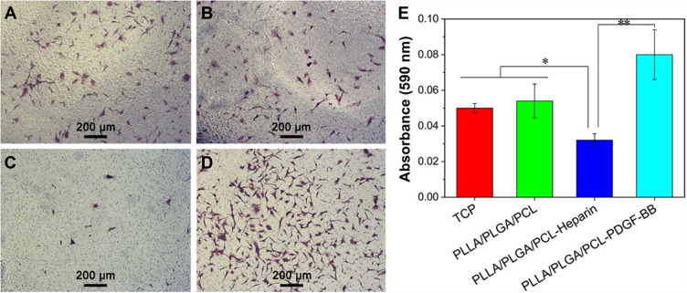
The authors have advised the representative image of the migratory HVSMCs for the TCP (Figure 13A) and PLLA/PLGA/PCL scaffold (Figure 13B) on Page 7017 were overlapped. Following a review of the original images, the authors found different sets of images were mixed when processing the data which resulted in the incorrect image to be used for PLLA/PLGA/PCL (Figure 13B).
The correct Figure 13 is shown below.
Figure 13.
(A–D) The representative image of the migratory HVSMCs located on the lower site of Transwell membrane for the TCP, PLLA/PLGA/PCL, PLLA/PLGA/PCL-Heparin, and PLLA/PLGA/PCL-PDGF-BB scaffolds, respectively. (E) The quantification of HVSMCs migration.
Notes: *P<0.05. **P<0.01.
Abbreviations: HVSMCs, human vascular smooth muscle cells; PCL, poly(ε-caprolactone); PDGF-BB, platelet-derived growth factor-BB; PLGA, poly(lactic-co-glycolic acid); PLLA, poly(L-lactic acid); TCP, tissue culture plate.
The authors apologize for this error and advise it does not affect the results and conclusions of the paper.


