ABSTRACT
Immune checkpoint inhibitors (ICIs) have revolutionized the management of various advanced-stage malignancies. The utilization of ICIs can be limited by their immune-related adverse events such as diarrhea and enterocolitis. Isolated ICI-induced enteritis is an uncommon presentation compared with ICI-induced colitis/enterocolitis. ICI withdrawal and systemic corticosteroids can lead to resolution of enterocolitis in up to 70% of patients. We present a rare case of isolated ICI-induced enteritis refractory to high-dose systemic corticosteroids and responsive to a course of open-capsule budesonide only.
INTRODUCTION
Immune checkpoint inhibitors (ICIs) have become an integral aspect of treatment of several advanced-stage malignancies. ICI mechanism of action includes releasing the inhibitory breaks of T cells, which allows for activation of the immune system and strong anti-tumor responses.1 In addition to anti-tumor response, overactivation of the immune system can lead to several adverse effects termed immune-related adverse events (irAEs), ranging from mild rash to severe colitis.2 Gastrointestinal adverse effects are the second most common after dermatologic irAEs due to ICI therapy.3 Enterocolitis is more likely to occur as a side effect of cytotoxic T-lymphocyte-associated protein 4 inhibitors compared with programmed cell death protein/ligand 1 inhibitors.4 The incidence of ICI-induced diarrhea and enterocolitis can approach 40%.5 Isolated ICI-induced enteritis is far less common than colitis, with an estimated incidence of approximately 10%.5
We present an uncommon case of isolated ICI-induced enteritis refractory high-dose systemic corticosteroids and eventually responsive to open-capsule budesonide only.
CASE REPORT
A 64-year-old woman presented with a history of metastatic lung adenocarcinoma status post wedge resection with subsequent recurrence treated with a full course of pembrolizumab for 2 years, resulting in treatment response. She did not receive other medical treatments for her lung adenocarcinoma. She subsequently presented with peripheral edema, ascites, abdominal bloating, and intermittent nonbloody diarrhea. She described up to 4 loose/watery bowel movements a day with urgency and nocturnal stools. This was also associated with an unintentional weight loss of 9 kg in 3 months. She was not taking nonsteroidal anti-inflammatory drugs or angiotensin receptor blockers.
Laboratory test results were notable for albumin 2.7 g/dL, hemoglobin 7.5 g/dL, and ferritin 16 ng/mL. Celiac serologies, including gliadin, endomysial, and tissue transglutaminase antibodies (normal total IgA), and human leukocyte antigens (HLA) DQ2/DQ8 testing were negative. Antienterocyte antibody testing was negative.
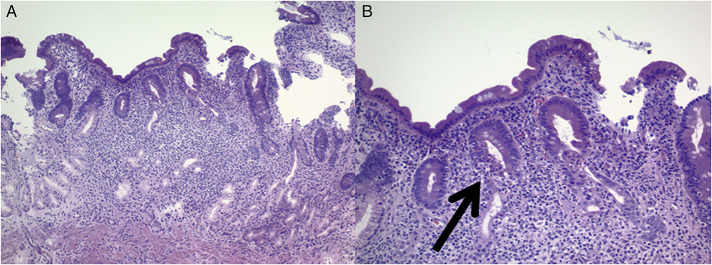
She underwent an upper endoscopy, which revealed complete loss of villi, erythema, congestion, and numerous ulcers from the bulb to the third part of the duodenum (Figure 1). Duodenal pathology showed villous blunting, increased lamina propria inflammation, and focal cryptitis (Figure 2). Cytomegalovirus staining was negative, and gastric biopsies were negative for Helicobacter pylori. Colonoscopy showed normal terminal ileum and no evidence of active inflammation throughout the colon. Biopsies throughout the colon were obtained and were normal. A video capsule endoscopy showed duodenal and proximal jejunal congestion, erythema, and villous blunting.
Figure 1.
Loss of villi, congestion, erythema, ulcerations, and fissuring at initial presentation.
Figure 2.
Duodenal biopsies reveal villous blunting and expansion of the lamina propria by inflammatory cells (A). A higher magnification, active epithelial injury is seen with neutrophils and lymphocytes infiltrating crypt epithelium (arrow). Note that there is not a significant increase in surface intraepithelial lymphocytes (B).
Workup also revealed mild diastolic dysfunction and myocarditis related to ICI therapy. She was treated with intravenous methylprednisolone 2 mg/kg and then transitioned to a prednisone taper starting at 80 mg per day. This resulted in resolution of the myocarditis, but she continued to have gastrointestinal symptoms including intermittent diarrhea, bloating, and weight loss. She was on systemic corticosteroids for 6 weeks before discontinuation.
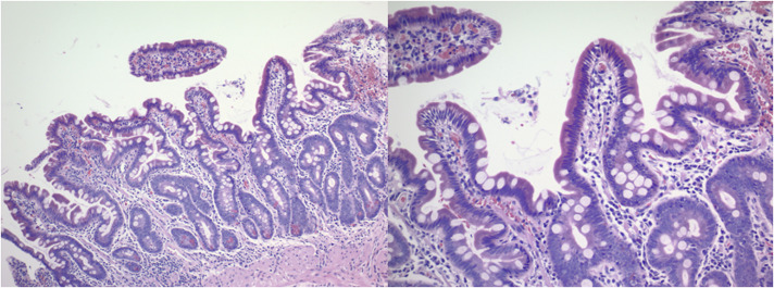
Three months later, given ongoing symptoms despite ICI withdrawal and systemic corticosteroids, she was started on budesonide 3 mg capsules 3 times daily as follows: 1 capsule was swallowed whole; 1 capsule was opened, granules mixed with applesauce, and chewed then swallowed; 1 capsule opened, granules mixed with applesauce, and then swallowed without chewing. A total dose of 9 mg of budesonide daily was given for 2 months and then tapered by 3 mg every 2 months. After 1 month of budesonide treatment, her abdominal bloating and diarrhea resolved, and her hemoglobin improved to 11.2 g/dL and albumin to 3.4 g/dL. Repeat upper endoscopy 4 months later (on 3 mg of budesonide) showed duodenal erythema and congestion but resolution of ulcerations (Figure 3). Duodenal biopsies were normal without villous atrophy or increase in lamina propria inflammation (Figure 4). Four months after completion of budesonide therapy, she remained symptom-free with normalization of her hemoglobin to 14.4 g/dL and albumin to 4.1 g/dL.
Figure 3.
Persistent erythema and congestion of duodenal bulb but resolution of ulcerations after treatment with budesonide.
Figure 4.
Duodenal biopsies reveal recovery of normal villous architecture and a diminution in lamina propria inflammation. No epithelial injury is noted.
DISCUSSION
ICI-induced enterocolitis is one of the most common irAEs associated with checkpoint therapy. The presentation of ICI-induced enterocolitis is variable, but colonic involvement is most commonly noted. Uncommon luminal gastrointestinal manifestations include microscopic colitis, celiac disease, and isolated enteritis.
The treatment of ICI-induced enterocolitis depends on the grade of the irAEs. For mild (grade 1) ICI-induced enterocolitis, symptomatic/supportive treatment while continuing ICI can be sufficient. For moderate-to-severe (grade 2 or more) ICI-induced enterocolitis, treatment generally involves withdrawal of the ICI and high-dose systemic corticosteroids. This strategy can lead to resolution of ICI-induced enterocolitis in up to 40%–70% of patients.6 Biologic agents such as infliximab and vedolizumab have been used as a next step in treating ICI-induced enterocolitis that is nonresponsive to ICI withdrawal and systemic corticosteroids.
Enteric-coated oral budesonide has a time-sensitive and pH-sensitive release mechanism to optimally deliver the drug to the terminal ileum and right colon. The time-sensitive and pH-sensitive release mechanisms relate to the pellets (contents inside the capsule) containing the active drug. The rationale for opening the capsule is to allow delivery of the pellets containing the active drug to the proximal small bowel. The rational for chewing/crushing the granules vs swallowing the granules whole without chewing is to deliver the active drug to the more proximal parts of the gastrointestinal tract (duodenum and proximal jejunum). Open-capsule budesonide has been described in the treatment of multiple different enteropathies including refractory celiac disease.7,8
In this case, we present an uncommon irAE of checkpoint therapy with isolated enteritis without colonic involvement. More importantly, our case is novel in that resolution of the ICI-induced enteritis was achieved only after open-capsule budesonide and not with high-dose systemic steroids and ICI withdrawal. Resolution of ICI enteritis in our case was confirmed with repeat endoscopic and histologic evaluation after treatment along with normalization of laboratory parameters including albumin and hemoglobin.
DISCLOSURES
Authors contributions: N. Hussain: manuscript drafting and manuscript revision. M. Robert: pathology review and pathology image acquisition and manuscript review. B. Al-Bawardy: manuscript drafting, manuscript revisions, final approval, and is the article guarantor.
Previous presentation: Abstract was presented at the American College of Gastroenterology Annual Scientific Meeting on October 26, 2021, in Las Vegas, NV.
Financial disclosure: Dr Al-Bawardy: Speaker honoraria—AbbVie, Takeda; Advisory board—Bristol Myers Squibb. Dr. Robert: Consultant—Takeda, Bristol Myers Squibb. All other authors have no disclosures.
Informed consent was obtained for this case report.
Contributor Information
Nadeen Hussain, Email: nadeen.hussain@yale.edu.
Marie Robert, Email: marie.robert@yale.edu.
REFERENCES
- 1.Bagchi S, Yuan R, Engleman EG. Immune checkpoint inhibitors for the treatment of cancer: Clinical impact and mechanisms of response and resistance. Annu Rev Pathol 2021;16:223–49. [DOI] [PubMed] [Google Scholar]
- 2.Martins F, Sofiya L, Sykiotis GP, et al. Adverse effects of immune-checkpoint inhibitors: Epidemiology, management and surveillance. Nat Rev Clin Oncol 2019;16:563–80. [DOI] [PubMed] [Google Scholar]
- 3.Kröner PT, Mody K, Farraye FA. Immune checkpoint inhibitor-related luminal GI adverse events. Gastrointest Endosc 2019;90:881–92. [DOI] [PubMed] [Google Scholar]
- 4.Tran AN, Wang M, Hundt M, et al. Immune checkpoint inhibitor-associated diarrhea and colitis: A systematic review and meta-analysis of observational studies. J Immunother 2021;44:325–34. [DOI] [PubMed] [Google Scholar]
- 5.Dougan M, Wang Y, Rubio-Tapia A, et al. AGA clinical practice update on diagnosis and management of immune checkpoint inhibitor colitis and hepatitis: Expert review. Gastroenterology 2021;160:1384–93. [DOI] [PubMed] [Google Scholar]
- 6.Collins M, Soularue E, Marthey L, et al. Management of patients with immune checkpoint inhibitor-induced enterocolitis: A systematic review. Clin Gastroenterol Hepatol 2020;18:1393–403.e1. [DOI] [PubMed] [Google Scholar]
- 7.Mukewar SS, Sharma A, Rubio-Tapia A, et al. Open-capsule budesonide for refractory celiac disease. Am J Gastroenterol 2017;112:959–67. [DOI] [PubMed] [Google Scholar]
- 8.Hartranft ME, Regal RE. “Triple phase” budesonide capsules for the treatment of olmesartan-induced enteropathy. Ann Pharmacother 2014;48:1234–7. [DOI] [PubMed] [Google Scholar]


