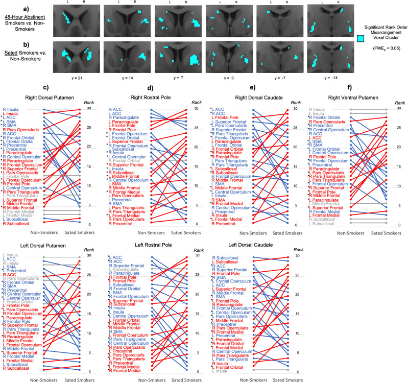
Fig. 2. Rank order rearrangement of striatal connectivity profiles in smokers relative to non-smokers.
Significant rank order rearrangement relative to non-smokers is present in smokers in both the a acutely abstinent state and b nicotine-sated state in the dorsal and lateral striatum (blue). c–f At the striatal sites where rank order significantly differed between non-smokers and sated smokers (Table S2), illustrations of how the rank order arrangement of non-smokers (left) differed from that of sated smokers (right). Cortical ROIs at the top (i.e., with high ranks) indicate those with stronger connectivity with the striatal site, while cortical ROIs at the bottom (i.e., with low ranks) indicate those with weaker connectivity with the striatal site. Red indicates cortical ROIs whose rank was higher in sated smokers than in non-smokers, while blue indicates cortical ROIs whose rank was lower in sated smokers than in non-smokers. Gray indicates cortical ROIs whose rank was the same in both groups. Star denotes cortical ROIs whose rank order difference between non-smokers and sated smokers was statistically significant (p < 0.001, rank order difference > 5).

