Abstract
Peripapillary choroidal neovascularization (PPCNV), a rare presentation of optic nerve sheath meningioma (ONSM), is associated with various ocular pathologies. Herein, we report a case with characteristics of age-related macular degeneration, PPCNV, optic disc edema, and a retinal–choroidal venous collateral. In addition to the recognition of an orbital base ONSM, magnetic resonance imaging revealed a distended perioptic subarachnoid space with the accumulation of cerebrospinal fluid anterior to the tumor. On the basis of these clinical findings, we postulated the pathogenesis of PPCNV-associated ONSMs.
Keywords: Optic nerve sheath compartment, optic nerve sheath meningioma, peripapillary choroidal neovascularization, retinal-choroidal venous collateral
Introduction
Peripapillary choroidal neovascularization (PPCNV) refers to the development of choroidal neovascularization within one disc diameter of the optic nerve head. A recent epidemiological study on the elderly UK population reported that PPCNV had a prevalence of only 0.29% and had a female preponderance.[1] A clinicopathological study revealed that although PPCNV accounts for only 14% of choroidal neovascular membranes, it is a critical condition because it could be attributed to several disorders.[2] Lopez and Green[3] classified PPCNV-associated conditions into five categories: (1) degenerations, including age-related macular degeneration (AMD), angioid streaks, myopia, trauma, and laser photocoagulation; (2) inflammations, including uveitis (e.g. sarcoidosis and ocular histoplasmosis syndrome) and choroiditis; (3) optic disc lesions, including optic nerve pit, drusen, morning glory syndrome, and coloboma; (4) peripapillary choroidal tumors; and (5) idiopathic conditions. In a series of 115 eyes from 96 patients, AMD and idiopathy (constituting 45% and 39% of the cases, respectively) were the most common etiologies.[4] In addition to the aforementioned ocular conditions, PPCNV has been reported to be associated with intracranial tumors and pseudotumor cerebri.[5,6,7] However, PPCNV is rarely present in optic nerve sheath meningiomas (ONSMs).[5,8] Herein, we report a case of a PPCNV-associated ONSM and discuss the possible pathogenesis of PPCNV.
Case Report
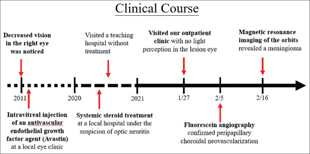
An 81-year-old woman with hyperopia presented to our outpatient department with concerns about progressive blurred vision in the right eye during the past 10 years. She had a medical history of hypertension. Approximately 10 years prior to her presentation to our department, she received an intravitreal injection of an antivascular endothelial growth factor (VEGF) agent (Avastin) in her right eye at a local eye clinic. However, owing to deteriorating vision, she visited a local hospital 1 year prior to her presentation, where she was treated with systemic steroids because of the suspicion of optic neuritis. Before visiting our outpatient department, she had previously visited an ophthalmologist at a teaching hospital but had not received treatment.
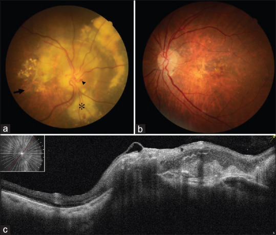
At presentation, her best-corrected visual acuity was no light perception in the right eye and 6/6 in the left eye. The intraocular pressure was normal. An examination of the anterior segment of her eyes revealed grade II nuclear sclerosis in her right eye and pseudophakia in her left eye. A funduscopic examination revealed disc edema, a subretinal hemorrhage, geographic atrophy over the foveal region, and extensive peripapillary lipid exudation in her right eye. Furthermore, a tortuous pre-papillary vessel was observed over the nasal optic disc [Figure 1a]. Her left eye had multiple drusen over the macula [Figure 1b]. Optical coherence tomography of the right eye revealed the thickening of the prepapillary retina with no indication of subretinal or intraretinal fluid over the macular region [Figure 1c].
Figure 1.

(a) Fundus photograph of the right eye revealed centrally involved geographic atrophy (arrow), retinal-choroidal venous collateral (arrow head), disc edema, subretinal hemorrhage, and peripapillary exudation (asterisk). (b) Fundus photograph of the left eye revealed multiple drusen. The optic disc was normal. (c) Optical coherence tomography centered on the right eye's optic disc revealed peripapillary retina thickening and a hyperreflective area adjacent to the optic nerve head, indicating peripapillary choroidal neovascularization
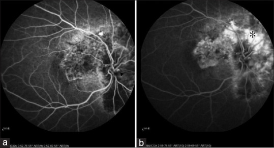
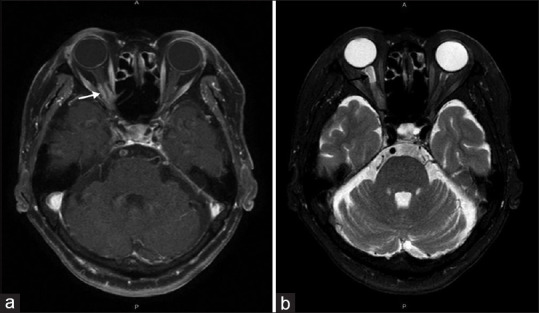
Because impaired optic nerve axoplasmic flow can cause optic disc edema for a variety of reasons, including increased intracranial pressure, local mechanical compression, ischemia, and inflammation, we subsequently scheduled fluorescein angiography.[9] During the arteriovenous phase, we discovered that her tortuous peri-papillary vessel was filled with fluorescence without leakage [Figure 2a], indicating a retinal-choroidal venous collateral. Moreover, we noted a large area of late leakage around the right optic disc and early hyperfluorescence, confirming PPCNV [Figure 2b]. In the left eye, no leakage point was identified. Other causes of PPCNV were considered because the patient's PPCNV was mainly located in the superior and nasal areas of the optic disc, rather than being located in the temporal area of the optic disc, which is the typical site of AMD-related PPCNV.[3] The patient had no history of ocular inflammation, ocular trauma or laser therapy, fundus manifestations of angioid streaks, congenital optic disc lesions, or peripapillary choroidal tumors. Therefore, we performed magnetic resonance imaging (MRI) on the orbits to detect any potential neoplasms. The MRI results revealed a right frontal base tumor extending into the right orbit as well as a thickened and enhanced right optic nerve sheath in the posterior orbit and an optic canal, consistent with an ONSM [Figure 3a]. We also noted increased T2-weighted hyperintensity anterior to the tumor, suggesting the accumulation of cerebrospinal fluid and the dilation of the perioptic subarachnoid space (SAS) [Figure 3b]. Considering the patient's age, the total loss of visual function in the lesion eye without proptosis, and the absence of intracranial tumor extension, we decided with her consent to monitor the progression of the disease. [Figure 4] illustrates the timeline of this patient's ocular history.
Figure 2.

(a) Fluorescein angiography demonstrated that the retinal-choroidal venous collateral (arrow head) was filled up during the arteriovenous phase in the patient's right eye. (b) Fluorescein angiography demonstrated that the peripapillary hyperfluorescence (asterisk) expanded from the superior to the nasal area of the optic disc consistent with peripapillary choroidal neovascularization
Figure 3.

(a) Aaxial T1-weighted magnetic resonance image revealed the thickening and enhancement (white arrow) of the right optic nerve sheath over the right eye's posterior orbit and optic canal, consistent with an optic nerve sheath meningioma. (b) Axial T2-weighted image revealed the increased hyperintensity (black arrow) anterior to the tumor in the right eye, indicating the accumulation of cerebrospinal fluid and distention of the perioptic subarachnoid space
Figure 4.
Timeline of the patient's clinical course and significant examination findings
The patient provided written informed consent to the publication of her case report and any accompanying images.
Discussion
Optociliary shunt vessels are also called retinal-choroidal shunt vessels of the optic disc or retinal-choroidal venous collaterals. However, the designation “optociliary shunt vessels” may be misleading because collateral vessels connect the retinal and choroidal circulations and are not shunts (direct communication between an artery and a vein). According to the Hoyt–Spencer triad, a primary ONSM is characterized by optociliary veins, disc atrophy, and painless visual loss.[10] In a previous study, retinal-choroidal collaterals were found in 25% of ONSM cases and were considered to be an indication of impaired retinal venous outflow.[11] Other causes of retinal-choroidal collaterals include sphenoid wing meningioma, optic nerve glioma, central retinal vein occlusion, and chronic papilledema.[12] The discovery of retinal-choroidal collaterals prompted us to schedule a neuroimaging session to rule out the possibility of an ONSM. In addition to confirming the presence of an ONSM, MRI scanning revealed the dilation of perioptic SAS anterior to the tumor in our patient. Previous studies have reported SAS enlargement associated with intraorbital meningiomas in 12 cases with 50% of the cases involving disc edema.[13,14,15,16] Although the posterior intraorbital tumors did not directly obstruct the central retinal vein, perioptic SAS compartmentalization along with a subsequent increased in pressure and/or accumulation of toxins could have been responsible for the reported optic disc edema.[14] Moreover, the reported disc edema could have been caused by PPCNV; this cause must be recognized to avoid any unnecessary neurological examination.[17]
PPCNV is a rare occurrence in ONSMs. Schatz et al. described three cases involving the coexistence of retinal-choroidal venous collaterals and PPCNV.[8] In their clinicopathological study, they discovered that one patient had an ONSM that directly compressed the retrolaminar portion of the optic nerve; they also observed the coexistence of four collateral veins and two PPCNV. Nevertheless, the causal relationship between these collaterals and PPCNV remains unclear. The shunt vessels may have redirected the obstructed retinal venous outflow to the choroidal circulation. Serial imaging for documenting the natural course of a condition could be highly helpful in identifying the pathomechanisms of the condition. However, our patient did not have such imaging information. Lee and Lessell presumed that PPCNV could be caused by subclinical intraocular tumor invasion.[5] Moreover, considering the association between PPCNV and increased intracranial pressure, Morse et al. proposed that pressure could deform the border of Bruch's membrane at the optic disc.[18] PPCNV was assumed to be formed by the anatomical dehiscence of the chorioretinal layers along with compromised circulation (ischemia or/and retinal nutrition impairment), which was believed to promote angiogenesis. In addition to the aforementioned postulations, the accumulation of VEGF from an ONSM in the compartmentalized perioptic SAS may play a role in the formation of PPCNV; this is because meningiomas are highly vascular tumors with increased microvascular density and elevated expression of VEGF.[19] Nevertheless, further research is required to confirm these hypotheses.
The visual outcome of conventional surgery for ONSMs would be disappointing owing to the proximity of the tumor to the optic nerve, and to the risk of pial vascular plexus damage during surgery. Stereotactic radiotherapy and radiosurgery (Gamma Knife) are suggested for patients with progressive visual deterioration or tumor growth. Surgical intervention is indicated only when intracranial tumor extension threatens the vision of the other eye or for cosmetic reasons. If the patient's vision remains stable, and the tumor size remains unchanged, observation is appropriate.[20]
Conclusion
PPCNV is an uncommon disorder that may be associated with several ocular or systemic conditions. We report the first case of PPCNV associated with an ONSM in Taiwan. The patient's vision may be preserved if the underlying disease is diagnosed and treated early.
Declaration of patient consent
The authors certify that they have obtained all appropriate patient consent forms. In the form, the patient has given her consent for her images and other clinical information to be reported in the journal. The patient understands that name and initials will not be published and due efforts will be made to conceal identity, but anonymity cannot be guaranteed.
Financial support and sponsorship
Nil.
Conflicts of interest
Dr. Cheng-Kuo Cheng, an editorial board member at Taiwan Journal of Ophthalmology, had no role in the peer review process of or decision to publish this article. The other authors decalared no conflicts of interest in writing this paper.
References
- 1.Wilde C, Poostchi A, Mehta RL, Hillman JG, MacNab HK, Messina M, et al. Prevalence of peripapillary choroidal neovascular membranes (PPCNV) in an elderly UK population-the Bridlington eye assessment project (BEAP): A cross-sectional study (2002-2006) Eye (Lond) 2019;33:451–8. doi: 10.1038/s41433-018-0232-y. [DOI] [PMC free article] [PubMed] [Google Scholar]
- 2.Sarks SH. New vessel formation beneath the retinal pigment epithelium in senile eyes. Br J Ophthalmol. 1973;57:951–65. doi: 10.1136/bjo.57.12.951. [DOI] [PMC free article] [PubMed] [Google Scholar]
- 3.Lopez PF, Green WR. Peripapillary subretinal neovascularization. A review. Retina. 1992;12:147–71. [PubMed] [Google Scholar]
- 4.Browning DJ, Fraser CM. Ocular conditions associated with peripapillary subretinal neovascularization, their relative frequencies, and associated outcomes. Ophthalmology. 2005;112:1054–61. doi: 10.1016/j.ophtha.2004.11.062. [DOI] [PubMed] [Google Scholar]
- 5.Lee MS, Lessell S. Choroidal neovascularisation associated with meningioma. Br J Ophthalmol. 2005;89:1384–6. doi: 10.1136/bjo.2005.071696. [DOI] [PMC free article] [PubMed] [Google Scholar]
- 6.Wendel L, Lee AG, Boldt HC, Kardon RH, Wall M. Subretinal neovascular membrane in idiopathic intracranial hypertension. Am J Ophthalmol. 2006;141:573–4. doi: 10.1016/j.ajo.2005.09.030. [DOI] [PubMed] [Google Scholar]
- 7.Sathornsumetee B, Webb A, Hill DL, Newman NJ, Biousse V. Subretinal hemorrhage from a peripapillary choroidal neovascular membrane in papilledema caused by idiopathic intracranial hypertension. J Neuroophthalmol. 2006;26:197–9. doi: 10.1097/01.wno.0000235583.10546.0a. [DOI] [PubMed] [Google Scholar]
- 8.Schatz H, Green WR, Talamo JH, Hoyt WF, Johnson RN, McDonald HR. Clinicopathologic correlation of retinal to choroidal venous collaterals of the optic nerve head. Ophthalmology. 1991;98:1287–93. doi: 10.1016/s0161-6420(91)32141-9. [DOI] [PubMed] [Google Scholar]
- 9.Tariq Bhatti M. 2019-2020 BCSC (Basic and Clinical Science Course), Section 05: Neuro-Ophthalmology. San Francisco, California: American Academy of Ophthalmology; 2019-2020. p. 108. [Google Scholar]
- 10.Frisèn L, Royt WF, Tengroth BM. Optociliary veins, disc pallor and visual loss. A triad of signs indicating spheno-orbital meningioma. Acta Ophthalmol (Copenh) 1973;51:241–9. doi: 10.1111/j.1755-3768.1973.tb03801.x. [DOI] [PubMed] [Google Scholar]
- 11.Saeed P, Rootman J, Nugent RA, White VA, Mackenzie IR, Koornneef L. Optic nerve sheath meningiomas. Ophthalmology. 2003;110:2019–30. doi: 10.1016/S0161-6420(03)00787-5. [DOI] [PubMed] [Google Scholar]
- 12.Tariq Bhatti M. 2019-2020 BCSC (Basic and Clinical Science Course), Section 05: Neuro-Ophthalmology. San Francisco, California: American Academy of Ophthalmology; 2019-2020. p. 157. [Google Scholar]
- 13.Arnold AC, Lee AG. Dilation of the perioptic subarachnoid space anterior to optic nerve sheath meningioma. J Neuroophthalmol. 2021;41:e100–2. doi: 10.1097/WNO.0000000000000908. [DOI] [PubMed] [Google Scholar]
- 14.Killer HE, Jaggi GP, Flammer J, Miller NR, Huber AR. The optic nerve: A new window into cerebrospinal fluid composition. Brain. 2006;129:1027–30. doi: 10.1093/brain/awl045. [DOI] [PubMed] [Google Scholar]
- 15.Lindblom B, Norman D, Hoyt WF. Perioptic cyst distal to optic nerve meningioma: MR demonstration. AJNR Am J Neuroradiol. 1992;13:1622–4. [PMC free article] [PubMed] [Google Scholar]
- 16.Mcnab AA, Wright JE. Cysts of the optic nerve three cases associated with meningioma. Eye (Lond) 1989;3:355–9. doi: 10.1038/eye.1989.51. [DOI] [PubMed] [Google Scholar]
- 17.Tagoe NN, Sharma RA, Biousse V. Optic disc edema due to peripapillary choroidal neovascularization. Taiwan J Ophthalmol. 2021;11:93–6. doi: 10.4103/tjo.tjo_77_20. [DOI] [PMC free article] [PubMed] [Google Scholar]
- 18.Morse PH, Leveille AS, Antel JP, Burch JV. Bilateral juxtapapillary subretinal neovascularization associated with pseudotumor cerebri. Am J Ophthalmol. 1981;91:312–7. doi: 10.1016/0002-9394(81)90282-8. [DOI] [PubMed] [Google Scholar]
- 19.Preusser M, Hassler M, Birner P, Rudas M, Acker T, Plate KH, et al. Microvascularization and expression of VEGF and its receptors in recurring meningiomas: Pathobiological data in favor of anti-angiogenic therapy approaches. Clin Neuropathol. 2012;31:352–60. doi: 10.5414/NP300488. [DOI] [PubMed] [Google Scholar]
- 20.Douglas VP, Douglas KA, Cestari DM. Optic nerve sheath meningioma. Curr Opin Ophthalmol. 2020;31:455–61. doi: 10.1097/ICU.0000000000000700. [DOI] [PubMed] [Google Scholar]


