Abstract
PURPOSE:
Dacryoliths of the canalicular pathway are classically attributed to Actinomyces species as the most common organism. However, global shifts toward Streptococcus and Staphylococcus species have been reported. The objective of this article is to update the American Midwest epidemiology of lacrimal system dacryoliths for targeted clinical treatment.
MATERIALS AND METHODS:
A retrospective chart review from January 2015 to 2021 of patients with a history of surgical procedure for lacrimal removal of dacryolith for canaliculitis, canalicular obstruction, dacryocystitis, and nasolacrimal duct obstruction was included. Specimens were sent for histopathological evaluation and microbial culture.
RESULTS:
A total of 48 specimens were included. The most common organism isolated for canalicular pathology was Actinomyces spp (23%), followed by Staphylococcus spp (21%) and Streptococcus spp (19%). Histopathological staining accounted for 45% of Actinomyces isolation when culture data inconclusive. In a subgroup analysis of lacrimal sac dacryoliths, the most common organism was Staphylococcus spp (29%). Actinomyces species were not isolated from the lacrimal sac or nasolacrimal duct.
CONCLUSION:
Actinomyces maintains a microbial predominance in canalicular dacryoliths and requires careful culture and histopathological analysis for its fastidious nature. Lacrimal sac and nasolacrimal duct dacryolith found no isolates of Actinomyces, and the most common organism was Staphylococcus.
Keywords: Canaliculitis, dacryolith, epidemiology, lacrimal system, nasolacrimal duct obstruction
Introduction
Lacrimal system dacryoliths are a well-known cause of primary canalicular and nasolacrimal duct obstruction.[1,2] Obstructive systems present with recurrent epiphora and discharge and cause lacrimal system stasis with subclinical microbial overgrowth. Prolonged obstruction can lead to clinically significant infections including canaliculitis and dacryocystitis.[3]
Clinical treatment is dependent on the surgical removal of the dacryolith obstruction, which is based on location. This is generally achieved with either punctoplasty with canalicular curettage, canaliculotomy, or dacryocystorhinostomy (DCR). Precedent or concurrent antibiotic usage is generally used to control an active infection.[1,4] The underlying microbial profile of dacryoliths can aid in empiric treatment for presenting infections of the lacrimal system. In general, the Gram-positive filamentous bacteria actinomyces has been considered the most common causative pathogen.[2,5]
However, in recent reports from Asia, there is a geographic change to streptococcus and staphylococcus as the most common pathogens.[4,6,7] Further, when subdividing out lacrimal sac from canalicular dacryoliths, there are fewer reports of Actinomyces as a pathogen.[1,3] The objective of this article is to update the American Midwest epidemiologic microbial profile of lacrimal system dacryoliths to optimize empiric antimicrobial therapy and improve clinical outcomes.
Materials and Methods
A single-center retrospective chart review from January 2015 to 2021 was conducted with IRB approval from Beaumont Health (#2021-126) and adhered to the tenets of the Declaration of Helsinki. Patients with a history of surgical removal of lacrimal or canalicular dacryolith were identified and included primary canaliculitis, canalicular obstruction, dacryocystitis, and nasolacrimal duct obstruction. Secondary canaliculitis, other foreign body causing nasolacrimal duct obstruction, or lack of culture data were excluded from the study.
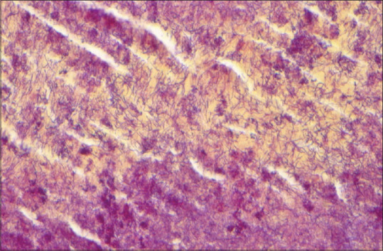
Pertinent clinical and demographic data were collected. All specimens were sent for microbial anaerobic and aerobic culture and submitted to ophthalmic pathology for histopathological diagnosis. All specimens were evaluated with Brown and Hopps Gram-stain. Gomori's methenamine-silver stain and periodic acid-Schiff stain were run as necessary. Isolated pathogens were recorded and cross-referenced to microbial culture data. Actinomyces was diagnosed presumptively if the ophthalmic pathologist determined gram-positive filamentous organisms on examination [Figure 1]. Statistical analysis was performed with R for mean, standard deviation, and Fischer exact test.
Figure 1.

Histopathology of canalicular dacryolith showing numerous branching filamentous gram-positive bacteria consistent with Actinomyces (Brown and Hopps stain, ×60 Insert: hematoxylin-eosin, ×2)
Results
Eighty-one patients were identified, 19 were removed for secondary canaliculitis or foreign body obstruction and 14 for incomplete culture data. Of the 48 patients included, the average age was 64 years (range 4–92 years) with a female-to-male ratio of 5:1. Forty-one (85%) were located in the canaliculus and 7 (15%) in the lacrimal sac. Pathogen frequency of canalicular dacryolith by culture and histopathology is shown in [Table 1]. The most common organism isolated was Actinomyces spp (23%), followed by Staphylococcus spp (21%) and Streptococcus spp (19%). Histopathological staining accounted for 45% of Actinomyces determination when culture data was not conclusive. Of lacrimal sac and nasolacrimal duct dacryolith subgroup [Table 2], the most common organism was Staphylococcus spp (29%) with no Actinomyces identified.
Table 1.
Frequency of pathogens isolated from dacryoliths removed for primary canalicular obstruction or nasolacrimal duct obstruction.
| Pathogen (n=48) | Number of cases (%) |
|---|---|
| Actinomyces spp | 11 (23)* |
| Staphylococcus spp | 10 (21) |
| Streptococcus spp | 9 (19) |
| Fusobacterium nucleatum | 4 (8) |
| Peptostreptococcus spp | 4 (8) |
| Parvimonas micra | 3 (6) |
| Propionibacterium spp | 3 (6) |
| Serratia marcescens | 3 (6) |
| Fungal spp+ | 3 (6) |
| Escherichia Coli | 2 (4) |
| Prevotella spp | 2 (4) |
| Pseudomonas aeruginosa | 2 (4) |
| Gemella morbillorum | 2 (4) |
*Histopathologic staining yielded 5/11 (45%) of positive cases. +Includes one Candida Albicans, one Aspergillus fumigatus, one non-specific yeast. The following bacteria were positive in only one case (2%): Proteus mirabilis, Klebsiella oxytoca, Enterobacter cloacae, Capnocytophaga sputigena, Aggregatibacter aphrophilus, Haemophilus influenzae, Stenotrophomonas maltophilia.
Table 2.
Frequency of pathogens isolated from lacrimal sac dacryolith obtained during dacryocystorhinostomy.
| Pathogen (n=7) | Number of cases (%) |
|---|---|
| Staphylococcus spp | 2 (29) |
| Peptostreptococcus spp | 1 (14) |
| Serratia marcescens | 1 (14) |
| Escherichia Coli | 1 (14) |
| Klebsiella oxytoca | 1 (14) |
| Fungal yeast forms | 1 (14) |
| Actinomyces spp | 0 (0) |
Discussion
Lacrimal system dacryoliths are commonly misdiagnosed and are generally recurrent if not surgically treated. They can be present in up to 1%–2% of patients presenting with epiphora and are reported to be present in 6%–18% of DCR surgeries.[1,2,3,4] Given the prevalence, understanding the microbial background when encountered can aid in adjunctive treatment. Antibiotic usage may help decrease bacterial load or control an active infection; however, the definitive treatment is surgical removal of the obstruction.
The results of this study support a continued prevalence of Actinomyces in American Midwest canalicular dacryoliths as previously described by Repp et al. 12 years prior.[3] The more recent studies supporting a shift to Staphylococcus and Streptococcus species in Asia may have a geographic difference in the microbiome although this reasoning is unlikely as the highest prevalence of Actinomyces species follows the equatorial belt specifically in the locations of prior publications.[8]
Actinomyces can be difficult to isolate on culture media. These bacteria are slow-growing, Gram-positive, anaerobic, Gram-positive branching filamentous rods. They are the most commonly isolated microbe, yet other viruses, bacteria, and fungae such as staphylococcus, streptococcus, and candida may also appear. Culture results can take between 5 and 20 days, and therefore, an incubation period of at least 10 days is required for conclusion of a negative culture.[9] As facultative anaerobes, they require a strictly anaerobic culture environment. Given the fastidious nature of such bacteria, culture data can be limited and a histopathological evaluation with bacterial and fungal evaluation can be critical in confirmation of the underlying cause.[5] However, with the appropriate staining, actinomyces species may be isolated in all histopathologic specimens. Therefore, H and E stains is necessary as a routine patient's care standard. Further investigations might include special stains for microorganisms, such as gram stain, Grocott, or Periodic acid Schiff. In our study, approximately 45% of the Actinomyces identified were on histopathology by an ocular pathologist when culture data was inconclusive. The main pathological characteristics of Actinomyces are blue-staining bacteria surrounded by an eosinophilic fibrillary coat, known as the Splendore-Hoeppli phenomenon, which was found by an ocular pathologist on the histopathological specimens in this study. This finding may explain prior study epidemiologic data using bacterial aerobic and anaerobic culture alone, limiting evaluation of Actinomyces without histopathological analysis.[4,6]
Many other bacteria have been pathologically reported in lacrimal excretory system dacryoliths. Myroides spp., Stenotrophomonas maltophilia and multi-drug resistant Escherichia coli have been reported in case-studies.[6,10,11,12] Pseudomonas aeruginosa was also found to be most common in secondary canaliculitis from punctal plugs.[7] Anatomically, there appears to be a microbial difference in lacrimal sac dacryoliths cultured on DCR with less Actinomyces isolated from the lacrimal sac.[1,3] The subgroup analysis of specimens is in concordance, with no Actinomyces found in our lacrimal sac subgroup. However, this small sample size is insufficiently powered for statistical significance. Given the numerous and sometimes rare organisms associated with dacryoliths, clinicians should maintain a broad differential.
Conclusion
In conclusion, the regional prevalence of Actinomyces in canalicular dacryoliths is maintained. This data can be used to guide empiric therapy in the cases where culture data may be pending or inconclusive. Careful anaerobic culture with histopathological analysis of dacryolith specimens including staining to differentiate other bacteria, fungi, and viruses that may grow alongside Actinomyces is recommended. These infections can cause serious morbidity without appropriate treatment including orbital cellulitis and vision loss. Although a broad differential should always be maintained, this epidemiologic data may help the clinician isolate the causative organism, tailor patient counseling, and provide targeted treatment for lacrimal system dacryoliths.
Financial support and sponsorship
Nil.
Conflicts of interest
The authors declare that there are no conflicts of interests of this paper.
References
- 1.Yazici B, Hammad AM, Meyer DR. Lacrimal sac dacryoliths: Predictive factors and clinical characteristics. Ophthalmology. 2001;108:1308–12. doi: 10.1016/s0161-6420(01)00596-6. [DOI] [PubMed] [Google Scholar]
- 2.Balıkoğlu Yılmaz M, Şen E, Evren E, Elgin U, Yılmazbaş P. Canaliculitis awareness. Turk J Ophthalmol. 2016;46:25–9. doi: 10.4274/tjo.68916. [DOI] [PMC free article] [PubMed] [Google Scholar]
- 3.Repp DJ, Burkat CN, Lucarelli MJ. Lacrimal excretory system concretions: Canalicular and lacrimal sac. Ophthalmology. 2009;116:2230–5. doi: 10.1016/j.ophtha.2009.04.029. [DOI] [PubMed] [Google Scholar]
- 4.Kim UR, Wadwekar B, Prajna L. Primary canaliculitis: The incidence, clinical features, outcome and long-term epiphora after snip-punctoplasty and curettage. Saudi J Ophthalmol. 2015;29:274–7. doi: 10.1016/j.sjopt.2015.08.004. [DOI] [PMC free article] [PubMed] [Google Scholar]
- 5.Freedman JR, Markert MS, Cohen AJ. Primary and secondary lacrimal canaliculitis: A review of literature. Surv Ophthalmol. 2011;56:336–47. doi: 10.1016/j.survophthal.2010.12.001. [DOI] [PubMed] [Google Scholar]
- 6.Kaliki S, Ali MJ, Honavar SG, Chandrasekhar G, Naik MN. Primary canaliculitis: Clinical features, microbiological profile, and management outcome. Ophthalmic Plast Reconstr Surg. 2012;28:355–60. doi: 10.1097/IOP.0b013e31825fb0cd. [DOI] [PubMed] [Google Scholar]
- 7.Huang YY, Yu WK, Tsai CC, Kao SC, Kau HC, Liu CJ. Clinical features, microbiological profiles and treatment outcome of lacrimal plug-related canaliculitis compared with those of primary canaliculitis. Br J Ophthalmol. 2016;100:1285–9. doi: 10.1136/bjophthalmol-2015-307500. [DOI] [PubMed] [Google Scholar]
- 8.van de Sande WW. Global burden of human mycetoma: A systematic review and meta-analysis. PLoS Negl Trop Dis. 2013;7:e2550. doi: 10.1371/journal.pntd.0002550. [DOI] [PMC free article] [PubMed] [Google Scholar]
- 9.Valour F, Sénéchal A, Dupieux C, Karsenty J, Lustig S, Breton P, et al. Actinomycosis: Etiology, clinical features, diagnosis, treatment, and management. Infect Drug Resist. 2014;7:183–97. doi: 10.2147/IDR.S39601. [DOI] [PMC free article] [PubMed] [Google Scholar]
- 10.Bansal O, Bothra N, Sharma S, Ali MJ. Multidrug-resistant Escherichia coli canaliculitis. Ophthalmic Plast Reconstr Surg. 2020;36:e122–4. doi: 10.1097/IOP.0000000000001623. [DOI] [PubMed] [Google Scholar]
- 11.Habib M, Saunders PJ, Rubinstein TJ. Stenotrophomonas maltophilia-associated dacryocystitis in leukemia-infiltrated lacrimal sacs: Case and review of literature. Ophthalmic Plast Reconstr Surg. 2021;37:e143–5. doi: 10.1097/IOP.0000000000001925. [DOI] [PubMed] [Google Scholar]
- 12.Ali MJ, Joseph J, Sharma S, Naik MN. Canaliculitis with isolation of myroides species. Ophthalmic Plast Reconstr Surg. 2017;33:S24–5. doi: 10.1097/IOP.0000000000000604. [DOI] [PubMed] [Google Scholar]


