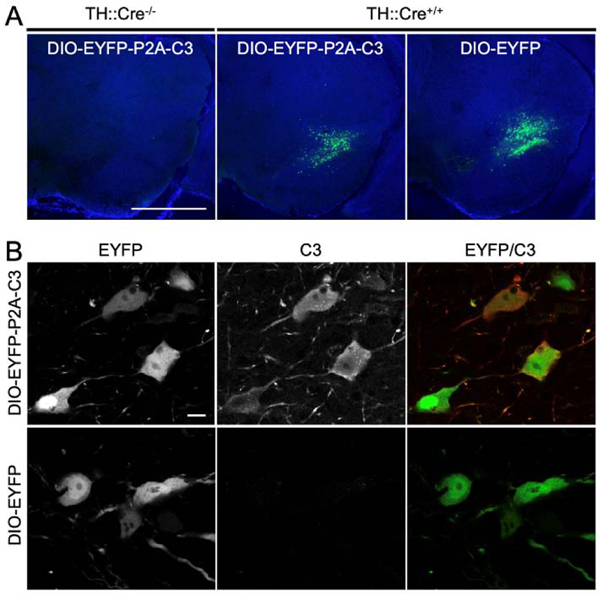
Figure 2.
Confirmation of C3 Expression In Vivo. (2A) Fluorescent micrographs demonstrating the presence of EYFP in the SNc of TH-Cre+/− mice infected with EYFP-P2A-C3 and those infected with EYFP only. In contrast, EYFP expression was not visualized in TH-Cre−/− mice. The specific expression of EYFP-P2A-C3 and EYFP in the SNc in TH-Cre+/− mice and its absence in other structures including the SNc of TH-Cre−/− mice demonstrated efficacy of the Cre-Lox system for cell-specific vector expression. (2B) Fluorescent micrographs of further C3-specific staining revealing that dopaminergic neurons infected with the floxed vectors containing the EYFP-P2A-C3 cassette were indeed expressing C3 while dopaminergic neurons infected with the EYFP only cassette were not. Scale bars: A) 1 mm; B) 10 μm.

