



Abstract
Human cognitive abilities are generally thought to arise from cortical expansion over the course of human brain evolution. In addition to increased neuron numbers, this cortical expansion might be driven by adaptations in the properties of single neurons and their local circuits. We review recent findings on the distinct structural, functional, and transcriptomic features of human cortical neurons and their organization in cortical microstructure. We focus on the supragranular cortical layers, which showed the most prominent expansion during human brain evolution, and the properties of their principal cells – pyramidal neurons. We argue that the evolutionary adaptations in neuronal features that accompany the expansion of the human cortex partially underlie interindividual variability in human cognitive abilities.
Keywords: Human pyramidal neurons, cortical neurons, human cortex, cognition, evolution, intelligence
Cortical neurons as the principal building blocks of cognition
Compared to other animals, human cognition clearly stands out. Many species can display very intelligent behavior, typically tuned to their ecological niche. However, human brains are capable of developing unique cognitive skills, e.g. sharing and storing information through symbolic language, without which human civilization would not be possible. For thousands of years, one of the most fundamental questions in philosophy and neuroscience has been the biological origin of human intelligence. What neurobiological principles underlie the cognitive abilities of the human species?
A major development in the evolution of the mammalian brain is the immense expansion of the neocortex [1]. The neocortex supports the higher cognitive functions that characterize humans. Relative to the total brain size, the human cerebral cortex is the largest among mammals, its grey and white matter occupies 75 to 82% of the entire brain mass or volume [2], and has a noticeably larger number and size of gyri [3]. Larger cortices also contain more neurons and thus more computational units to process and store information. A comparison of the cognitive abilities of 75 species of mammals and birds demonstrated that the number of neurons in the cortex or avian pallium (see Glossary) correlates with measurements of cognition [4].
However, compared to other primates, the increase in both brain size and the number of neurons in humans is “remarkable, yet not extraordinary” [2], and the key to the puzzle of human cognition should not be sought solely in the overall brain or cortical anatomy. In fact, the human brain has just the number of neurons that would be expected for a primate brain of the same size [2]. Furthermore, frontal cortices that are often linked to human cognitive advantage (although relatively and non-allometrically expanded compared to most mammals [5]) are not disproportionately large in humans [6,7]. Moreover, across most mammalian species including humans, cortex invariably contains 20–25% of all brain neurons, even though the relative size of the cortex can reach 82% in humans [2].
Hence, cortical expansion and associated increases in cognitive ability might reflect changes in neuronal and non-neuronal properties rather than changes in the sheer number of neurons. Our brains function through the activity of almost one hundred billion neurons and their connections, which form the principal building blocks for the coding, processing, and storage of information in the brain, ultimately giving rise to cognition. Given the astronomic number of neuronal connections, in the order of 1014 to 1015 [4,8], even the slightest changes in the efficiency of information transfer by neurons can translate into a large impact on the overall computational power of the brain.
Accelerated evolution of the human brain took place at all scales of brain organization: at the scale of genes [9], genetic regulation [10], synaptic proteins [11], and lipids [10], influencing how neurons [12], networks [13], and brain connectivity [14] evolve. The overall result of this multi-scale evolutionary process is not only increased cortical volume but increased diversity and complexity in the structure and function of cortical neurons and their synaptic connections. Thus, the speed and efficiency with which we accomplish cognitive tasks might be rooted deeper in the hierarchy of brain structure and arise from the characteristics of its basic elements – cortical neurons [15–18].
Do human neurons have specialized properties? Recent advances in large-scale single-cell transcriptomics of human cortical neurons, as well as the identification of human neuron types, physiological, morphological, molecular, and connectivity properties, allow us to answer this question. [19–21]. By comparing human cortical neurons to those of other species, evolutionarily conserved neuronal cell types and features can be distinguished from those unique to humans.
In this review, we discuss the evolution of the human brain from the vantage point of single cortical neurons and their local circuits. We attempt to answer how the evolutionary increase in brain size and complexity is linked to adaptations in the structure and function of human cortical neurons. We put forward the hypothesis that neuronal properties may partially explain the observed gap in mental ability between humans and other species, and provide an overview of the recent evidence for this. We focus primarily on the adult pyramidal neurons, the most abundant cortical neuron type, but acknowledge that other cell types and neurotransmitter systems [22,23] likely also contribute to human cognition. We also acknowledge that evolutionary changes in the developmental timing of cortical neurons and synapses play an important role in the evolution of human cognition. Although this topic falls outside the scope of this review, we would like to point the reader to excellent recent reviews [24–26]. Finally, the focus of our review is on the pyramidal neurons of supragranular layers, as the morpho-electric properties of these neurons are more extensively studied in human cortex compared to deeper layers.
Cortical expansion is accompanied by changes in pyramidal neuron structure
Comparing cortical structure across multiple orders of mammalian species [27–29], including primates [30], reveals that not only does the cortical surface and gyrencephalization increase in larger animals, but also cortical thickness tends to increase with brain size, while neuron densities decrease [1,31,32]. Although the mammalian neocortex has a similar six-layered structure (Box 1), across the neocortex there are substantial variations in the thickness of these layers and overall cytoarchitectonic organization. This variation reflects the functional role of cortical areas [33,34]. As sensory information flows from primary sensory areas to progressively more integrative areas in temporal and parietal lobes [35,36], more complex features of sensory stimuli are combined along the way to finally create a complex mental representation. Similarly, cortical and laminar structure follows this functional hierarchy [33]. From sensory to more integrative cortical areas, there is a gradient of increasingly thicker cortex [37] (supported mostly by layers 3 and 5 which contain large pyramidal neurons [38]), larger neurons with more complex dendritic arbors [39,40] and greater number of dendritic spines for synaptic contacts [41] (Figure 1). The larger pyramidal neuron size in larger brains across species is particularly pronounced in higher-order association areas [12,40,42]. Even the gigantopyramidal Betz cells in primary motor cortex, although having a larger soma and covering a larger cortical volume in big cats, have the most complex dendrites and longest total dendritic length in primates [20,28]. Yet, cortical synapse densities remain relatively constant, resulting in humans having the highest number of synapses per neuron [12,30,43].
Box 1. Cortical organization and its neurons.
In mammalian species, including humans, cortical neurons are arranged into a six-layered structure [1,8,47]. Cortical layers 2 to 6 contain predominantly (70–80%) pyramidal neurons, the principal projection neurons of the cortex. They accumulate and integrate synaptic information on their vast dendrites and pass it on locally or to other cortical and subcortical areas through excitatory synapses. Although the principal load of information transmission lies on pyramidal neurons, their activity is strongly regulated and shaped by a diverse population of local inhibitory interneurons [8].
The broad structural characteristics of pyramidal neurons are similar across species: these neurons consist of a pyramidal-shaped soma that gives rise to apical and basal dendrites. The apical dendrite is orientated towards the cortical surface and ends in a distal tuft, while basal dendrites extend radially towards deeper layers.
The stereotypical organization of the cortex in a laminar structure has long tempted neuroscientists to derive generalized rules for connectivity. Layer 1 (L1) contains mainly dendrites and axons of neurons from other layers and a sparse population of interneurons to regulate the inputs [101]. Based on neuroanatomical and physiological evidence, the upper supragranular layers – layers 2 and 3 (L2/L3) – are thought to be the place where most recurrent computation happens: L2/L3 pyramidal cells have many re-excitatory connections, pre-amplify information and send it to pyramidal cells in deeper output L5/L6 [102]. L3 pyramidal cells are also the main source of the feedforward projections to other cortical areas and receive recurrent input from other local superficial and deep pyramidal cells. L5 pyramidal cells are part of both direct thalamocortical and cortico-cortical microcircuits and dictate output to subcortical structures [103].
Supragranular layers seem to play an especially important role in cortico-cortical connectivity in the human brain (Figure 2). Relative to monkeys and other primates, in humans there is a progressive shift of the laminar origin of connections from deeper (L5/L6) to upper layers (L2/L3). The result is that within human cortex a larger proportion of cortico-cortical connections originate from the supragranular layers [44]. These layers are also proportionately thicker in the human cortex compared to other primates [46] supporting a more important role of these layers in the integration of cortical information.
Figure 1. Gradients in the complexity of pyramidal neurons and their spine numbers.
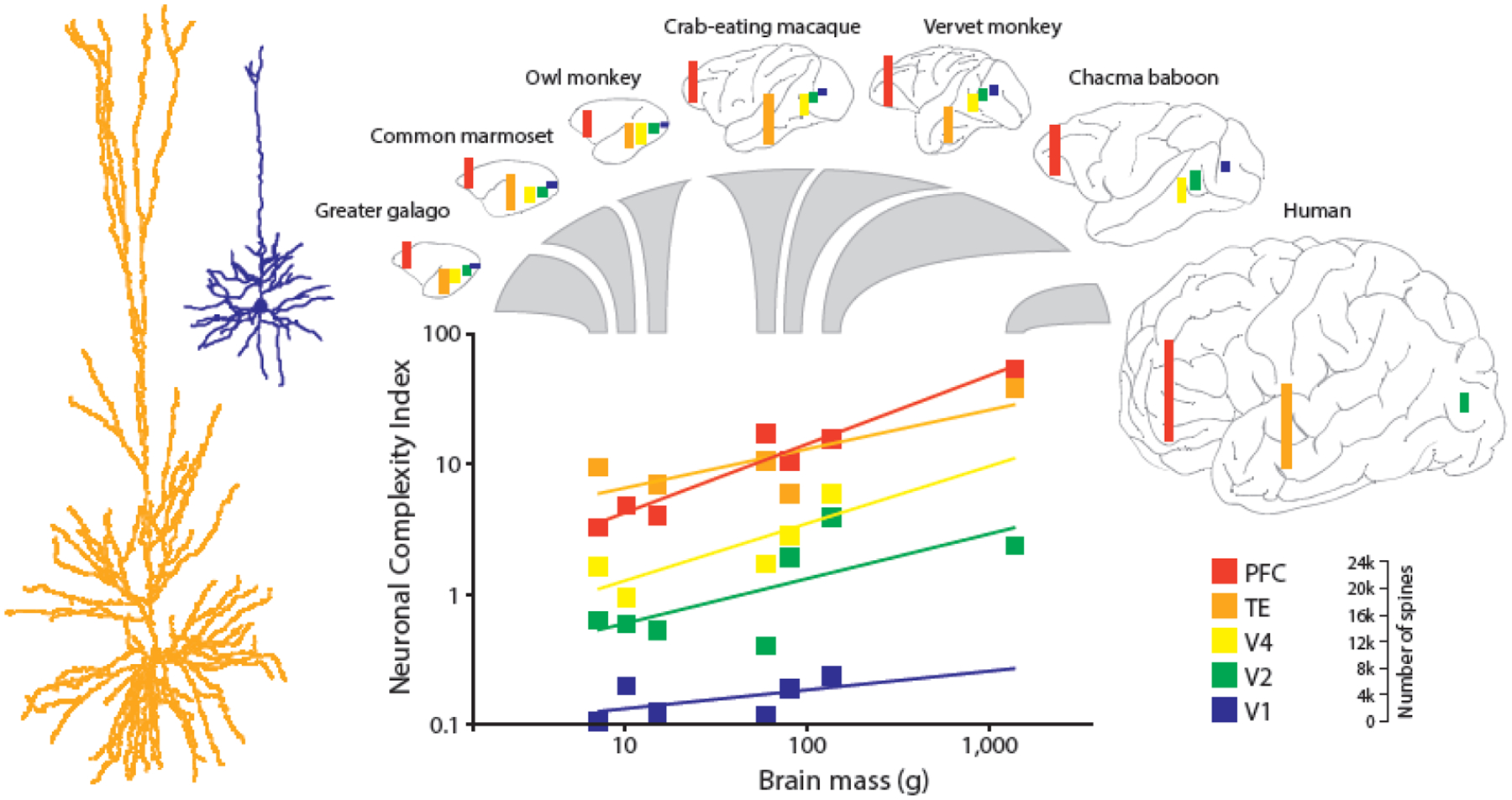
Gradients follow the functional hierarchy of cortical areas in primate species: neurons at the top of the hierarchy in prefrontal cortex and temporal lobe have the highest complexity and spine number. Neuronal complexity increases both across brain areas within a primate species, and with brain mass across species. Two example L3 pyramidal neurons from the primary visual area (blue) and the inferotemporal cortical area (orange) from macaque monkey are shown left (modified from [39,40], image from Verhoog, M.B, PhD thesis, Vrije Universiteit Amsterdam, 2016). The height of the colored bars drawn inside the various brain areas in the brain cartoons reflects the number of spines found on neurons, scale bar depicted in the bottom right corner. PFC - prefrontal cortex; TE – inferiotemporal cortex; V1 - primary visual cortex; V2 – secondary visual cortex; V4 – visual area 4.
Such gradients most likely represent increases in intracortical connectivity. In the human cortex, supragranular cortical layers - layer 2 (L2) and especially layer 3 (L3)– contain large pyramidal neurons that send and receive predominantly cortico-cortical connections [44,45]. These layers are proportionally the thickest in humans (~50%) followed by other primates (46%), carnivores (36%), and then rodents (19%)[46], suggesting distinctions between species in the proportion of cortex devoted to cortico-cortical connectivity (Figure 2).
Figure 2. Cortical expansion of the human brain.

A. Human and mouse brains differ in size and cortical thickness: histology images of two comparable cortical areas in human (middle temporal gyrus, MTG) and mouse (temporal association area, TeA) show proportionally larger supragranular layer. Images from © 2010 Allen Institute for Brain Science. Allen Human Brain Atlas. Available from: human.brain-map.org). B. Supragranular layer thickness is larger in human cortex than other species, both in absolute size (top) as well as relative to the other cortical layers (bottom) (adapted from [46]).
What effects do cortical expansion and increased connectivity have on the morphology of pyramidal cells? Neurons connect by making synaptic contacts on their dendrites, the larger the dendrites the more physical space for potential connections. The most distinctive feature of human pyramidal neurons compared to mouse and macaque pyramidal neurons is the size and complexity of their dendrites [19,47]. On average, human supragranular neurons are three times larger compared to mouse [47] (Figure 3, Key Figure). Importantly, the larger human neurons are not simply up-scaled rodent neurons: they have a more complex structure with longer dendritic terminals [47,48]. The elongation of terminals might reflect the extended period of cortical development and dendritic growth in humans, whose L3 pyramidal dendrites continue to branch and grow throughout childhood and adolescence [49].
Figure 3, Key Figure. Cortical expansion is accompanied by adaptations in pyramidal neuron morphology.
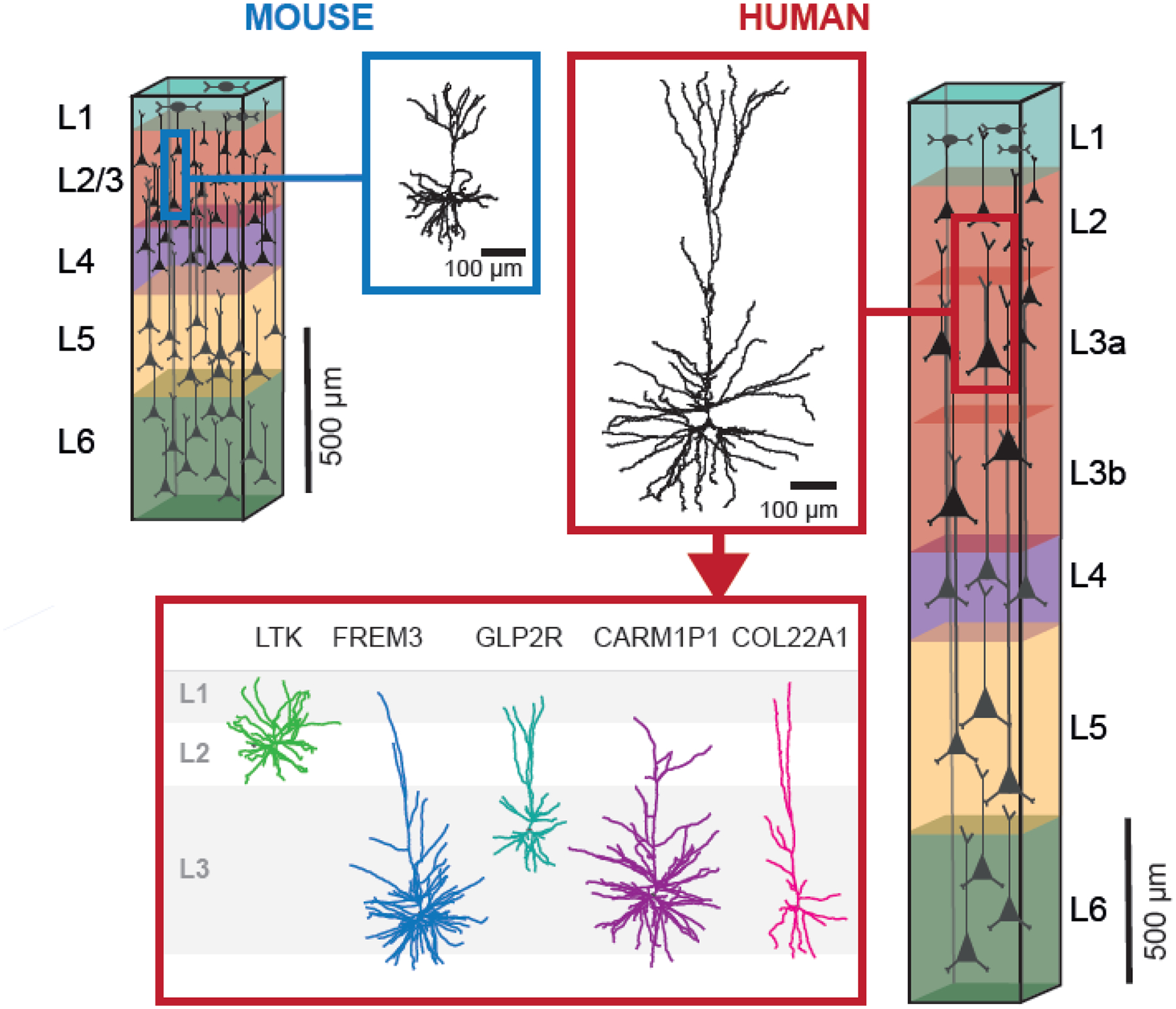
Increased L2/L3 size is accompanied by changes in cytoarchitecture of cortical layers and morphological properties of neurons. Human L2/L3 neurons have 3-fold larger and more complex dendrites than mouse or macaque and contain a larger diversity in transcriptomic excitatory neuron types. In mouse cortex, L2/3 contain three distinct transcriptomic types with relatively compact dendritic morphology, and cell body location strictly adhere to layer boundaries. In contrast, human cortical layers 2 and 3 contain five transcriptomic types that show strong diversity in dendrite morphology, with several large dendrite neuron types that are not found in rodents (based on the data from [1,19,47]).
Recent advances in single-cell transcriptomics make it possible to combine gene expression analysis (transcriptomics) of neuron types with detailed morphological and electrophysiological characterization [19,50]. Such analysis revealed another distinctive feature of L2/L3 pyramidal neurons: the increased diversity in dendritic structure, gene expression, and physiology between and within neuron types [19]. Using transcriptomic signature, L2/L3 pyramidal neurons in human temporal cortex can be divided into five neuron types, named after their most distinctively expressed gene: LTK, GLP2R, FREM3, CARM1P1 and COL22A1 [19] (Figure 3). Deep L3 neurons – FREM3, CARM1P1, and COL22A – are of particular interest. Firstly, FREM3 is the most abundant type in human cortex, and shows the highest diversity in molecular, physiological, and structural properties that follow the gradient from more superficial to deeper cortical neurons [19]. Secondly, CARM1P1 and COL22A1 do not have homologues in mouse and might be a more recent evolutionary addition to the human cortex. Finally, large L3 neuron types CARM1P1 and FREM3 are immune-responsive to neurofilament protein marker SMI-32 that marks mammalian pyramidal neurons preferentially forming long-range cortico-cortical projections. These SMI-32-positive neurons were found to be selectively lost in Alzheimer’s disease [19,51]. The fact that these cells are only found in human cortex and are vulnerable to a disease marked by cognitive deterioration emphasizes the link of the large L3 neurons to human cognition.
Furthermore, on the subcellular level, human dendrites also show more variability in spine size and density compared to other primate and rodent species [40,43,52]. Human spines are larger, longer, and more abundant along the dendrite [43,52]. In particular, the apical dendritic tree shows more complex morphology, carrying bigger spines distributed more densely over the dendrite [53]. Because the apical tree is the biggest contributor to the total dendritic length of a neuron, the absolute number of spines is consequently also larger [53]. The morphology of dendritic spines is important since it directly influences the function of neurons [54] – the size of the spine corresponds to the number of receptors and therefore the ability of the neuron to receive inputs from surrounding cells. In particular, presynaptic active zones and postsynaptic densities have been reported to be larger in human neurons [55–57], suggesting the synaptic connections are also functionally stronger. Finally, since larger spines are more stable and long-lasting, fluctuations in spine volumes might mirror the psychological properties of complex behaviors such as memorizing and forgetting [58].
Thus, human neurons have more complex and larger dendrites compared to other mammalian species, and also more diversity within neuron types. Long dendrites not only physically contain more space for potential synaptic contacts but also have more abundant and larger spines. In addition, dendritic complexity and spine numbers increase in brain areas with higher cognitive load, suggesting that more complex neuronal morphology reflects a more complex cortical function. Taken together, the morphological features of human neurons point to a larger repertoire of information processing patterns that human dendrites can process.
Complex dendrites increase the computational repertoire of single neurons
Neurons process information by integrating thousands of synaptic inputs on their dendrites. The increased size and complexity of dendritic trees offer a greater diversity for computation. Dendritic branches may act as individual processing units and perform distinct computations independently. Thereby, a single neuron can perform similarly to a network of several neurons. Indeed, detailed models of hippocampal and cortical pyramidal neurons demonstrate that these neurons behave computationally similarly to a network with multiple hidden layers. [59,60].
Direct recordings from distal dendrites in human pyramidal neurons in neurosurgery brain tissue have recently confirmed these theoretical predictions and directly demonstrated that human dendrites act as separate processing compartments [61–63]. Human dendrites in supragranular pyramidal neurons were shown to be readily excitable, which was not observed in rodent L2/3 pyramidal neurons [64], and this excitability was mediated by dendritic calcium potentials [62]. Importantly, the dendritic responses only occurred in response to a highly selective window of input strength. If that window was exceeded, the response to additional synaptic input dropped off. This mechanism acts as an anti-coincidence detector: a single, sufficiently large dendritic input is amplified and passed on to the soma, while more coincident (and thus larger when summed) inputs are canceled. In this way, single human pyramidal neurons can perform logical operations such as ‘exclusive or’ (XOR) – converting one or the other input, but not both - previously thought to require multiple neurons [62]. Thus, human pyramidal neuron dendrites may possess a different logical operator repertoire, which could alter cortical computation by allowing dendrites to process inputs more independently. Finally, dendritic electrogenesis has also been observed in L5 human pyramidal neurons [61,63,65], though underlying mechanisms might deviate for different cell types [63] (Box 2).
Box 2. Properties of human L2/L3 versus L5 pyramidal neurons.
The diversity of human neurons and their properties is not limited to supragranular layers of cortex [19] but extends to deeper layers such as L5 [63,104]. In fact, for most physiological features, human L5 neurons show even larger heterogeneity across neurons and types than across species [63]. Several studies have recently compared the physiological properties of human neurons across and within layers [19,104,105]. Human L5 pyramidal neurons were the most excitable and exhibited the most prominent HCN-channel-related membrane properties, such as increased Ih, sag current, and resonance gain at delta and theta frequencies [104].
L5 pyramidal neurons are not homogenous. Based on their output targets they can be divided into two broad classes: Intratelencephalic-projecting (IT), which send axons to targets within telencephalon, and Extratelencephalic-projecting (ET), which also project to subcerebral targets and thus control cortical output [106]. Using transcriptomic information to identify human IT and ET types, ET neurons were revealed to be surprisingly sparse in human cortex (2–6%) compared to rodents (20–30%), but their morpho-electric properties and gene expression profiles are as distinctive as their rodent counterparts [105]. Morphologically, similar to rodent, human L5 ET neurons have thick apical dendritic tufts, whereas L5 IT neurons are thin-tufted [63,107]. Electrophysiologically, in both rodent and human L5 ET neurons, HCN-channel-related membrane properties are enhanced compared to L5 IT neurons, especially at distal dendritic recording sites [61,63]. Moreover, human L5 ET neurons have fast AP kinetics and respond to near-threshold current injections with high-frequency AP bursts, a phenomenon that has been associated with dendritic Ca2+ electrogenesis. Indeed, dendrites of L5 ET neurons show strong electrogenesis, suggesting that human L5 ET neurons possess active mechanisms capable of generating dendritic spikes. In contrast, other reports showed that human L5 neurons have lower electrogenesis in distal dendrites compared to L5 neurons of other mammalian species, including rodents, rabbits, marmosets, and macaques, and thus increased computational compartmentalization [61,65]. In particular, potassium and HCN currents were lower than expected based on the size of the human L5 neurons. The discrepancy between the studies can partly be explained by the recordings at different dendritic locations and possible differences in targeted subtypes of L5 neurons, but other mechanisms might also be involved.
Thus, like supragranular layers, human L5 neurons demonstrate both conserved functional properties across species as well as high phenotypic diversity that could reflect increased functional specialization of human cortex.
Human neurons receive strong and reliable excitatory inputs
The computational advantages of large human dendrites bring significant challenges to neuron physiology. Because human supragranular pyramidal neurons have twice the number of synapses compared to mouse or rat neurons [8], many simultaneous inputs must be integrated and relayed over longer dendritic distances to generate output in the distant cell body. How do human neurons cope with this challenge and process multiple signals without information loss?
Apart from dendritic amplification by calcium action potentials (APs), human synaptic connections differ from those of rodents in their strength and dynamics. For instance, human excitatory synapses have three-fold larger synaptic vesicle pools and presynaptic active zones compared to rodents [55,56] resulting in larger synaptic currents in individual connections [55]. Excitatory synapses are not only stronger in humans, but they are also more resilient to repeated activation [66]. In synaptically connected pyramidal neurons that fire repeatedly, the postsynaptic response in the receiving cell typically decreases as a result of synaptic depression. During such repetitive activity, depressing human synapses were shown to recover three times faster than those of mouse [66]. Rapid recovery from depression increases synaptic resolution and allows neurons to distinguish fast incoming inputs with substantially larger amplitudes for computation. Models based on rodent synapses predicted that depressing synapses perform optimally in response to very low frequencies of inputs [67]. However, because recovery from depression is so fast in human synapses, they can actually also transfer substantial amounts of information during trains of APs and high-frequency bursts [66]. As a result, human synapses can transfer up to nine times more information compared to rodents [66].
These findings were recently confirmed in several connectivity studies investigating connected pairs of neurons in mouse and human [68–70]. L2/L3 excitatory connections were stronger and more reliable in humans, with fast recovery from depression [68,70]. In particular, L3 neurons have the highest excitatory responses even compared to pyramidal neurons in other layers [68]. Notably, there is a gradient in the type of short-term plasticity: connections of more superficial L2 neurons facilitate (second response is greater than the first) while deeper L3 neurons show depression (second response is smaller) [68]. The facilitating connections have the highest impact during sustained high-frequency activity, while depressing synapses are the strongest during sparse firing. Thus, such a gradient may suggest different activity patterns of L2 and L3 neurons, with L3 neurons favoring sparse but strong activation. Finally, the observed connectivity rates and specific patterns of activity indicate a preferential one-directional routing of information from superficial (L2) to deeper layers (L3). In human cortex, this information flow can even be triggered by single APs in a subset of pyramidal neurons, which can initiate a cascade of complex, precisely timed events and recruit neuronal ensembles relevant to cognition [68,71,72].
Thus, synaptic dynamics in human pyramidal neurons are characterized by unique patterns of activity that can be triggered through highly reliable and strong excitatory synapses.
Mechanisms for overcoming large dendritic distance
Human pyramidal neurons need to convey synaptic inputs from very distal dendrites to soma. How do they achieve this? The shape of the dendritic tree and dendritic diameter might reduce the resistance in the dendrites, and this might help to decrease the attenuation of the incoming synaptic signal, similar to water flowing with less resistance through a wider tube. Another mechanism was predicted by fitting cable models to short subthreshold membrane potential responses of human large L3 pyramidal neurons [73]. These neurons have reduced specific membrane capacitance compared to rodent pyramidal neurons, which facilitates dendritic signaling to the soma due to reduced capacitive charging currents. This property may be explained by differences in lipid composition of human cortical neurons [10]. Apart from the structural effects, dendrites might have active mechanisms to counteract the filtering of synaptic inputs: ion channels that are activated by changes in voltage and influence input transfer [74].
One of the most striking physiological differences between human and rodent neurons is the presence of a prominent hyperpolarization-activated non-specific cation current, Ih, in human neurons [19,68,75]. This current is carried by HCN channels, which have high and ubiquitous expression in human supragranular excitatory neurons, but not in mouse neurons [75]. Computational modeling demonstrates that in the presence of Ih, synaptic inputs arrive at the soma faster, with distal inputs in large L3 neurons benefiting the most from this mechanism. [75]. Thus, HCN channels with their high density in the distal apical dendrites [76] may help large neurons to accelerate the transfer of distal inputs to the soma.
Converting inputs to outputs: fast and stable signaling helps to encode more information
The integration of synaptic inputs on human dendrites will ultimately determine whether the neuron will generate output: the firing of APs. Theoretical studies predict that the key determinant of computational speed in neuronal networks is the time neurons need to process inputs and convert them into neuronal output - AP signaling [77]. Recordings from human L2/L3 neurons show they are better able to maintain fast AP generation compared to rodents, and that this might help them encode very dense synaptic information content [66]. AP generation speed is influenced by the large dendritic compartments of human pyramidal neurons, which act as a current sink and result in faster AP onset [78], but differences in voltage-gated ion channels might also play a role.
The ability of neurons to maintain faster AP speed may be especially important during high-frequency firing when neurons are engaged in cognitive tasks. Studies where single neuron activity was recorded in human subjects (usually by implanting recording electrodes in epilepsy patients as part of their treatment) typically report very low average baseline firing rates in cortical neurons (<1 Hz). However, during cognitive tasks, such as reading or learning, single neurons might increase their activity up to 30 Hz, which can last for minutes [79–81]. Human supragranular pyramidal neurons seem to be especially adapted to such activity and can maintain fast AP signaling at these frequencies much better than mouse neurons [82].
Furthermore, the combination of faster AP onset and faster recovery from synaptic depression allows human pyramidal neurons to track synaptic information at considerably higher frequencies. Even when inputs have a frequency of 1000 Hz, human neurons can reliably encode this information by timing their APs to the input, compared to the 200–300 Hz limit in rodent neurons [66]. Fast active properties allow human supragranular neurons to respond to inputs more quickly and therefore encode information at higher bandwidths and with higher temporal resolution [66].
Taken together, human neurons show physiological adaptations that might be linked to their increased size and complexity. Larger dendrites can act as separate computational compartments, but can also lead to faster action potential onsets, have more HCN channels and Ih current, and can generate dendritic calcium action potentials. These characteristics enable neurons to boost and accelerate the transfer of inputs, allowing them to function similarly to a multi-layered network. The increased computational power of single neurons may lead to advantages on the network level: fewer neurons are needed to encode a certain concept. Such sparse coding has enormous cognitive advantages because the same number of neurons can process more information [83]. Indeed, human concept neurons in the temporal lobe show an extraordinarily low sparseness of coding, in the range of 0.2–1% [84], compared to 33% for monkey temporal neurons [85,86]. Therefore, a shift in computation from networks to single neurons and even to dendrites might be a defining feature of human cortical evolution.
Cellular correlates of human intelligence
The evolution of cognitive functions that distinguish humans from other species is accompanied by cortical expansion and increased neuronal complexity. One might ask: are these neurobiological adaptations also relevant to human cognition? And could the same neurobiological differences also explain differences in cognitive ability between human individuals?
One of the strongest correlates of general intelligence that emerges from brain-imaging studies is increased cortical thickness and volume [87]. In multiple high-order association areas in temporal and frontal lobes, cortical thickness correlates with the outcomes of cognitive tests [87–92]. Since cortical function relies on the activity of neurons and connections on neuronal dendrites, interindividual variation in cognitive ability could express itself first and foremost at the cellular level. Do human neurons perform differently in individuals with higher or lower cognitive ability?
One way to approach this question is by measuring neuronal parameters in brain tissue from neurosurgery, and correlating the findings to the results of pre-operative cognitive tests. Before surgery, IQ tests are taken to assess various aspects of the patient’s general cognitive ability, or general intelligence ‘g’, a measure that strongly correlates with educational attainment and socioeconomic status [93,94]. Using this method, it was recently shown that high IQ scores are associated with larger temporal cortical thickness containing pyramidal neurons with larger, more complex dendrites [16]. Based on the experimentally measured dendritic structures, model neurons showed that larger dendrites process rapid synaptic inputs with higher temporal precision [16], achieved by faster AP speed. Indeed, experimental recordings of AP firing in human supragranular pyramidal neurons showed that neurons from individuals with higher IQ scores were better at maintaining fast AP speed during neuronal activity [16]. These findings provide evidence that human intelligence is associated with specific neuronal parameters such as larger, more complex dendrites, faster Aps, and more efficient synaptic information transfer.
In a follow-up study [95], the authors provided a more detailed microstructural analysis of cortical layers and showed that thicker cortex in subjects with higher general and verbal intelligence is due to the increased thickness of supragranular cortical layers (L2/L3) only, while other cortical layers remain unchanged. The thicker supragranular layers did not contain more neurons, but rather larger cells at lower density (Figure 4).
Figure 4. Cellular and cortical properties underlying interindividual differences in human intelligence.
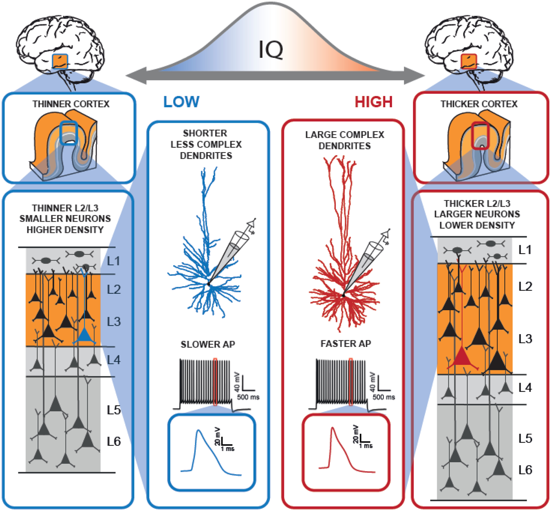
The well-documented association of cortical thickness of MTG with IQ scores in healthy individuals [88,92,93] is driven by interindividual differences in the thickness of layers 2 and 3 in human cortex that also associate with IQ scores. Subjects with higher IQ scores (right column in red boxes) have thicker L2/L3 that contains larger neurons with larger cell bodies at lower neuron densities, than subjects with lower IQ scores (left column in blue boxes). Pyramidal neurons in subjects with higher IQ scores (center red box) have larger and more complex dendritic trees, and these neurons can maintain faster action potential rise speed during repeated firing than neurons in cortical L2/L3 of subjects with lower IQ scores (center blue box). Larger dendrites have more dendritic spines and have physically more room to receive synaptic inputs, thereby larger neurons can integrate more information from other neurons. Action potential shape stability supports fast neuronal information processing. Both factors are potentially contributing to cognitive ability and possibly drive the association of IQ scores with activity of MTG areas in fMRI ([93]; Figure based on the data from [16,95]).
Taken together, these studies provide evidence that higher cognitive ability in human individuals is supported by several cortical and neuronal features that are also favored by human evolution: thicker supragranular layers, larger neurons, complex dendrites, and faster signaling properties [95].
Concluding remarks
One of the primary driving motivations of neuroscience is to understand how the human brain processes information. Compared to other species, the human brain consists of large and complex neurons [47] that are capable of efficient information processing and fast-tracking of incoming inputs [73,82]. Moreover, the increased size of the dendritic tree can result in decompartmentalization between soma and distal dendrites, where dendritic branches become separate computational compartments processing diverse input patterns [61,62,96]. The human brain contains human-specialized pyramidal neuron types [19,21,97,98], but even types homologous to mouse show a larger diversity in physiology, morphology, and gene expression features [19,21]. In addition, microcircuit properties differ: human neurons show strong and reliable local connectivity with synapses that recover quickly from synaptic depression [55,69,82] and display unique forms of network activity [71].
Evolution-driven adaptations of the human cortex are remarkably similar to those that underlie differences in cognitive ability between human subjects. Overall brain volume and cortical thickness of association cortices, the hallmark of human evolution, are larger in subjects with higher cognitive performance [93,99,100]. Similarly, supragranular cortical layers, which expanded disproportionately during human brain evolution [45,46] are thicker in subjects with higher IQ [95]. The size and complexity of human dendrites are larger in human neurons [19,47] and also associate with IQ scores [16]. Finally, increased cognitive performance in humans might result from the ability of large human neurons to perform faster computations and maintain fast AP speed during high-frequency firing [66], and similarly, AP speed is faster in individuals with high IQ scores [16].
In conclusion, we are only beginning to understand how the broad spectrum of human cognitive abilities relates to neuronal function. New insights indicate that the evolution of human cognition and differences in cognitive ability might be driven by similar mechanisms at primarily the neuronal and microcircuit levels: increased cell size and complexity, more diverse computational repertoire, and distinct expression profiles of various genes within and between neuron types. Identifying conserved and specialized features of cellular organization and function in the human brain and building theories of how they support human cognition is only the first step. The critical and challenging next direction is to formulate testable predictions (see Outstanding questions). Novel high-resolution techniques applied to human neurons might provide the necessary tools to face this challenge and directly test theories of neurons supporting human cognition.
Outstanding questions.
Many comparisons described here are based on rodent to human comparisons, but how do neuronal features compare across primate species? Are there truly human-specific neuron types and features?
How do neuronal features of different neuron types, especially human-specific types, vary across different cortical areas? Are there gradients in gene expression profiles and physiology (similar to gradients in neuron and cortical morphology) from sensory to higher-order association areas?
Can specific neuronal features be manipulated to establish causal relationships between gene expression profiles, specific cellular phenotypes, neuron and network function, and ultimately cognitive function? Until now the relationship between neuronal features and cognitive ability remains correlational.
How do neuronal connectivity motifs within cortex relate to macroscale connectivity? Where are the projection targets of e.g. cortico-cortical layer 3 pyramidal neurons? Where do they receive inputs from?
We only focused here on cortical pyramidal neurons, but cortex contains many more neuronal and non-neuronal types such as inhibitory interneurons and glia cells. How do the properties of these cells compare across species?
Morphological and physiological diversity within and between neuron types may be caused by differential gene expression. Are there transcriptomic correlates of these specific neuronal features?
What role do specialized neuron types play in human cortex? How do these neurons, such as the large, spindle-shaped Von Economo neurons found in restricted cortical regions of large-brained animals - humans, great apes, macaques, cetaceans, cows, and elephants, - contribute to cortical processing? Do the characteristic double-bouquet cells facilitate cortical computation by organizing pyramidal neuron information processing in the primate cortex in ‘microcolumns’?
Highlights.
Cortical expansion in humans might be driven by changes in neuronal properties and connectivity rather than sheer increase in neuronal numbers.
Recent breakthroughs in large scale transcriptomic analyses of cortical neurons make it now possible to identify genetic signature of neuron types and compare neuronal properties across species.
Human upper cortical layers contain neurons with large and complex dendrites, that allow a single human neuron to perform computations similar to a multi-layered network.
Strong and reliable synapses that recover quickly from synaptic activity, and fast signaling properties help these neurons efficiently process information.
Several neuronal features of human neurons, such as dendritic size and complexity and fast signaling properties, also associate with cognitive ability in human individuals, supporting the link between neurons and cognitive function not only between but also within species.
Acknowledgements
We thank Matthijs Verhoog for providing the image displayed in Figure 1. The work was supported by several grant awards including award U01MH114812 from National Institute of Mental Health, grant no. 945539 (Human Brain Project SGA3) from the European Union’s Horizon 2020 Framework Programme for Research and Innovation, and NWO Gravitation program BRAINSCAPES: A Roadmap from Neurogenetics to Neurobiology (NWO: 024.004.012) and VI.Vidi.213.014 grant from Netherlands Organization for Scientific Research (NWO).
Glossary
- Apical
The main dendrite extending to L1 and the surface of the cortex from neurons located in deeper layers
- Avian pallium
The layers of grey and white matter that cover the upper surface of the cerebrum in birds; in mammals pallium is referred to as cerebral cortex
- Axons
long projections that originate at the cell body of a neuron and conduct electrical signals (action potentials) to other neurons
- Cortical (micro)circuit
two or more synaptically connected neurons that can collectively produce a functional output
- Dendrites
Tree-like processes of neurons that are specialized in receiving synaptic information through synapses
- Dendritic excitability
the readiness of synapses and branches in a dendritic tree to respond with a positive change in membrane potential (depolarization)
- Dendritic (calcium) potential
depolarization of the dendrites caused by the influx of ions through the activation of voltage-gated channels or receptors
- Depression
the progressive reduction of the postsynaptic response
- Facilitate
show facilitation, an increase in synaptic efficacy
- Gigantopyramidal Betz cells
giant pyramidal neurons located within L5 of the primary motor cortex, the largest pyramidal neurons within cortex
- Gyrencephalization
cortical folding
- Non-allometrically
deviating from the expected relationship of volume to body mass
- Presynaptic
the neuron that releases neurotransmitters
- Postsynaptic
the neuron that receives neurotransmitters
- Pyramidal neurons
principal excitatory neurons of cerebral cortex and hippocampus
- Short-term plasticity
a temporary change in synaptic efficacy caused by the history of presynaptic activity
- Soma
cell body
- Spine
a small protrusion on the dendritic membrane where synaptic connection (synapse) is formed
- Synapse
junctions that allow a neuron to electro-chemically transmit a signal from axon of one cell to the dendrite of another cell. Inhibitory synapses decrease the likelihood of firing action potential, while excitatory synapses increase its likelihood
Footnotes
Publisher's Disclaimer: This is a PDF file of an unedited manuscript that has been accepted for publication. As a service to our customers we are providing this early version of the manuscript. The manuscript will undergo copyediting, typesetting, and review of the resulting proof before it is published in its final form. Please note that during the production process errors may be discovered which could affect the content, and all legal disclaimers that apply to the journal pertain.
Bibliography
- 1.DeFelipe J (2011) The evolution of the brain, the human nature of cortical circuits, and intellectual creativity. Front. Neuroanat 5, 29. [DOI] [PMC free article] [PubMed] [Google Scholar]
- 2.Herculano-Houzel S (2012) The remarkable, yet not extraordinary, human brain as a scaled-up primate brain and its associated cost. Proc. Natl. Acad. Sci. U. S. A 109 Suppl, 10661–10668 [DOI] [PMC free article] [PubMed] [Google Scholar]
- 3.Fernández V et al. (2016) Cerebral cortex expansion and folding: what have we learned? EMBO J. 35, 1021–1044 [DOI] [PMC free article] [PubMed] [Google Scholar]
- 4.Herculano-Houzel S (2017) Numbers of neurons as biological correlates of cognitive capability. Curr. Opin. Behav. Sci 16, 1–7 [Google Scholar]
- 5.Preuss TM and Wise SP (2022) Evolution of prefrontal cortex. Neuropsychopharmacology 47, 3–19 [DOI] [PMC free article] [PubMed] [Google Scholar]
- 6.Semendeferi K et al. (2002) Humans and great apes share a large frontal cortex. Nat. Neurosci 5, 272–276 [DOI] [PubMed] [Google Scholar]
- 7.Smaers JB et al. (2017) Exceptional Evolutionary Expansion of Prefrontal Cortex in Great Apes and Humans. Curr. Biol 27, 714–720 [DOI] [PubMed] [Google Scholar]
- 8.DeFelipe J et al. (2002) Microstructure of the neocortex: comparative aspects. J. Neurocytol 31, 299–316 [DOI] [PubMed] [Google Scholar]
- 9.Florio M et al. (2018) Evolution and cell-type specificity of human-specific genes preferentially expressed in progenitors of fetal neocortex. Elife 7, [DOI] [PMC free article] [PubMed] [Google Scholar]
- 10.Bozek K et al. (2015) Organization and Evolution of Brain Lipidome Revealed by Large-Scale Analysis of Human, Chimpanzee, Macaque, and Mouse Tissues. Neuron 85, 695–702 [DOI] [PubMed] [Google Scholar]
- 11.Grant SGN (2009) , A general basis for cognition in the evolution of synapse signaling complexes. , in Cold Spring Harbor Symposia on Quantitative Biology, 74, pp. 249–257 [DOI] [PubMed] [Google Scholar]
- 12.Elston GN et al. (2001) The Pyramidal Cell in Cognition: A Comparative Study in Human and Monkey. J. Neurosci 21, RC163 [DOI] [PMC free article] [PubMed] [Google Scholar]
- 13.Ardesch DJ et al. (2019) Evolutionary expansion of connectivity between multimodal association areas in the human brain compared with chimpanzees. Proc. Natl. Acad. Sci. U. S. A 116, 7101–7106 [DOI] [PMC free article] [PubMed] [Google Scholar]
- 14.Changeux JP et al. (2021) A Connectomic Hypothesis for the Hominization of the Brain. Cereb. Cortex 31, 2425–2449 [DOI] [PMC free article] [PubMed] [Google Scholar]
- 15.Dorus S et al. (2004) Accelerated evolution of nervous system genes in the origin of Homo sapiens. Cell 119, 1027–1040 [DOI] [PubMed] [Google Scholar]
- 16.Goriounova NA et al. (2018) Large and fast human pyramidal neurons associate with intelligence. Elife 7, e41714. [DOI] [PMC free article] [PubMed] [Google Scholar]
- 17.Lee K et al. (2020) Human in vitro systems for examining synaptic function and plasticity in the brain. J. Neurophysiol 123, 945–965 [DOI] [PubMed] [Google Scholar]
- 18.Hill RS and Walsh CA (2005) Molecular insights into human brain evolution. Nature 437, 64–67 [DOI] [PubMed] [Google Scholar]
- 19.Berg J et al. (2021) Human neocortical expansion involves glutamatergic neuron diversification. Nature 598, 151–158 [DOI] [PMC free article] [PubMed] [Google Scholar]
- 20.Bakken TE et al. (2021) Comparative cellular analysis of motor cortex in human, marmoset and mouse. Nature 598, 111–119 [DOI] [PMC free article] [PubMed] [Google Scholar]
- 21.Hodge RD et al. (2019) Conserved cell types with divergent features in human versus mouse cortex. Nature 536, 171–178 [DOI] [PMC free article] [PubMed] [Google Scholar]
- 22.Berto S et al. (2019) Accelerated evolution of oligodendrocytes in the human brain. Proc. Natl. Acad. Sci. U. S. A 116, 24334–24342 [DOI] [PMC free article] [PubMed] [Google Scholar]
- 23.Oberheim NA et al. (2006) Astrocytic complexity distinguishes the human brain. Trends Neurosci. 29, 547–553 [DOI] [PubMed] [Google Scholar]
- 24.Doan RN et al. (2018) Annual Review of Neuroscience Evolutionary Changes in Transcriptional Regulation: Insights into Human Behavior and Neurological Conditions. DOI: 10.1146/annurev-neuro-080317 [DOI] [PubMed]
- 25.Espinós A et al. (2022) Evolution of genetic mechanisms regulating cortical neurogenesis. Dev. Neurobiol DOI: 10.1002/DNEU.22891 [DOI] [PMC free article] [PubMed] [Google Scholar]
- 26.Fenlon LR (2022) Timing as a Mechanism of Development and Evolution in the Cerebral Cortex. Brain. Behav. Evol 97, 8–32 [DOI] [PubMed] [Google Scholar]
- 27.Butti C et al. (2015) The neocortex of cetartiodactyls: I. A comparative Golgi analysis of neuronal morphology in the bottlenose dolphin (Tursiops truncatus), the minke whale (Balaenoptera acutorostrata), and the humpback whale (Megaptera novaeangliae). Brain Struct. Funct 220, 3339–3368 [DOI] [PubMed] [Google Scholar]
- 28.Jacobs B et al. (2018) Comparative morphology of gigantopyramidal neurons in primary motor cortex across mammals. J. Comp. Neurol 526, 496–536 [DOI] [PubMed] [Google Scholar]
- 29.Jardim-Messeder D et al. (2017) Dogs Have the Most Neurons, Though Not the Largest Brain: Trade-Off between Body Mass and Number of Neurons in the Cerebral Cortex of Large Carnivoran Species. Front. Neuroanat 11, [DOI] [PMC free article] [PubMed] [Google Scholar]
- 30.Sherwood CC et al. (2020) Invariant synapse density and neuronal connectivity scaling in primate neocortical evolution. Cereb. Cortex 30, 5604–5615 [DOI] [PMC free article] [PubMed] [Google Scholar]
- 31.Balaram P and Kaas JH (2014) Towards a unified scheme of cortical lamination for primary visual cortex across primates: Insights from NeuN and VGLUT2 immunoreactivity. Front. Neuroanat DOI: 10.3389/fnana.2014.00081 [DOI] [PMC free article] [PubMed] [Google Scholar]
- 32.Semendeferi K et al. (2011) Spatial Organization of Neurons in the Frontal Pole Sets Humans Apart from Great Apes. Cereb. Cortex 21, 1485–1497 [DOI] [PubMed] [Google Scholar]
- 33.Elston GN (2003) Cortex, Cognition and the Cell: New Insights into the Pyramidal Neuron and Prefrontal Function. Cereb. Cortex 13, 1124–1138 [DOI] [PubMed] [Google Scholar]
- 34.Elston GN (2007) Specialization of the Neocortical Pyramidal Cell during Primate Evolution. In Evolution of Nervous Systems 4pp. 191–242, Elsevier [Google Scholar]
- 35.Maunsell JHR (1992) Functional visual streams. Curr. Opin. Neurobiol 2, 506–510 [DOI] [PubMed] [Google Scholar]
- 36.Bizley JK and Cohen YE (2013) The what, where and how of auditory-object perception. Nat. Rev. Neurosci 14, 693–707 [DOI] [PMC free article] [PubMed] [Google Scholar]
- 37.Wagstyl K et al. (2015) Cortical thickness gradients in structural hierarchies. Neuroimage 111, 241–250 [DOI] [PMC free article] [PubMed] [Google Scholar]
- 38.Wagstyl K et al. (2020) BigBrain 3D atlas of cortical layers: Cortical and laminar thickness gradients diverge in sensory and motor cortices. PLOS Biol. 18, e3000678. [DOI] [PMC free article] [PubMed] [Google Scholar]
- 39.Behav B et al. (2013) The evolutions of large brain size in mammals: the “over-700-gram club quartet”. Brain. Behav. Evol 82, 68–78 [DOI] [PubMed] [Google Scholar]
- 40.Elston GN and Fujita I (2014) Pyramidal cell development: postnatal spinogenesis, dendritic growth, axon growth, and electrophysiology. Front. Neuroanat 8, 78. [DOI] [PMC free article] [PubMed] [Google Scholar]
- 41.Jacobs B et al. (2001) Regional Dendritic and Spine Variation in Human Cerebral Cortex: a Quantitative Golgi Study. Cereb. Cortex 11, 558–571 [DOI] [PubMed] [Google Scholar]
- 42.Bianchi S et al. (2013) Dendritic morphology of pyramidal neurons in the chimpanzee neocortex: regional specializations and comparison to humans. Cereb. Cortex 23, 2429–2436 [DOI] [PMC free article] [PubMed] [Google Scholar]
- 43.Benavides-Piccione R et al. (2002) Cortical area and species differences in dendritic spine morphology. J. Neurocytol 31, 337–346 [DOI] [PubMed] [Google Scholar]
- 44.Goulas A et al. Cortical Gradients and Laminar Projections in Mammals. , Trends in Neurosciences, 41. (2018) , 775–788 [DOI] [PubMed] [Google Scholar]
- 45.Semendeferi K et al. (2001) Prefrontal cortex in humans and apes: A comparative study of area 10. Am. J. Phys. Anthropol 114, 224–241 [DOI] [PubMed] [Google Scholar]
- 46.Hutsler JJ et al. (2005) Comparative analysis of cortical layering and supragranular layer enlargement in rodent carnivore and primate species. Brain Res. 1052, 71–81 [DOI] [PubMed] [Google Scholar]
- 47.Mohan H et al. (2015) Dendritic and axonal architecture of individual pyramidal neurons across layers of adult human neocortex. Cereb. Cortex 25, 4839–4853 [DOI] [PMC free article] [PubMed] [Google Scholar]
- 48.Deitcher Y et al. (2017) Comprehensive Morpho-Electrotonic Analysis Shows 2 Distinct Classes of L2 and L3 Pyramidal Neurons in Human Temporal Cortex. Cereb. Cortex 27, 5398–5414 [DOI] [PMC free article] [PubMed] [Google Scholar]
- 49.Petanjek Z et al. (2008) Lifespan alterations of basal dendritic trees of pyramidal neurons in the human prefrontal cortex: a layer-specific pattern. Cereb. Cortex 18, 915–929 [DOI] [PubMed] [Google Scholar]
- 50.Cadwell CR et al. (2016) Electrophysiological, transcriptomic and morphologic profiling of single neurons using Patch-seq. Nat. Biotechnol 34, 199–203 [DOI] [PMC free article] [PubMed] [Google Scholar]
- 51.Hof PR et al. (1990) Quantitative analysis of a vulnerable subset of pyramidal neurons in Alzheimer’s disease: I. Superior frontal and inferior temporal cortex. J. Comp. Neurol 301, 44–54 [DOI] [PubMed] [Google Scholar]
- 52.Benavides-Piccione R et al. (2013) Age-based comparison of human dendritic spine structure using complete three-dimensional reconstructions. Cereb. Cortex 23, 1798–1810 [DOI] [PMC free article] [PubMed] [Google Scholar]
- 53.Anton-Sanchez L et al. (2017) Three-dimensional spatial modeling of spines along dendritic networks in human cortical pyramidal neurons. PLoS One 12, e0180400. [DOI] [PMC free article] [PubMed] [Google Scholar]
- 54.Cornejo VH et al. (2022) Voltage compartmentalization in dendritic spines in vivo. Science (80-.) 375, 82–86 [DOI] [PMC free article] [PubMed] [Google Scholar]
- 55.Molnár G et al. (2016) Human pyramidal to interneuron synapses are mediated by multi-vesicular release and multiple docked vesicles. Elife 5, 223. [DOI] [PMC free article] [PubMed] [Google Scholar]
- 56.Yakoubi R et al. (2019) Quantitative Three-Dimensional Reconstructions of Excitatory Synaptic Boutons in Layer 5 of the Adult Human Temporal Lobe Neocortex: A Fine-Scale Electron Microscopic Analysis. Cereb. Cortex 29, 2797–2814 [DOI] [PubMed] [Google Scholar]
- 57.Yakoubi R et al. (2019) Ultrastructural heterogeneity of layer 4 excitatory synaptic boutons in the adult human temporal lobe neocortex. Elife 8, e48373. [DOI] [PMC free article] [PubMed] [Google Scholar]
- 58.Kasai H et al. (2010) Structural dynamics of dendritic spines in memory and cognition. Trends Neurosci. 33, 121–129 [DOI] [PubMed] [Google Scholar]
- 59.Beniaguev D et al. (2021) Single cortical neurons as deep artificial neural networks. Neuron 109, 2727–2739.e3 [DOI] [PubMed] [Google Scholar]
- 60.Poirazi P et al. (2003) Pyramidal neuron as two-layer neural network. Neuron 37, 989–999 [DOI] [PubMed] [Google Scholar]
- 61.Beaulieu-Laroche L et al. (2018) Enhanced Dendritic Compartmentalization in Human Cortical Neurons. Cell 175, 643–651.e14 [DOI] [PMC free article] [PubMed] [Google Scholar]
- 62.Gidon A et al. (2020) Dendritic action potentials and computation in human layer 2/3 cortical neurons. Science 367, 83–87 [DOI] [PubMed] [Google Scholar]
- 63.Kalmbach BE et al. (2021) Signature morpho-electric, transcriptomic, and dendritic properties of human layer 5 neocortical pyramidal neurons. Neuron 109, 2914–2927.e5 [DOI] [PMC free article] [PubMed] [Google Scholar]
- 64.Larkum ME (2022) Are Dendrites Conceptually Useful? Neuroscience 489, 4–14 [DOI] [PubMed] [Google Scholar]
- 65.Beaulieu-Laroche L et al. (2021) Allometric rules for mammalian cortical layer 5 neuron biophysics. Nature 600, 274–278 [DOI] [PMC free article] [PubMed] [Google Scholar]
- 66.Testa-Silva G et al. (2014) High Bandwidth Synaptic Communication and Frequency Tracking in Human Neocortex. PLoS Biol. 12, e1002007. [DOI] [PMC free article] [PubMed] [Google Scholar]
- 67.Fuhrmann G et al. (2002) Coding of Temporal Information by Activity-Dependent Synapses. J. Neurophysiol 87, 140–148 [DOI] [PubMed] [Google Scholar]
- 68.Campagnola L et al. (2022) Local connectivity and synaptic dynamics in mouse and human neocortex. Science (80-.) 375, eabj5861 [DOI] [PMC free article] [PubMed] [Google Scholar]
- 69.Seeman SC et al. (2018) Sparse recurrent excitatory connectivity in the microcircuit of the adult mouse and human cortex. Elife 7, 221. [DOI] [PMC free article] [PubMed] [Google Scholar]
- 70.Hunt S et al. (2022) Strong and reliable synaptic communication between pyramidal neurons in adult human cerebral cortex. Cereb. Cortex DOI: 10.1093/cercor/bhac246 [DOI] [PMC free article] [PubMed] [Google Scholar]
- 71.Molnár G et al. (2008) Complex events initiated by individual spikes in the human cerebral cortex. PLoS Biol. 6, e222. [DOI] [PMC free article] [PubMed] [Google Scholar]
- 72.Szegedi V et al. (2017) High-Precision Fast-Spiking Basket Cell Discharges during Complex Events in the Human Neocortex. eneuro 4, ENEURO.0260-17.2017 [DOI] [PMC free article] [PubMed] [Google Scholar]
- 73.Eyal G et al. (2016) Unique membrane properties and enhanced signal processing in human neocortical neurons. Elife 5, [DOI] [PMC free article] [PubMed] [Google Scholar]
- 74.Stuart GJ and Sakmann B (1994) Active propagation of somatic action potentials into neocortical pyramidal cell dendrites. Nature 367, 69–72 [DOI] [PubMed] [Google Scholar]
- 75.Kalmbach BE et al. (2018) h-Channels Contribute to Divergent Intrinsic Membrane Properties of Supragranular Pyramidal Neurons in Human versus Mouse Cerebral Cortex. Neuron 100, 1194–1208.e5 [DOI] [PMC free article] [PubMed] [Google Scholar]
- 76.Harnett MT et al. (2015) Distribution and function of HCN channels in the apical dendritic tuft of neocortical pyramidal neurons. J. Neurosci 35, 1024–1037 [DOI] [PMC free article] [PubMed] [Google Scholar]
- 77.Volgushev M Cortical Specializations Underlying Fast Computations. , Neuroscientist, 22. (2016) , 145–164 [DOI] [PMC free article] [PubMed] [Google Scholar]
- 78.Eyal G et al. (2014) Dendrites impact the encoding capabilities of the axon. J. Neurosci 34, 8063–8071 [DOI] [PMC free article] [PubMed] [Google Scholar]
- 79.Ojemann GA and Schoenfield-McNeill J (1999) Activity of Neurons in Human Temporal Cortex during Identification and Memory for Names and Words. J. Neurosci 19, 5674–5682 [DOI] [PMC free article] [PubMed] [Google Scholar]
- 80.Haglund MM et al. (1994) Neuronal activity in human lateral temporal cortex during serial retrieval from short-term memory. J. Neurosci 14, 1507–15 [DOI] [PMC free article] [PubMed] [Google Scholar]
- 81.Ojemann GA et al. (1988) Neuronal activity in human lateral temporal cortex related to short-term verbal memory, naming and reading. Brain 111, 1383–1403 [DOI] [PubMed] [Google Scholar]
- 82.Testa-Silva G et al. (2010) Human synapses show a wide temporal window for spike-timing-dependent plasticity. Front. Synaptic Neurosci 2, 12. [DOI] [PMC free article] [PubMed] [Google Scholar]
- 83.Olshausen BA and Field DJ (2004) Sparse coding of sensory inputs. Curr. Opin. Neurobiol 14, 481–487 [DOI] [PubMed] [Google Scholar]
- 84.Waydo S et al. (2006) Sparse representation in the human medial temporal lobe. J. Neurosci 26, 10232–10234 [DOI] [PMC free article] [PubMed] [Google Scholar]
- 85.Rolls ET and Tovee MJ (1995) The responses of single neurons in the temporal visual cortical areas of the macaque when more than one stimulus is present in the receptive field. Exp. Brain Res 103, 409–420 [DOI] [PubMed] [Google Scholar]
- 86.Young MP and Yamane S (1992) Sparse Population Coding of Faces in the Inferotemporal Cortex. Science (80-.) 256, 1327–1331 [DOI] [PubMed] [Google Scholar]
- 87.Jung RE and Haier RJ (2007) The Parieto-Frontal Integration Theory (P-FIT) of intelligence: converging neuroimaging evidence. Behav. Brain Sci 30, 135–187 [DOI] [PubMed] [Google Scholar]
- 88.Choi YY et al. (2008) Multiple bases of human intelligence revealed by cortical thickness and neural activation. J. Neurosci 28, 10323–10329 [DOI] [PMC free article] [PubMed] [Google Scholar]
- 89.Narr KL et al. (2007) Relationships between IQ and regional cortical gray matter thickness in healthy adults. Cereb. Cortex 17, 2163–2171 [DOI] [PubMed] [Google Scholar]
- 90.Colom R et al. (2009) Gray matter correlates of fluid, crystallized, and spatial intelligence: Testing the P-FIT model. Intelligence 37, 124–135 [Google Scholar]
- 91.Colom R et al. (2006) Distributed brain sites for the g-factor of intelligence. Neuroimage 31, 1359–1365 [DOI] [PubMed] [Google Scholar]
- 92.Karama S et al. (2009) Positive association between cognitive ability and cortical thickness in a representative US sample of healthy 6 to 18 year-olds. Intelligence 37, 145–155 [DOI] [PMC free article] [PubMed] [Google Scholar]
- 93.Deary IJ et al. (2010) The neuroscience of human intelligence differences. Nat. Rev. Neurosci 11, 201–211 [DOI] [PubMed] [Google Scholar]
- 94.Strenze T (2007) Intelligence and socioeconomic success: A meta-analytic review of longitudinal research. Intelligence 35, 401–426 [Google Scholar]
- 95.Heyer DB et al. (2021) Verbal and General IQ Associate with Supragranular Layer Thickness and Cell Properties of the Left Temporal Cortex. Cereb. Cortex DOI: 10.1093/cercor/bhab330 [DOI] [PMC free article] [PubMed] [Google Scholar]
- 96.Fişek M and Häusser M Are Human Dendrites Different? , Trends in Cognitive Sciences, 24. Jun-(2020) , Elsevier Ltd, 411–412 [DOI] [PMC free article] [PubMed] [Google Scholar]
- 97.Boldog E et al. (2018) Transcriptomic and morphophysiological evidence for a specialized human cortical GABAergic cell type. Nat. Neurosci 21, 1185–1195 [DOI] [PMC free article] [PubMed] [Google Scholar]
- 98.Oberheim NA et al. (2009) Uniquely hominid features of adult human astrocytes. J. Neurosci 29, 3276–3287 [DOI] [PMC free article] [PubMed] [Google Scholar]
- 99.McDaniel M (2005) Big-brained people are smarter: A meta-analysis of the relationship between in vivo brain volume and intelligence. Intelligence 33, 337–346 [Google Scholar]
- 100.Pietschnig J et al. (2015) Meta-analysis of associations between human brain volume and intelligence differences: How strong are they and what do they mean? Neurosci. Biobehav. Rev 57, 411–432 [DOI] [PubMed] [Google Scholar]
- 101.Genescu I and Garel S (2021) Being superficial: a developmental viewpoint on cortical layer 1 wiring. Curr. Opin. Neurobiol 66, 125–134 [DOI] [PubMed] [Google Scholar]
- 102.Shepherd GM (2011) The Microcircuit Concept Applied to Cortical Evolution: from Three-Layer to Six-Layer Cortex. Front. Neuroanat. 5, 30. [DOI] [PMC free article] [PubMed] [Google Scholar]
- 103.Takahashi N et al. (2020) Active dendritic currents gate descending cortical outputs in perception. Nat. Neurosci 23, 1277–1285 [DOI] [PubMed] [Google Scholar]
- 104.Moradi Chameh H et al. (2021) Diversity amongst human cortical pyramidal neurons revealed via their sag currents and frequency preferences. Nat. Commun 12, 1–15 [DOI] [PMC free article] [PubMed] [Google Scholar]
- 105.Kalmbach BE et al. (2021) Signature morpho-electric, transcriptomic, and dendritic properties of human layer 5 neocortical pyramidal neurons. Neuron 109, 2914–2927.e5 [DOI] [PMC free article] [PubMed] [Google Scholar]
- 106.Baker A et al. (2018) Specialized Subpopulations of Deep-Layer Pyramidal Neurons in the Neocortex: Bridging Cellular Properties to Functional Consequences. J. Neurosci 38, 5441–5455 [DOI] [PMC free article] [PubMed] [Google Scholar]
- 107.Ramaswamy S et al. (2015) Anatomy and physiology of the thick-tufted layer 5 pyramidal neuron. DOI: 10.3389/fncel.2015.00233 [DOI] [PMC free article] [PubMed] [Google Scholar]