Arquivos Brasileiros de Cardiologia logoLink to Arquivos Brasileiros de Cardiologia
letter
. 2022 Oct 5;119(4):635–637. [Article in Portuguese] doi: 10.36660/abc.20220143
View full-text in English

Ablação Septal com Radiofrequência e Uso de Novas Tecnologias em Pacientes com Cardiomiopatia Hipertrófica no Laboratório de Eletrofisiologia

Muhieddine Chokr 1, Marina Mayrink 1, Pedro Mario Pinto Vandoni 1, Pedro Vieira Linhares 1, Italo Bruno dos Santos Sousa 1, Hélio José Castello Júnior 2, Mauricio Scanavacca 1
PMCID: PMC9563876  PMID: 36287419

Prezado Editor,

Lemos com grande interesse o artigo “Ablação septal com cateteres e radiofrequência guiada pela ecocardiografia para tratamento de pacientes com cardiomiopatia hipertrófica obstrutiva (CHO): Experiência inicial”, publicado recentemente por Valdigem et al. 1 nos Arquivos Brasileiros de Cardiologia.

Nesse estudo, os autores avaliaram os efeitos da ablação endocárdica por radiofrequência (RF) do septo interventricular com redução do gradiente ventrículo-arterial e melhora de classe funcional em 12 pacientes com CHO. Cateteres com pontas sólidas de 8 mm de comprimento foram utilizados para aplicação de RF termo controlada. A intensidade de energia foi de 80 Watts com temperatura máxima de 60 0 C. A região de maior gradiente na via de saída do ventrículo esquerdo foi o alvo para ablação e identificada pelo ecocardiograma transesofágico. Os autores observaram uma redução média dos gradientes obtidos de 96,8±34 mmHg para 36,1±23 mmHg (p=0,0001) no seguimento de 1 ano, com melhora clínica em todos os pacientes da série. Concluíram que a ablação septal com RF é uma estratégia eficaz, segura e uma nova opção para tratamento de pacientes com CHO com gradiente elevados e sintomáticos. Parabenizamos os autores pelos bons resultados ao utilizar tecnologia de fácil acessibilidade e ao trazer novas informações sobre um procedimento ainda em desenvolvimento.

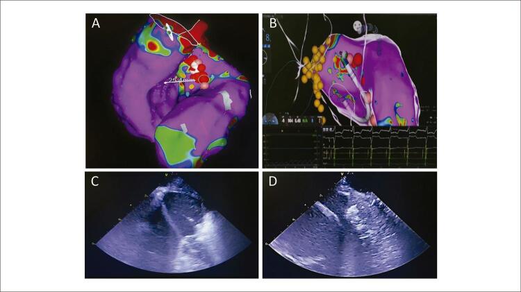
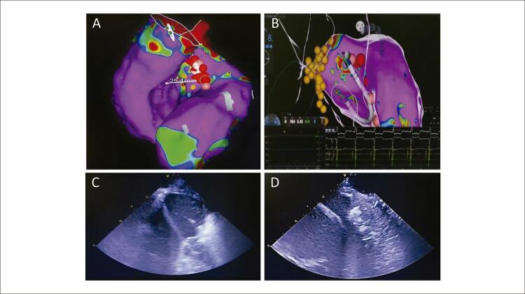
No período de agosto de 2020 a janeiro de 2021, realizamos ablação com RF do septo interventricular em dois pacientes (homem de 44 anos e mulher de 38 anos de idade) com CHO sintomática, refratários ao tratamento clínico, ambos com seguimento superior a 12 meses. Entretanto, diferentemente da técnica descrita por Valdigem et al., 1 utilizamos novas tecnologias de imagem, como mapeamento eletroanatômico (MEA) e o ecocardiograma intracardíaco ( Figura 1 ). O MEA permitiu delimitar a localização do sistema de condução intraventricular e conferiu maior segurança na aplicação de RF (evitar o bloqueio do ramo esquerdo ou atrioventricular total). A construção da geometria pelo MEA dos ventrículos esquerdo e direito também forneceu informação importante na delimitação da área a ser abordada. O ecocardiograma intracardíaco (ICE) permitiu acompanhar a produção das lesões de RF no septo interventricular e a evolução do edema próximo à via de saída do ventrículo esquerdo durante o procedimento, sem a necessidade de um ecocardiografista. Adicionalmente, a ablação com radiofrequência foi otimizada com a utilização de cateteres com ponta irrigada, e as lesões foram controladas pelo software VISITAG SURPOINT (J&J) 2 para uniformizar sua profundidade.

Figura 1. A) Mapa eletroanatômico Carto 3 do ventrículo direito e esquerdo. Pode-se observar o ponto de maior espessamento septal (25 mm). Os pontos em vermelho representam a região onde foi aplicada radiofrequência. B) Início da aplicação de radiofrequência. Os pontos amarelos representam as áreas a serem evitadas em que foi identificado sistema de condução. C) Hiperecogenicidade da região septal, avaliada continuamente com ecocardiografia intracardíaca durante a aplicação de radiofrequência. É possível identificar cateter em alça apoiado sobre a região septal. D) Ao término do procedimento, observado intenso edema na região septal, associado a hiperecogenicidade próximo a via de saída do ventrículo esquerdo.

Figura 1

O critério de interrupção do procedimento utilizado por Valdigem et al. 1 foi a queda aguda de 25% no gradiente ventrículo-arterial. No entanto, alguns autores sugerem que a ablação septal excessiva para atingir esses índices agudamente pode provocar aumento paradoxal e agudo do gradiente com risco de congestão pulmonar importante após a ablação. 3 Nossa impressão é que o uso de uma estratégia puramente anatômica, com aplicações septais logo acima do ramo esquerdo, tendo como alvo um Ablation Index entre 600 e 700, usando cateter irrigado de 3,5 mm (50 Watts e 43 0 C) e avaliação contínua do edema da via de saída do ventrículo esquerdo com o ICE, pode tornar o procedimento mais seguro.

As diversas séries publicadas até o momento não valorizam o gradiente imediato sugerindo que o maior benefício na redução do gradiente ocorre entre 9 e 12 meses da ablação. 4 , 5 Nossos pacientes tiveram uma redução significativa do gradiente intraventricular, com redução média de 91±22 mmHg para 27±14 mmHg cerca de 12 meses após o procedimento índice, e redução no primeiro dia de pós-operatório de 22±6 mmHg, ambos com melhora significativa dos sintomas e atualmente em classe funcional II. A utilização de cateter irrigado, permite a realização de lesões mais previsíveis, mas pode contribuir com quadros de congestão pulmonar como descrito pelos autores. A utilização simultânea do ICE para acompanhar as aplicações de RF também pode evitar a ocorrência de “Stem Pops” , fato comum em aplicações prolongadas e com alta energia. Adicionalmente, o ecocardiograma intracardíaco auxilia na monitoração do risco de aplicações excessivas ao acompanhar a formação de edema septal. Apesar disso, um dos nossos pacientes apresentou quadro de congestão pulmonar imediatamente após a ablação, que foi resolvido com uso de diuréticos e ventilação não invasiva. Tanto a utilização de cateter irrigado como o edema importante na via de saída podem ter contribuído para o quadro apresentado pela paciente. Novos estudos são necessários a fim de comparar diferentes técnicas bem como padronizar qual seria a forma ideal de criar as lesões, que minimizem o risco de aumentos agudos de gradiente ventrículo-arterial após a ablação.

Referências

  • 1.Valdigem BP, Correia EB, Moreira DAR, Bihan DL, Pinto IMF, Abizaid AAC, et al. Septal Ablation with Radiofrequency Catheters Guided by Echocardiography for Treatment of Patients with Obstructive Hypertrophic Cardiomyopathy: Initial Experience. 10.36660/abc.20200732.Arq Bras Cardiol. 2022;118(5):861–872. doi: 10.36660/abc.20200732. [DOI] [PMC free article] [PubMed] [Google Scholar]
  • 2.Okumura Y, Watanabe I, Iso K, Nagashima K, Sonoda K, Sasaki N, et al. Clinical Utility of Automated Ablation Lesion Tagging Based on Catheter Stability Information (VisiTag Module of the CARTO 3 System) with Contact Force-Time Integral During Pulmonary Vein Isolation for Atrial Fibrillation. 10.1007/s10840-016-0156-z.J Interv Card Electrophysiol. 2016;47(2):245–252. doi: 10.1007/s10840-016-0156-z. [DOI] [PubMed] [Google Scholar]
  • 3.Lawrenz T, Lawin D, Radke K, Stellbrink C. Acute and Chronic Effects of Endocardial Radiofrequency Ablation of Septal Hypertrophy in HOCM. 10.1111/jce.15203.J Cardiovasc Electrophysiol. 2021;32(10):2617–2624. doi: 10.1111/jce.15203. [DOI] [PubMed] [Google Scholar]
  • 4.Cooper RM, Shahzad A, Hasleton J, Digiovanni J, Hall MC, Todd DM, et al. Radiofrequency Ablation of the Interventricular Septum to Treat Outflow Tract Gradients in Hypertrophic Obstructive Cardiomyopathy: A Novel Use of CARTOSound® Technology to Guide Ablation. 10.1093/europace/euv302.Europace. 2016;18(1):113–120. doi: 10.1093/europace/euv302. [DOI] [PubMed] [Google Scholar]
  • 5.Crossen K, Jones M, Erikson C. Radiofrequency Septal Reduction in Symptomatic Hypertrophic Obstructive Cardiomyopathy. 10.1016/j.hrthm.2016.04.018.Heart Rhythm. 2016;13(9):1885–1890. doi: 10.1016/j.hrthm.2016.04.018. [DOI] [PubMed] [Google Scholar]
Arq Bras Cardiol. 2022 Oct 5;119(4):635–637.

Carta-resposta

Bruno Valdigem, Andrea de Andrade Vilela, Antonio Tito Paladino Filho, David Le Bihan

Parabéns pelo interesse no procedimento,1 acreditamos que novos protocolos de ablação e em especial dos cuidados de pós-operatório devem ser estimulados, visto que mesmo a forma de liberação de energia e a localização do ponto de aplicação da radiofrequência não são um consenso entre os autores (note que Lawrenz em publicação de agosto de 2021, apresentou dados com aplicação bilateral e apenas em septo direito).2

Após revisão cuidadosa dos primeiros 40 casos realizados por nosso grupo (publicação pendente) pudemos observar que diferenças relacionadas a morfologia do septo interventricular podem ser importantes na escolha do local de início da ablação. Septos com hipertrofia mais homogênea tendem a apresentar migração do gradiente no sentido apical. Assim, ablação guiada apenas pela anatomia pode resultar em lesões mais extensas sem necessidade. Também podem ocorrer gradientes medioventriculares residuais que implicariam numa segunda abordagem.

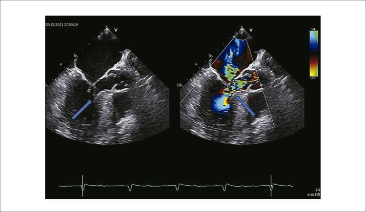
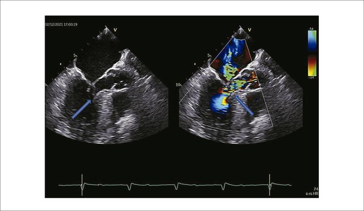
Outro benefício do uso do ecocardiograma transesofágico (ETE) seria a localização do ponto de real início do gradiente. Não raramente, o gradiente se inicia mais na região apical que do que na região mais espessa do septo, marcado pelo ponto de maior aliasing , evidenciado pelo mapeamento de fluxo a cores ao ecocardiograma ( Figura 1 ). O ETE apresenta imagens com melhor visualização da via de saída do ventrículo esquerdo do que o ecocardiograma intracardíaco, informação que é extremamente importante para adequado alinhamento do cateter.

Figura 1. ETE intraprocedimento com e sem color (a 150°): avaliação da valva mitral, identificação do SAM e sua localização. Nessa angulação conseguimos identificar mais facilmente o local de maior aceleração de fluxo e o grau do refluxo mitral.

Figura 1

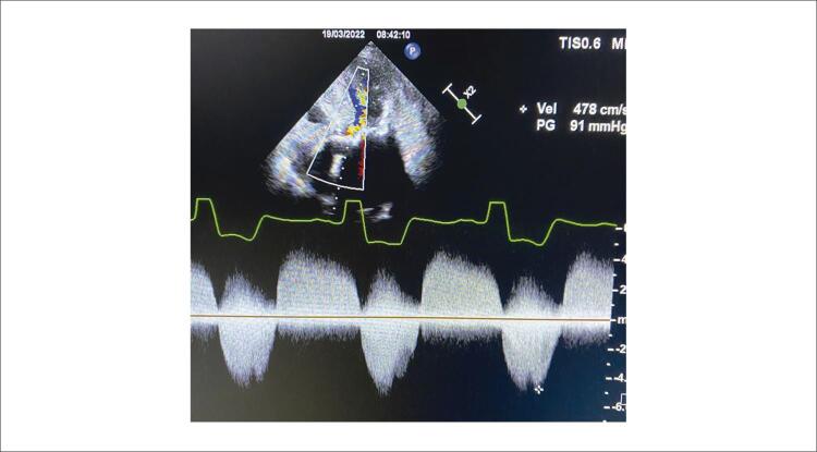
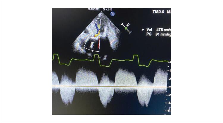
A avaliação ecocardiográfica diferencia as curvas de obstruções valvares (na estenose aórtica) da subvalvar, por meio da imagem espectral do Doppler contínuo ( Figura 2 ). Tal informação é fundamental nos casos de estenoses sequenciais, como ocorre nos casos de ablação septal por radiofrequência pré-TAVI (em que esse método tem demonstrado um papel promissor).

Figura 2. Registro do Doppler contínuo com as curvas relacionadas à estenose aórtica e à obstrução subvalvar.

Figura 2

Adicionalmente, o ETE fornece informações sobre estruturas adjacentes ao ponto de aplicação da radiofrequência, permitindo a pronta identificação de intercorrências possíveis como lesões em cordas tendíneas, ou cúspide anterior da valva mitral.

Um dos parâmetros indicadores de sucesso no intraprocedimento é a redução do refluxo mitral, decorrente da redução no deslocamento sistólico anterior do aparato valvar mitral devido à redução da obstrução na via de saída do ventrículo esquerdo.

Mais uma vez parabenizamos os autores pelas suas considerações e pelo esforço em simplificar o procedimento e aumentar sua propagação. Colocamo-nos à disposição para análise conjunta de dados e trocas de experiências.

References

  • 1.Chokr M, Mayrink M, Vandoni PMP, Linhares PV, Sousa IBS, Castello HJ, Júnior, et al. Septal Ablation with Radiofrequency and the Use of New Technologies in Patients with Hypertrophic Cardiomyopathy in an Electrophysiology Laboratory. Arq Bras Cardiol. 2022;119(4):634–637. doi: 10.36660/abc.20220143. [DOI] [PMC free article] [PubMed] [Google Scholar]
  • 2.Lawrenz T, Lawin D, Radke K, Stellbrink C. Acute and Chronic Effects of Endocardial Radiofrequency Ablation of Septal Hypertrophy in HOCM. 10.1111/jce.15203.J Cardiovasc Electrophysiol. 2021;32(10):2617–2624. doi: 10.1111/jce.15203. [DOI] [PubMed] [Google Scholar]
Arq Bras Cardiol. 2022 Oct 5;119(4):635–637. [Article in English]

Septal Ablation with Radiofrequency and the Use of New Technologies in Patients with Hypertrophic Cardiomyopathy in an Electrophysiology Laboratory

Muhieddine Chokr 1, Marina Mayrink 1, Pedro Mario Pinto Vandoni 1, Pedro Vieira Linhares 1, Italo Bruno dos Santos Sousa 1, Hélio José Castello Júnior 2, Mauricio Scanavacca 1

Dear Editor,

We have read, with great interest, the article “Septal ablation with catheters and radiofrequency guided by echocardiography for treatment in patients with obstructive hypertrophic cardiomyopathy (OHC): First experience”, published recently by Valdigem et al.1 in the journal Arquivos Brasileiros de Cardiologia .

In this study, the authors evaluated the effects of endocardial ablation by radiofrequency (RF) of the interventricular septum with reduction of the ventricular-arterial gradient and improvement in functional class in 12 patients with OHC. Catheters with solid 8-mm tips were used to apply the thermo-controlled RF. The energy intensity was of 80 Watts, with a maximum temperature of 60ºC. The target for ablation was the region with the highest gradient in the left ventricle outflow tract and was identified by the transesophageal echocardiogram. The authors observed an average reduction of the gradients obtained from 96.8±34 mmHg to 36.1±23 mmHg (p=0.0001) during a 1-year follow-up, with a clinical improvement in all patients of the series. They concluded that the septal ablation with RF is an effective, safe strategy and represents a new option to treat OHC patients with high and symptomatic gradients. We would like to congratulate the authors for their fine results in using a technological device that is of easy access, as well as for bringing new and relevant information about the procedure, which is still under development.

From August 2020 to January 2021, our team conducted an ablation with RF of the interventricular septum in two patients (a 44-year-old man and a 38-year-old woman) with symptomatic OHC, who were refractory to clinical treatment, both undergoing follow-up for more than 12 months. However, in contrast to the technique described by Valdigem et al.,1 we used new imaging techniques, including electroanatomic mapping (EAM) and intracardiac echocardiography (ICE) ( Figure 1 ). The EAM allowed us to define the localization of the intraventricular conduction system and conferred greater safety in the application of RF (avoiding the left bundle branch block or complete atrioventricular block). The geometric construction produced by the EAM of the left and right ventricles also provided important information on the definition of the area to be treated. The ICE allowed us to follow the production of the lesions of RF in the interventricular septum and the evolution of the edema near the left ventricle outflow track during the procedure, without the need for an echocardiographer. Additionally, the ablation with radiofrequency was optimized with the use of irrigated-tipped catheters, and the lesions were controlled by the VISITAG SURPOINT (J&J)2 software in order to standardize its depth.

Figure 1. Carto 3-guided electroanatomic maps of the right and left ventricle. This picture shows the point with the largest septal thickness (25 mm). The points in red represent the region where the radiofrequency was applied. B) Beginning of the application of radiofrequency. The yellow points represent the areas to be avoided in which the conduction system was identified. C) Hyperechogenicity of the septal region, evaluated continually using the intracardiac echocardiogram during the application of radiofrequency. It is possible to identify the catheter in the strap resting on the septal region. D) At the end of the procedure, an intense edema is observed in the septal region, associated with hyperechogenicity near the left ventricle outflow tract.

Figure 1

The procedure’s interruption criteria used by Valdigem et al.1 was an acute 25% drop in the ventricular-arterial gradient. However, some authors suggest that the excessive septal ablation to reach these indexes can acutely provoke a paradoxical and acute increase of the gradient with the risk of significant pulmonary congestion after the ablation.3 Our impression is that the use of a purely anatomic strategy, with septal applications directly above the left branch, with an Ablation Index target of between 600 and 700, using an 3.5-mm irrigated catheter (50 Watts and 43ºC) and a continuous evaluation of the edema of the left ventricle outflow tract with the ICE, may well make the procedure safer.

The wide range of series published to date do not give value to the immediate gradient, suggesting that the greatest benefit in the reduction of the gradient occurs between 9 and 12 months of ablation.4 , 5 Our patients witnessed a significant reduction of the interventricular gradient, with an average drop from 91±22 mmHg to 27±14 mmHg, approximately 12 months after the initial procedure, and a further reduction in the first post-operative day of 22±6 mmHg, both with a significant improvement in the symptoms. The patient is currently at the functional class II level. The use of an irrigated catheter can cause more predictable lesions, but it can also contribute in treating pulmonary congestion, as described by the authors. The simultaneous use of the ICE to follow up on the RF applications can also prevent the occurrence of “Stem Pops” , a common fact in prolonged applications and with high energy. Additionally, the intracardiac echocardiogram aids in monitoring the risk of excessive applications by accompanying the formation of a septal edema. Nevertheless, one of our patients presented a medical condition of immediate pulmonary congestion after the ablation, which was resolved with the use of diuretics and non-invasive ventilation. Both the use of the irrigated catheter as well as the significant edema in the outflow tract may have contributed to the medical condition presented by the patient. Further studies are warranted in order to compare different techniques, as well as to standardize what would be the ideal means through which to create the lesions, which minimize the risk of acute increases in the ventricular-arterial gradient after the ablation.

Arq Bras Cardiol. 2022 Oct 5;119(4):635–637.

Reply


Congratulations for your interest in the procedure,1 we believe that new protocols for ablation and especially for post-operative care should be encouraged, given that even the way of releasing the energy and the localization of the point where the radiofrequency is applied are not a consensus among authors (note that Lawrenz, in a publication in August 2021, presented data with a bilateral application and only in the right septum).2

After a careful review of the first 40 cases carried out by our research group (publication pending), we observed that differences related to the morphology of the interventricular septum can be significant in the choice of the location to begin the ablation. More homogeneous hypertrophic septa tend to present a migration from the gradient in the apical direction. Thus, the ablation guided only by the anatomy can result in more extensive lesions unnecessarily. Moreover, residual medioventricular gradients can also occur, which would require a second approach.

Another benefit for the use of the transesophageal echocardiogram (TEE) would be the localization of the real point where the gradient began. More often, the gradient begins in the apical region than in the thicker region of the septum, marked by the point of greater aliasing, illustrated by the color flow mapping echocardiogram ( Figure 1 ). The TEE presents images with a better visualization of the left ventricle outflow tract than does the intracardiac echocardiogram, information that is of utmost importance for the adequate alignment of the catheter.

Figure 1. Intra-procedure TEE with and without color (at 150°): evaluation of the mitral valve, identification of the SAM and its localization. From this angle, we were able to more easily identify the location of greater acceleration of the flow and the degree of mitral valve regurgitation.

Figure 1

The echocardiographic evaluation differentiates the shapes of valvular obstructions (in aortic stenosis) from the subvalvar obstructions, through the spectral image of the Continuous Doppler ( Figure 2 ). This information is essential in cases of sequential stenosis, as occurs in the cases of septal ablation by pre-TAVI radiofrequency (in which this method has shown a promising role).

Figure 2. Continuous Doppler image with the shapes related to aortic stenosis and subvalvar obstruction.

Figure 2

Furthermore, the TEE provides information about the adjacent structures, including the application of radiofrequency, enabling the ready identification of possible complications, such as lesions in tendinous cords, or the anterior cusp of the mitral valve.

One of the main parameters of success in the intra-procedure is the reduction in mitral regurgitation, resulting from the reduction in the systolic anterior motion (SAM) of the mitral valve due to the reduction in the obstruction of the left ventricle outflow tract.

Once again, we would like to congratulate the authors for their considerations and for their efforts to simplify the procedure and increase its diffusion. If it should be of your interest, we are willing to participate in joint analyses and sharing of experiences.


Articles from Arquivos Brasileiros de Cardiologia are provided here courtesy of Sociedade Brasileira de Cardiologia

RESOURCES