Abstract
目的
分析多节段胸椎后纵韧带骨化症(ossification of the posterior longitudinal ligament, OPLL)术中超声辅助下环形减压术的手术疗效和术后神经功能改善情况。
方法
选择2016年1月至2021年1月北京大学第三医院多节段胸椎OPLL患者的病例资料进行回顾性分析, 所有病例均完成后壁切除后行术中超声检查确定环形减压节段, 并进行环形减压。纳入研究的30例患者男性14例, 女性16例, 平均年龄(49.3±11.4)岁。首发症状以下肢麻木无力为主(83.3%), 平均症状持续时间为(33.9±42.9)个月(1~168个月)。神经功能通过术前及末次随访时改良日本骨科协会(modified Japanese Orthopedic Association, mJOA)评分(0~11分)评估, 神经功能改善率根据Harabayashi法计算。根据神经功能改善率是否大于25%将患者分为较优改善组和较差改善组, 收集两组患者的年龄、体重指数(body mass index, BMI)、病程时间、手术时间、出血量、mJOA评分、手术节段、脑脊液漏并进行分析比较。
结果
病例平均手术时间为(137.4±33.8) min(56~190 min), 平均出血量为(653.7±534.2) mL(200~3 000 mL); 术前mJOA评分为(6.0±2.1)分(2~9分), 末次随访时mJOA评分为(7.6±1.9)分(4~11分), 所有患者神经功能均较术前改善(P < 0.001)。神经功能改善率平均为(38.1±24.4)%(14.3~100.0%), 其中神经功能改善率75%~100% 4例, 50%~74% 3例, 25%~49% 14例, 0~24% 9例。较优改善组与较差改善组相比较, 术中出血量差异具有统计学意义(P=0.047)。
结论
通过术中超声辅助下胸椎环形减压术可以对长节段OPLL患者进行有效的减压, 并显著改善患者的神经功能, 控制患者术中出血量有助于术后神经功能的改善。
Keywords: 后纵韧带骨化症, 胸椎, 环形减压术, 术中超声, 胸椎管狭窄
Abstract
Objective
To analyze the effect of short-segment circumferential decompression and the nerve function improvement in 30 cases of multilevel thoracic OPLL assisted by intraoperative ultrasound.
Methods
A total of 30 patients with multilevel thoracic OPLL from January 2016 to January 2021 were enrolled, all of whom were located by intraoperative ultrasound and underwent circumferential decompression. There were 14 males and 16 females, with an average age of (49.3±11.4) years. The initial symptoms were mainly numbness and weakness of lower limbs (83.3%), and the mean duration of symptoms was (33.9±42.9) months (1-168 months). Neurological function was assessed by the Modified Japanese Orthopedic Association (mJOA) score (0-11) preoperative and at the last follow-up, in which the rate of neurological improvement was calculated by the Harabayashi method. The patients were divided into excellent improved group and poor improved group according to the improvement of neurological function. The age, body mass index (BMI), duration of symptoms, operation time, blood loss, mJOA score, surgical level, and cerebrospinal fluid leakage of the two groups were collected and analyzed for statistical differences. The factors influencing the improvement of neurological function were analyzed by univariate and multivariate Logisitic regression analysis.
Results
The mean operation time was 137.4±33.8 (56-190) min, and the mean blood loss was (653.7±534.2) mL (200-3 000 mL). The preoperative mJOA score was 6.0±2.1 (2-9), and the last follow-up mJOA score was 7.6±1.9 (4-11), which was significantly improved in all the patients (P < 0.001). The average improvement rate of neurological function was 38.1%±24.4% (14.3%-100%), including 75%-100% in 4 cases, 50%-74% in 3 cases, 25%-49% improved in 14 cases, and 0%-24% in 9 cases. There was significant difference in intraoperative blood loss between the excellent improved group and the poor improved group (P=0.047). Intraoperative blood loss was also an independent risk factor in regression analysis of neurological improvement.
Conclusion
Thoracic circumferential decompression assisted with intraoperative ultrasound can significantly improve the neurological function of patients with multilevel OPLL and achieve good efficacy. The improvement rate of nerve function can be improved effectively by controlling intraoperative blood loss.
Keywords: Ossification of the posterior longitudinal ligament, Thoracic vertebra, Circumferential decompression, Intraoperative ultrasound, Thoracic spinal stenosis
胸椎后纵韧带骨化症(ossification of the posterior longitudinal ligament, OPLL)是引起胸椎管狭窄症的病因之一,在东亚人群中并不少见。OPLL通常为慢性进行性加重的过程,一旦出现临床症状,引起脊髓损害,保守治疗往往无效且预后较差,需要手术减压[1-2]。对于多节段(≥3)胸椎OPLL的手术治疗通常有不同的手术入路,如单纯后入路、前外侧入路、后外侧入路、前后联合入路等,但这些术式难以取得令人满意的临床效果[3-4]。单纯的经后路胸椎后壁切除减压术是一种较为安全的手术入路,但对于前方压迫较重、鸟喙型OPLL以及重度后凸无法进行彻底减压。前路手术理论上可以直接去除脊髓前方的压迫,但手术技术要求较高,且该入路只能减压1~2个节段,无法处理多节段的胸椎OPLL压迫。胸椎环形减压术可以通过后路消除对脊髓造成压迫的所有致压因素,然而,该入路由于创伤较大、手术时间较长、出血量较多、脊髓前方无法直视下操作等,往往会引起术后不良事件的发生,因此,短节段的环形减压可以显著减少手术时间与相关风险。本研究旨在报告多节段胸椎OPLL减压的手术策略和实施技巧,借助术中神经电生理检测以及超声骨刀的帮助,完成胸椎后壁切除的同时,通过术中超声确定需要环形减压的节段,利用“涵洞塌陷法”以达到短节段环形减压的目的。
1. 资料与方法
1.1. 资料来源
选择2016年1月至2021年1月,在北京大学第三医院骨科手术治疗的胸椎OPLL患者病例资料进行回顾性分析。本研究开始前已经北京大学第三医院医学科学伦理委员会审查批准(M2019410)。
1.2. 纳入和排除标准
1.2.1. 纳入标准
(1) 多节段OPLL引起的胸椎管狭窄症,需要手术治疗者;(2)手术方案为胸椎环形减压术者。
1.2.2. 排除标准
(1) 缺少完整的病历资料,或随访时间较短(< 1年)者;(2)有胸椎外伤史、手术或有创操作史(如椎体穿刺活检等)者;(3)合并原发或继发性脊柱肿瘤者;(4)合并胸椎感染性疾病者;(5)合并胸椎先天性或后天性畸形者。
1.3. 一般资料
根据纳入和排除标准,共纳入胸椎OPLL患者30例,其中男性14例,女性16例。平均年龄(49.3±11.4)岁(26~72岁)。所有患者均成功随访,平均随访时间(42.8±18.7)个月(19~76个月)。
记录患者信息:(1)基本资料:包括性别、年龄、体重指数(body mass index,BMI)、发病时间、临床症状、术前及术后改良日本骨科协会(modified Japanese Orthopedic Association,mJOA)评分(0~11分)等;(2)术前影像学资料:OPLL的节段、部位以及是否合并黄韧带骨化(ossification of ligamentum flavum, OLF)等;(3)手术资料:包括手术时间、出血量、后壁切除节段以及环形减压节段、术中及术后并发症等;(4)末次随访及神经功能评估,包括影像学评估内固定情况、mJOA评分等。
1.4. 手术方法
1.4.1. 显露及置钉
麻醉后连接神经电生理极片,俯卧位后正中入路,显露术野,置入椎弓根螺钉。
1.4.2. 后壁切除
咬除减压节段的棘突,咬骨钳在双侧关节突内侧做骨槽,并用高速磨钻消磨松质骨至内侧皮质,然后用超声骨刀切透椎板内侧皮质,此时椎板游离,采用“揭盖式”整块取出椎管后壁,完成脊髓背侧减压。
1.4.3. 术中超声检查
采用“浸入法”行术中超声检查,将无菌镜套末端充满超声耦合剂,并完全包裹探头,向术野注满生理盐水作为超声成像介质,去除探头周边气泡以及伤口内凝血块,分别平行及垂直于脊髓长轴扫描,获得脊髓长轴及横截图像。根据超声检查结果显示腹侧脊髓仍有压迫的节段,确定胸脊髓腹侧需环形减压节段[5]。
1.4.4. 环形减压
沿椎弓根至椎体用磨钻进行削切,至椎体后壁水平后,探查脊髓硬膜的粘连情况,分离并保护肋间神经,用磨钻、超刀刮匙从椎体后壁两侧深层斜向内60°挖去椎体后1/3的松质骨,形成一个“涵洞”,此时脊髓硬膜前方为残留的椎体后壁和悬空的OPLL。根据粘连的程度采用锐性或钝性分离,严重者可以切除部分硬脊膜,使OPLL与脊髓硬脊膜分离,于OPLL的顶端将未骨化的后纵韧带切开,用神经剥离子压塌OPLL构成的涵洞壁,从侧后方取出OPLL块,完成脊髓前方的减压[6]。
1.4.5. 术中评估
再次行术中超声检查,评估脊髓减压情况,显示脊髓减压充分,安放连接棒及尾帽,测量体感诱发电位(somatosensory evoked potential,SEP)及运动诱发电位(motor evoked potential,MEP)较基线改善情况[7]。
1.5. 神经功能评估及分组
神经功能恢复情况根据Harabayashi法计算神经功能改善率,即改善率(%)=[(末次随访的mJOA评分)-(术前mJOA评分)]/(11-术前mJOA评分)。根据神经功能改善率将患者分为神经功能较优改善组(改善率≥25%)和神经功能较差改善组(改善率 < 25%)。
1.6. 统计学分析
采用SPSS 25.0(SPSS公司,美国)软件,连续变量(如年龄、BMI、mJOA评分、病程时间、手术时间、出血量、手术节段等)服从正态分布,以均数和标准差表示,组间比较采用t检验;分类变量(如性别、脑脊液漏等)以频率表示,并应用Fisher检验进行分析,神经功能(mJOA评分)改善的影响因素通过分组比较进行分析,P < 0.05认为差异有统计学意义。
2. 结果
2.1. 一般情况
30例患者中,始发症状以下肢麻木无力为主(25例,83.3%),其余5例始发症状为胸背部疼痛。平均病程时间(出现症状到手术时间)为(33.9± 42.9)个月(1~168个月),13例患者(43.3%)合并胸椎黄韧带骨化。平均OPLL节段数为(3.5±0.9)个(3~6个)。患者平均住院时间为(8.0±1.5) d(5~13 d),术后住院时间为(5.3±1.0) d(4~8 d)。
2.2. 手术情况
所有30例患者手术过程顺利,均在神经电生理监测及超声骨刀辅助下,先行“揭盖法”后壁切除,并通过术中超声检查脊髓受压情况以确定环形减压节段,进行经椎弓根胸椎环形减压术。患者平均手术时间为(137.4±33.8) min(56~190 min),平均出血量为(653.7±534.2) mL(200~3000 mL)。后壁切除节段数为(3.5±0.9)个(3~6个),所有患者环形减压均为单节段,其中上胸椎6例,中胸椎16例,下胸椎8例(表 1)。
表 1.
30例患者的一般资料
General data of 30 patients
| Items | Value |
| Gender, n (%) | |
| Male | 14 (46.7) |
| Female | 16 (53.3) |
| Age/years, x±s (range) | 19.3±11.4 (26-72) |
| BMI /(kg/m2), x±s (range) | 28.0±4.8 (17.0-41.0) |
| Disease duration/months, x±s (range) | 33.9±42.9 (1-168) |
| Initial symptoms, n (%) | |
| Limbs numbness and weakness | 25 (83.3) |
| Chest and back pain | 5 (16.7) |
| Ossification of the ligamentum flavum, n (%) | 13(43.3) |
| Follow-up time/months, x±s (range) | 42.8±18.7 (18.5-75.9) |
| Hospital stay/d, x±s (range) | 8.0±1.5 (5-13) |
| Postoperative hospital stay/d, x±s (range) | 5.3±1.0 (4-8) |
| OPLL segment (vertebrae), x±s (range) | 3.5±0.9 (3-6) |
| Posterior thoracic wall resection segments (vertebrae), x±s (range) | 3.5±0.9 (3-6) |
| Circumferential decompression segments (vertebrae) | 1 |
| Circumferential decompression site,n (%) | |
| Upper thoracic spine | 6 (20) |
| Middle thoracic spine | 16 (53.3) |
| Lower thoracic spine | 8 (26.7) |
| Operation time/min, x±s (range) | 137.4±33.8 (56-190) |
| Operation blood loss/mL, x±s (range) | 653.7±534.2 (200-3000) |
| Preoperative mJOA score, x±s (range) | 6.0±2.1 (2-9) |
| The last follow-up mJOA score, x±s (range) | 7.6±1.9 (4-11) |
| Neurological improvement rate, x±s (range) | 38.1±24.4 (14.3-100.0) |
| 75%-100% | 4 (13.3) |
| 50%-74% | 3 (10.0) |
| 25%-49% | 14 (46.7) |
| 0%-24% | 9 (30.0) |
| < 0% | 0 (0) |
| Postoperative cerebrospinal fluid leakage,n (%) | 16 (53.3) |
| Postoperative transient loss of muscle strength, n (%) | 8 (26.7) |
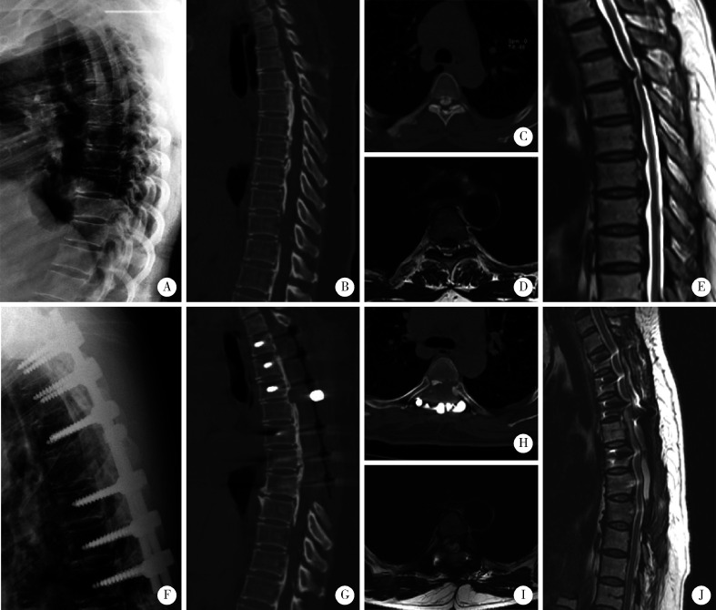
术后18例患者出现并发症,其中6例患者合并两种并发症。术后脑脊液漏16例,均延长至术后第3~5天拔除引流管,缝合引流口,采用加压包扎及俯卧体位维持24 h,未出现切口感染、不愈合等。8例患者出现术后短暂肌力下降,术后予以大剂量激素冲击,均在术后有不同程度恢复,随访时神经功能均优于术前。术后无感染性发热、肺炎、下肢静脉血栓、切口不良等并发症。随访时内固定良好,环形减压节段脊髓前方骨化块切除彻底(图 1)。
图 1.
60岁女性的影像学资料,术前mJOA评分6分,末次随访时mJOA评分8分,神经功能改善率为40%
Imaging data of a 60-year-old woman with mJOA score of 6 before surgery and 8 at the last follow-up, and neurological improvement rate was 40%
A, preoperative lateral thoracic X-ray; B, preoperative sagittal CT of thoracic vertebra, ossification of the posterior longitudinal ligament (OPLL) of T2-T9 could be seen, in which OPLL at T4-T5 was beak-like protrusion; C, preoperative axial CT of thoracic vertebrae at T4-T5 showed OPLL mass protruding into the spinal canal, with irregular shape and compression on the spinal cord; D and E, MRI of thoracic vertebrae before surgery showed multiple segments of spinal cord compression, with severe compression at T4-T5; F, lateral thoracic X-ray at the last follow-up showed good internal fixation; G and H, at the last follow-up, thoracic CT showed that the ossification mass was completely removed after circumferential decompression at the T4-T5 level, OPLL ossification was smooth at other segments, and intraspinal compression was effectively relieved; I and J, at last follow-up, MRI of the thoracic vertebra showed complete decompression of spinal cord compression at the operative segment, and no significant compression at the T4-T5 circumferential decompression segment.
2.3. 神经功能改善情况
术前mJOA评分为(6.2±2.2)分(3~10分),末次随访时mJOA评分为(7.5±2.2)分(3~11分),所有患者神经功能均较术前改善(P < 0.001)。神经功能改善率平均为(38.1±24.4)%(14.3%~100%),其中改善率75%~100% 4例,50%~74% 3例,25%~49% 14例,0%~24% 9例(表 1)。
30例患者中,神经功能较优改善组21例,神经功能较差改善组9例。在对两组患者的比较分析中,较优改善组的术中出血量为(467.1±203.9) mL,较差改善组的术中出血量为(1 088.9±791.3) mL,组间差异有统计学意义(P=0.047)。而年龄、BMI、病程、术前mJOA评分、手术时间、手术节段、是否合并脑脊液漏组间差异均无统计学意义(表 2)。
表 2.
神经功能较优改善组和神经功能较差改善组间资料比较
Comparison of data between the excellent neurological improved group and the poor neurological improved group
| Items | Excellent improved group | Poor improved group | P |
| *P < 0.05. | |||
| Case number | 21 | 9 | |
| Age/years, x±s | 49.5±11.5 | 49.0±12.1 | 0.921 |
| BMI/(kg/m2), x±s | 28.6±4.2 | 26.6±6.1 | 0.394 |
| Disease duration/months, x±s | 31.3±40.1 | 39.8±51.2 | 0.667 |
| Operation time/min, x±s | 136.9±28.0 | 138.4±46.7 | 0.911 |
| Operation blood loss/mL, x±s | 467.1±203.9 | 1088.9±791.3 | 0.047* |
| Preoperative mJOA score, x±s | 6.5±2.2 | 4.9±1.3 | 0.052 |
| The last follow-up mJOA score, x±s | 8.3±1.8 | 6.0±1.1 | 0.002* |
| Neurological improvement rate/%, x±s | 46.6±24.6 | 18.2±28.4 | < 0.001* |
| Operation segments, x±s | 3.4±0.7 | 4.0±1.0 | 0.121 |
| Cerebrospinal fluid leakage,n | 11 | 5 | 0.873 |
3. 讨论
胸椎管狭窄症的手术治疗一直是脊柱外科医生关注的重点之一,其中胸椎多节段OPLL是一种罕见且具有挑战性的疾病。胸椎后壁切除术是治疗多节段胸椎OPLL的常用方式,其操作难度较低且安全性较高,但其为间接减压,对于前方压迫较重、鸟喙型OPLL以及重度后凸无法进行彻底减压。胸椎存在生理性后凸,经后壁切除后脊髓向后移位受到限制,在前方压迫仍然存在的情况下减压有效率较低。为此本课题组建立了“涵洞塌陷法”环形减压术,可以从后方对脊髓前方的压迫进行直接减压,但由于其手术操作难度高等因素,每多操作一个节段,手术时间平均将延长1 h,进行多节段的环形减压手术风险较大,一般超过3个节段的环形减压为相对禁忌。
本研究30例胸椎长节段OPLL患者的手术治疗,在完成长节段后壁切除减压后,被挤压的硬膜囊和脊髓有了后撤的空间,脊髓膨起后移,再通过术中超声检查直接地检查脊髓腹侧的压迫情况,这时大部分OPLL的节段都可获得有效减压,而硬膜囊前方仍存在脊髓压迫的节段即需要环形减压的节段,术中超声表现为血流灌注减少和延迟;进行环形减压后,再次行术中超声显示脊髓恢复血液灌注,即确认减压彻底(图 2)。在既往研究中,环形减压的术后神经功能恢复情况良好,但其手术时间较长,术中出血量较多,术后并发症发生率较高[8]。近年来由于术中超声骨刀的使用,手术时间、术中出血量较前都显著减少(表 1)[9]。本研究所有患者均进行了单节段的环形减压,通过术中超声明确需要环形减压的部位,平均可减少1.6个环形减压的节段,降低手术创伤,进一步减少了手术时间与术中出血[10-11]。对本研究的30例患者在末次随访时可以观察到环形减压节段的OPLL骨化块切除彻底,获得了良好的手术效果(图 1)。
图 2.
环形减压节段术中超声影像
Intraoperative ultrasound images of circumferential decompression segments
A and B, intraoperative ultrasound before circumferential decompression showed severe anterior spinal cord compression with reduced and delayed blood perfusion; C and D, intraoperative ultrasound after circumferential decompression showed decompression of anterior spinal cord compression, spinal cord re-expansion, and blood perfusion recovery.
8例患者术后24 h内存在短暂的肌力下降,即刻在质子泵抑制剂保护下予以大剂量激素冲击,其肌力均在术后随访时间内不同程度恢复,且好于术前水平,出院时可以行走,这可能是由于脊髓缺血再灌注损伤所致,当被压迫的脊髓恢复膨起及血液供应后,会引起术后一过性的肌力下降,这时大剂量的糖皮质激素冲击治疗可以有效地缓解这一损伤。由于OPLL骨化块与硬膜囊粘连较重,其中16例合并脑脊液漏,但均在一周内拔除引流管,并在引流管拔除后24 h内绝对卧床以及切口敷料加压覆盖,未见切口不良事件发生。尽管在手术过程中硬膜囊的撕裂并不常见,但由于在前路操作时部分视野被遮挡,这种情况不可避免,因此需要术中谨慎预防并及时修复[12-13]。
本研究所有患者的神经功能末次随访时均较术前相对改善,其中术中出血量与神经功能改善情况显著相关,当术中出血量增多,神经功能往往改善较差,这可能与供应脊髓的节段血管大量缺血,进而造成脊髓功能恢复缓慢有关(表 2)。此外,神经功能较优改善组和较差改善组的术前mJOA评分差异无统计学意义,这说明无论术前神经功能状况如何,均可以获得较好的神经功能改善。手术可以有效地解除脊髓各个角度的压迫,即使对术前神经功能较差的患者仍具有重要的意义。
综上所述,术中超声辅助下的胸椎环形减压术可以有效地解除长节段OPLL患者的脊髓压迫,通过短节段的环形减压可以显著提高患者的神经功能并获得较好的疗效;同时在超声骨刀、超刀刮匙的使用下可以有效地减少手术时间、出血量及手术并发症的发生,控制术中出血量可以有效地提高术后神经功能的改善率。
Biography
刘晓光, 医学博士, 北京大学第三医院骨科教授, 博士生导师。中国康复医学会常务理事、颈椎病专业委员会名誉主委、脊柱脊髓专业委员会副主委、微创学组副主委。中华医学会骨科分会委员、疼痛分会常委、微创学组副组长。中华预防医学会卫生应急分会副主委, 中国医师协会骨科分会常委、教育委员会副主委, 中国中西医结合学会骨科专业委员会副主委, 北京医学会骨科分会副主委、微创学组组长, 北京中西医结合学会骨科分会副主委等。主持国家自然科学基金、科技部"十五攻关"课题、国家重点研发计划、首都医学发展科研基金、教育部"985青年基金"、中央保健委基金等多项课题, 作为分项目负责人承担了国家卫生健康委员会(原卫生部)、教育部创新团队等多项重点基金课题的研究, 资金总额超千万元。发表SCI及核心期刊论文百余篇, 主编、主译专著3部, 参与编写和翻译专著8部, 获得国家实用新型专利8项。研究成果获教育部高校科技创新二等奖(两项), 北京市科学技术奖一等奖及二等奖, 北京市优秀中青年医师"名医奖"。获得中国医师奖提名奖, 全国劳动模范, 首都劳动奖章, 中共中央、国务院、中央军委联合表彰的全国抗震救灾先进个人。 主要研究方向为胸椎管狭窄症、颈椎病、腰椎管狭窄、腰椎间盘突出、脊柱畸形、脊柱陈旧骨折、脊柱肿瘤、脊柱结核、脊柱微创技术治疗各类脊柱疾患(脊柱内镜手术、微创减压融合术、穿刺活检、椎体成形、射频、粒子植入等), 擅长脊柱病变的穿刺活检和复杂手术治疗。完成CT监测下经皮穿刺脊柱肿瘤活检1 000余例, 数量和准确率均在国内外领先。经皮穿刺寰枢椎侧块关节植骨融合术、经皮穿刺放射性粒子植入治疗脊柱肿瘤及颈椎椎管内病变经皮穿刺活检术为国际首创。完成国内首例经皮穿刺射频腰间盘髓核成型术, 并在国内率先开展经皮穿刺椎体成型术、颈椎病的经皮镜下手术等。国际上独创"涵洞塌陷法"胸椎360度脊髓环形减压术, 解决了以往手术高致瘫率的难题
Funding Statement
国家自然科学基金(81972103)
Supported by the National Natural Science Foundation of China (81972103)
References
- 1.Chen G, Fan T, Yang X, et al. The prevalence and clinical characteristics of thoracic spinal stenosis: A systematic review. Eur Spine J. 2020;29(9):2164–2172. doi: 10.1007/s00586-020-06520-6. [DOI] [PubMed] [Google Scholar]
- 2.Hou X, Sun C, Liu X, et al. Clinical features of thoracic spinal stenosis-associated myelopathy: A retrospective analysis of 427 cases. Clin Spine Surg. 2016;29(2):86–89. doi: 10.1097/BSD.0000000000000081. [DOI] [PubMed] [Google Scholar]
- 3.Ando K, Nakashima H, Machino M, et al. Postoperative progression of ligamentum flavum ossification after posterior instrumented surgery for thoracic posterior longitudinal ligament ossification: Long-term outcomes during a minimum 10-year follow-up [J/OL]. J Neurosurg Spine, 2021(2021-12-24)[2022-06-01]. https://pubmed.ncbi.nlh.gov/34952516/.
- 4.Kato S, Murakami H, Demura S, et al. Indication for anterior spinal cord decompression via a posterolateral approach for the treatment of ossification of the posterior longitudinal ligament in the thoracic spine: A prospective cohort study. Eur Spine J. 2020;29(1):113–121. doi: 10.1007/s00586-019-06047-5. [DOI] [PubMed] [Google Scholar]
- 5.刘 晓光. 胸椎管狭窄症的手术技术要点. 中国脊柱脊髓杂志. 2017;27(7):670–672. doi: 10.3969/j.issn.1004-406X.2017.07.17. [DOI] [Google Scholar]
- 6.刘 晓光, 刘 忠军, 陈 仲强, et al. "涵洞塌陷法"360°脊髓环形减压术治疗胸椎管狭窄症. 中华骨科杂志. 2010;30(11):1059–1062. doi: 10.3760/cma.j.issn.0253-2352.2010.11.010. [DOI] [Google Scholar]
- 7.Zheng C, Zhu Y, Lyu F, et al. Motor-evoked potentials in the intraoperative decision-making of circumferential decompression via posterior approach for treating thoracic posterior longitudinal ligament ossification. Spine J. 2021;21(7):1168–1175. doi: 10.1016/j.spinee.2021.01.023. [DOI] [PubMed] [Google Scholar]
- 8.Hu P, Yu M, Liu X, et al. A circumferential decompression-based surgical strategy for multilevel ossification of thoracic posterior longitudinal ligament. Spine J. 2015;15(12):2484–2492. doi: 10.1016/j.spinee.2015.08.060. [DOI] [PubMed] [Google Scholar]
- 9.Yang P, Ge R, Chen ZQ, et al. Treatment of thoracic ossification of posterior longitudinal ligament with one-stage 360 degree circumferential decompression assisted by piezosurgery. J Invest Surg. 2022;35(2):249–256. doi: 10.1080/08941939.2020.1839149. [DOI] [PubMed] [Google Scholar]
- 10.Gao A, Yu M, Wei F, et al. One-stage posterior surgery with intraoperative ultrasound assistance for thoracic myelopathy with simultaneous ossification of the posterior longitudinal ligament and ligamentum flavum at the same segment: A minimum 5-year follow-up study. Spine J. 2020;20(9):1430–1437. doi: 10.1016/j.spinee.2020.05.097. [DOI] [PubMed] [Google Scholar]
- 11.Yang X, Liu X, Liu X, et al. Clinical outcomes of intraoperative contrast-enhanced ultrasound compared with intraoperative neurophysiological monitoring during circumferential decompression for myelopathy associated with thoracic-ossification of the posterior longitudinal ligament [J/OL]. Med Sci Monit, 2020, 26: e921129(2020-04-29)[2022-06-22]. http://pubmid.ncbi.nlh.gov/32345957/.
- 12.Hu P, Yu M, Liu X, et al. Cerebrospinal fluid leakage after surgeries on the thoracic spine: A review of 362 cases. Asian Spine J. 2016;10(3):472–479. doi: 10.4184/asj.2016.10.3.472. [DOI] [PMC free article] [PubMed] [Google Scholar]
- 13.Hu PP, Liu XG, Yu M. Cerebrospinal fluid leakage after thoracic decompression. Chin Med J (Engl) 2016;129(16):1994–2000. doi: 10.4103/0366-6999.187854. [DOI] [PMC free article] [PubMed] [Google Scholar]


