Abstract
Background
Flavonoids are necessary for plant growth and resistance to adversity and stress. They are also an essential nutrient for human diet and health. Among the metabolites produced in Cannabis sativa (C. sativa), phytocannabinoids have undergone extensive research on their structures, biosynthesis, and biological activities. Besides the phytocannabinoids, C. sativa is also rich in terpenes, alkaloids, and flavonoids, although little research has been conducted in this area.
Results
In this study, we identified 11 classes of key enzyme-encoding genes, including 56 members involved in the flavonoid biosynthesis in C. sativa, from their physical characteristics to their expression patterns. We screened the potentially step-by-step enzymes catalyzing the precursor phenylalanine to the end flavonoids using a conjoin analysis of gene expression with metabolomics from different tissues and chemovars. Flavonol synthase (FLS), belonging to the 2-oxoglutarate-dependent dioxygenase (2-ODD) superfamily, catalyzes the dihydroflavonols to flavonols. In vitro recombinant protein activity analysis revealed that CsFLS2 and CsFLS3 had a dual function in converting naringenin (Nar) to dihydrokaempferol (DHK), as well as dihydroflavonols to flavonols with different substrate preferences. Meanwhile, we found that CsFLS2 produced apigenin (Api) in addition to DHK and kaempferol when Nar was used as the substrate, indicating that CsFLS2 has an evolutionary relationship with Cannabis flavone synthase I.
Conclusions
Our study identified key enzyme-encoding genes involved in the biosynthesis of flavonoids in C. sativa and highlighted the key CsFLS genes that generate flavonols and their diversified functions in C. sativa flavonoid production. This study paves the way for reconstructing the entire pathway for C. sativa’s flavonols and cannflavins production in heterologous systems or plant culture, and provides a theoretical foundation for discovering new cannabis-specific flavonoids.
Supplementary Information
The online version contains supplementary material available at 10.1186/s12934-022-01933-y.
Keywords: Cannabis sativa, Flavonoid metabolic pathway, flavonol, FLS, Gene family
Background
Flavonoids are widely distributed throughout the plant kingdom [1]. They function as copigments in flowers [2] and antibiotics in plant defense responses [3], and act as signal molecules in plant–microbe interactions [4]. They also establish an inevitable link with human diet and health [5] because of their antioxidative [6], anti-inflammatory [7], and strong anticancer activities [8]. Flavonoids are subdivided into six groups, including flavanols, flavanone, flavonols, flavones, anthocyanidins, and isoflavonoids [9]. To date, over 4000 different flavonoids have been identified [10], many of which have been testified to have pharmacological activities, such as catechin, rutin [11], apigenin (Api) [12], liquirigenin [13], and cannflavins [14].
Cannabis sativa L. is an annual herb of the Cannabinaceae family, the genera Cannabis [15]. C. sativa has been historically cultivated and utilized for 6000 years for food, textiles, and medicine [16]. Much attention has been primarily given to major phytocannabinoids, whereas besides cannabinoids, C. sativa also produces various non-cannabinoids constituents, including lignanamides, alkaloids, spiroindans, dihydrophenanthrenes, and flavonoids [17]. Flavonoids in C. sativa are currently understudied [18]. Over 20 flavonoids have been identified in C. sativa [19], belonging mainly to two classes, flavonols (kaempferol (K) and quercetin (Q)) and flavones (Api and luteolin) glycosides and aglycones [20]. Three geranylated/prenylated canniflavones, cannflavin A (geranyl), B (prenyl), and C (geranyl), unique to C. sativa (cannflavin A has also been detected in Mimulus bigelovii) [21, 22] {Rea, 2019 #19;Rea, 2019 #125}, have exhibited potent anti-inflammatory, anti-leishmania, and antioxidant activities, respectively [17, 23].
Many studies have reported the biosynthesis of the core flavonoid skeleton in medicinal plants [14]. For example, there are two distinct pathways from the root and aerial parts of Scutellaria baicalensis [24] responsible for synthesizing flavones [25]. The pathway of flavonoid biosynthesis, particularly the early phenylpropanoid biosynthetic steps in plants, show the commonness. Briefly, phenylalanine is used as the precursor substrate to generate the intermediate metabolite naringenin (Nar) through a series of enzymatic reactions, including phenylalanine ammonia lyase (PAL), cinnamic acid 4-hydroxylase (C4H), p-coumaroyl: CoA ligase (4CL), chalcone synthase (CHS), and chalcone isomerase (CHI). Subsequently, Nar is flowed to the biosynthesis of either flavonols such K, Q, and myricetin (M) by flavanone 3-hydroxylase (F3H) and flavonol synthase (FLS) or flavones such as Api, luteolin, and cannflavin A, B, and C in C. sativa via the catalytic action of flavone synthase (FNS) and other cytochrome P450 enzymes and transferases.
Two gene-encoding enzymes in the early phenylpropanoid biosynthetic pathway, CsPAL and Cs4CL [26], as well as both candidate O-methyltransferase (CsOMT21) and prenyltransferases (CsPT3) that form cannflavin A, B, or C [14, 21], have been identified in C. sativa. However, the key enzyme-encoding genes involved in flavonol biosynthesis, have not been systematically identified in C. sativa. FLS, a member of the 2-oxoglutarate and Fe(II)-dependent dioxygenases (2-ODD) superfamily, converts dihydroflavonols to flavonols [27]. In this study, we explored and characterized 56 enzymatic genes throughout the flavonoid biosynthetic pathway from a reference C. sativa genome assembly. We also identified potentially encoding candidate genes by combining their expression patterns with flavonoid content in different tissues and chemovars of C. sativa. We eventually focused on the CsFLS2 and CsFLS3 genes, and found that they retained the conservative function of FLS and exhibited additional enzymatic properties in vitro. These results systematically constitute step-by-step genes involved in the biosynthesis of flavonoids in C. sativa and entitle a potential function of CsFLS different from FLS in other higher plants.
Results
Identification and expression of genes involved in flavonoid biosynthesis in C. sativa and determination of flavonoid content in different cannabis chemovars
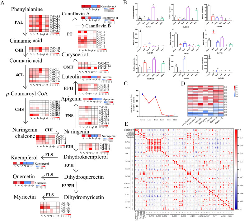
To identify the key structural genes related to flavonoid biosynthesis in C. sativa, we screened and explored 56 enzyme genes involved in flavonoid biosynthesis, including seven CsPALs, two CsC4Hs, six Cs4CLs, seven CsCHSs, four CsCHIs, eight CsFNSs, three CsF3'Hs, three CsF3Hs, five CsFLSs, three CsOMTs, and eight CsPTs using the BLASTP search and SwissProt database. The physical characteristics, including the amino acid (aa) length, molecular protein weight, isoelectric point, and their predicted subcellular localization, were also investigated (Additional file 1: Table S1). These proteins were located in different organelles, indicating that flavonoid biosynthesis in hemp is a complex and synergetic process. The expression patterns of these enzyme genes (Fig. 1A) were investigated based on the RNA-seq (RNA-sequencing) data of six different tissues (flower, bract, seed, root, leaf, and stem) of DiKu. Early genes (PAL, C4H, and 4CL) were highly expressed in root, flower, and bract. The CsCHS, CsCHI, CsFNS, CsF3'H, CsOMT, CsPT, and CsFLS gene families were mostly expressed in flowers, bracts, and leaves, while they showed relatively low transcriptional levels in roots, seeds, and stems, which were consistent with the distribution of flavonoids, including Nar, K, Q, Api, and cannflavins A and B. The candidate genes, including CsPAL7, CsC4H1, Cs4CL4, CsCHS9, CsCHI1, CsOMT21, CsPT3, and CsFLS2, were used to perform qRT-PCR (Fig. 1B and Additional file 2: Table S2), where they were largely consistent with the transcriptome data.
Fig. 1.
Expression of gene-related flavonoid biosynthesis and flavonoid content in different tissues in C. sativa. A Schematic representation of gene expression in the flavonoid biosynthesis pathway and accumulation of flavonoids in six tissues of DiKu. Data of the transcriptional level was present by log2 (FPKM + 1). The content of flavonoids was presented by the average of peak area. B: Bract, F: Flower, L: Leaf, St: Stem, Se: Seed, R: Root. PAL: phenylalanine ammonia lyase, C4H: cinnamate 4-hydroxylase, 4CL: 4-coumaric acid: CoA ligase, CHS: chalcone synthase; CHI: chalcone isomerase, F3H: flavanone 3-hydroxylase, FNS: flavone synthase; F3′H: flavonoid 3′-hydroxylase, F3′5′H: flavonoid 3′5′-hydroxylase, FLS: flavonol synthase, OMT: O-methyltransferase, and PT: prenyltransferase. B Relative expression of selected genes in different tissues of C. sativa. n = 3. Different letter indicates significance (p < 0.05). C Accumulation of major flavonoids of C. sativa. in different tissues of different chemovars. Data are presented as the average of the peak area.TI: Terra Italia, SD: Swiss Dream, PK: Pain killer, GG: Gorilla Glue, RP: Red Pure, and DK: Dinamed Kush. D Variation in total flavonoids in different tissues of C. sativa. Results were Mean ± SE (n = 3). E Correlation analysis of the transcription abundance of genes participating in the flavonol biosynthetic pathway with contents of cannflavin A, cannflavin B, luteolin, naringenin, apigenin, kaempferol, and quercetin in C. sativa
We also determined the content of total flavonoids in different tissues of DiKu (Fig. 1C). The total flavonoids were primarily distributed in flowers, bracts, and leaves, and they were hardly detected in seeds. Meanwhile, the content of different flavonoids in the six chemovars was detected (Fig. 1D). Data of the undetected flavonoids like M and its derivatives were not shown. Generally, flavonoids are inclined to be reserved in flowers and bracts compared with leaves and stems regardless of chemovars, although the content of different flavonoids varied in chemovars.
To characterize key genes involved in flavonoid biosynthesis, correlation analysis was performed based on the expression of key enzyme genes and the flavonoid content in different tissues of six different chemovars (Fig. 1E). Thereinto, CsPAL2–3 and 5, Cs4CL3–4, CsFNS1–3, CsFNS8, CsF3’H2, CsF3H1, and CsFLS1 and 3–5 showed a relatively high correlation with flavanone and flavonols, whereas Cs4CL6, CsCHS4–7, CsCHI1, CsFNS6–7, CsF3’H1, CsOMT12 and 21, CsPT1 and 3–6, CsF3H2, and CsFLS2 correlated highly with flavone accumulation in C. Sativa.
Identification of CsFLS orthologs and characterization of CsFLS genes and their encoding enzymes
FLS is the key enzyme that catalyzes dihydroflavonols to form flavonols [28]. Here, five potential CsFLS that may perform the function described above were investigated depending on the correlation analysis and gene expression. CsFLS genes were unevenly distributed on three chromosomes (Chr3, Chr5, and Chr7), where CsFLS2 and CsFLS3 were located at Chr3 and Chr5, respectively, while CsFLS4 and CsFLS5 were presented on Chr7 in tandem duplication (Additional file 3: Fig. S1A). The structure of the five CsFLS genes was investigated in that they contained one to three introns, and the whole gene spanned from 1600 bp to 9000 bp. Meanwhile, the length of the protein encoded by CsFLS genes ranged from 332 to 364 aa, and four motifs were highly conserved in CsFLS proteins (Additional file 3: Fig. S1B). The predicted protein secondary structure showed that the random coiling consisting of 40.95% to 57.22% of the protein, α-helix represented 32.49% to 35.01%, and the extended strand (16.30–18.40%) and β-turn (4.42–6.02%) accounted for less of the whole secondary structure (Additional file 4: Table S3).
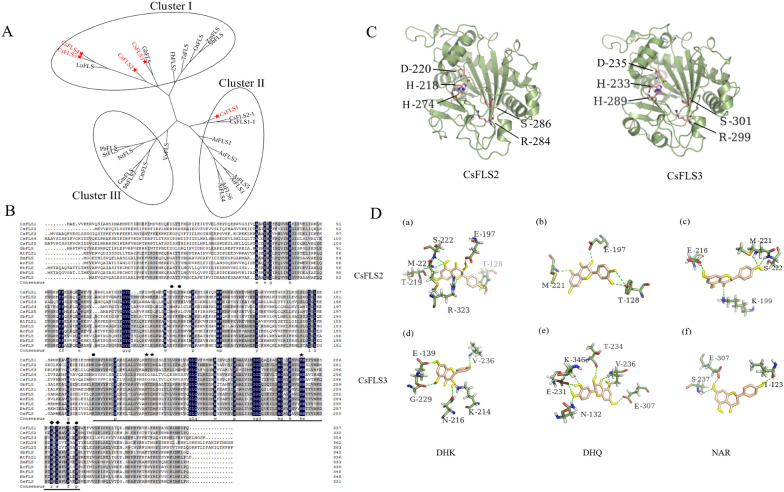
FLS belongs to the superfamily of 2-ODDs, and is broadly found in flowering plants regardless of monocots or dicots [29]. To identify orthologous FLS proteins in hemp, we performed a phylogenetic analysis of 22 FLSs in 16 different plant species (Fig. 2A and Additional file 5: Table S4). CsFLS proteins were classified into three clades, where CsFLS2–5 were clustered with Ginkgo biloba and linum usitatissimum, while CsFLS1 was grouped with FLSs of Citrus sinensis and Arabidopsis. To further analyze the differences in CsFLS at the amino acid sequence level, we conducted a sequence alignment with functionalized FLSs in other seven plant species. CsFLS1–3 had the conserved domian of 2-ODDs with binding sites of the Fe2+ and 2-oxoglutarate (2-OG) (Fig. 2B and C) [30], as well as the predicted active sites to bond to dihydroquercetin like other reported FLSs [34]. Nevertheless, CsFLS4–5 lacked the binding sites of Fe2+ (H233D and D235S) and dihydroquercetin (K214N and F146A). We also established a model for CsFLS2 (Fig. 2D, a–c) and CsFLS3 (Fig. 2D, d–f) docking to their potential substrates, dihydroflavonol (DHK and DHQ) and dihydroflavone (Nar), respectively. The results showed that hydrogen bonds formed between CsFLS2 and these three substrates (e.g., M221, S222, and E197) were close to the Fe2+ and substrate binding sites. Similarly, hydrogen bonds formed from V236, S237, and E307 to CsFLS3 may help the substrates bind more tightly to enzymes.
Fig. 2.
Catalytic characteristics of CsFLS. A Phylogenic analysis of CsFLS. The neighbor-joining method was used to construct this tree with 1000 replicate bootstraps using MEGA6.0. The CsFLSs characterized in this work are marked with red stars. Accession numbers of proteins from other plants are shown in Additional file 5. Table S4. B Sequence alignment of CsFLSs with functionally verified FLSs in Apiaceae. The proposed residues involved in binding the dihydroquercetin substrate are marked with circles. The conserved domain of 2-ODD was underlined. Inferred amino acids for binding the ferrous iron and 2-oxoglutarate (2-OG) were marked with pentagram and lozenge, respectively. C Predicted three-dimensional structures of CsFLS2 and CsFLS3. CsFLS2 binding sites for Fe2+ and 2-OG were H218, D220, and H274, and R284 and S286, respectively. Meanwhile, H233, D235, and H289, as well as R299 and S301 are the Fe2+ and 2-OG binding sites of CsFLS3, respectively. D Prediction of dihydrokaempferol (DHK), dihydroquercetin (DHQ), and naringenin (Nar) interacted with modeling CsFLS2 (a–c) and CsFLS3 (d–f) by docking simulation
Cloning and analysis of the catalytic activity of CsFLS
To analyze the catalytic activity of CsFLS2 and CsFLS3, each gene’s full open reading frame was cloned into the pET28a vector. Recombinant CsFLS2 and CsFLS3 proteins were independently expressed in Escherichia coil BL21 (DE3) strain as N/C-terminal proteins fusion with two His-6 tags. Purified proteins were verified using Western blotting at approximately 45 kDa, which was consistent with the predicted molecular weights of 43.86 kDa for CsFLS2 and 45.65 kDa for CsFLS3 (Additional file 6: Fig. S2).
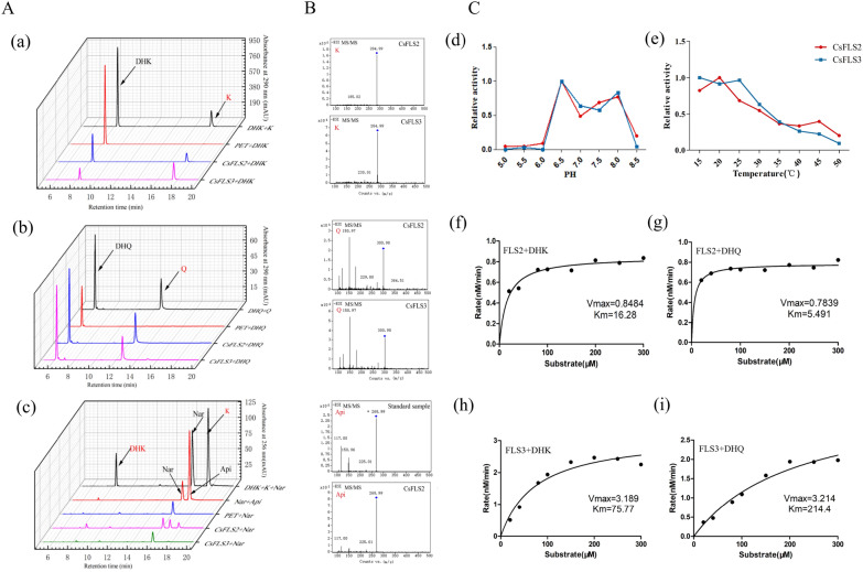
Both CsFLS2 and CsFLS3 converted dihydroflavonols (DHQ and DHK) to flavonols (Q and K), respectively (Fig. 3A, a and b). Additionally, when Nar was used as the substrate, we detected the Nar, DHK, and K in the products. This indicated that CsFLS2 and CsFLS3 had an additional F3H hydroxylation function, which can catalyze the 3-position hydroxylation of Nar to produce DHK (Fig. 3A, c). These data were further verified using LC–MS/MS (Fig. 3B). Interestingly, in addition to DHK and K, another peak occurred in the reaction product of CsFLS2 catalyzing Nar, and the retention time on HPLC, as well as LC–MS/MS, was consistent with the corresponding parameters of the Api standard (Fig. 3B). To better understand this phenomenon, we performed a phylogenetic analysis of 68 genes of DOXC 28/47 subgroups (including F3H, FLS, ANS, and FNS I) of the 2-ODD superfamily in 25 different plants with CsFLS2 and CsFLS3 [31] (Additional file 7: Fig. S3A). We subsequently compared the amino acid alignment of CsFLS2 and CsFLS3 with five Apiaceae FNS I in different plant species (Additional file 7. Fig. S3B). The active sites of FNS I consisted of seven amino acid residues [32], of which CsFLS2 possessed three, while CsFLS3 had only one. Together, these data indicated the promiscuous function of the ancestral forms of 2-ODD enzymes and more expansive substrate selectivity [33].
Fig. 3.
Enzyme activity of CsFLS2 and CsFLS3. A HPLC chromatogram of dihydroflavonols (a and b) and flavanone (c) catalyzed by recombinant CsFLS2 and CsFLS3 in vitro. B Validation of catalytic products using Q-TOF. C Analysis of catalytic activity and preference of CsFLS substrates. (d and e) The activity of CsFLS2 and CsFLS3 proteins was determined at various temperatures (15–50 ℃) and pH (5.0–8.5). (f–i) Michaelis–Menten plots for both dihydroflavonols of recombinant CsFLS2 and CsFLS3 enzymes. K, Kaempferol; Q, Quercetin; DHQ, dihydroquercetin; DHK, dihydrokaempferol; Nar, Naringenin; Api, Apigenin
To investigate the activity and preference of CsFLS2 and CsFLS3 with different substrates, the optimum pH and temperature of the enzymatic reactions were first determined using DHK as the substrate. The optimal pH for CsFLS2 and CsFLS3 was pH 6.5, with the highest catalytic activity at 20 ℃ and 15 ℃, respectively (Fig. 3C, d and e). Detailed kinetic studies conducted at the optimum and kinetic parameters were calculated using nonlinear regression analysis with Michaelis–Menten plots [34]. The Michaelis constant (Km) of DHK and DHQ for CsFLS2 was 16.28 µM and 5.49 µM, respectively, indicating that CsFLS2 had a higher affinity for DHQ than DHK (Fig. 3C, f and g). Conversely, CsFLS3 had a larger affinity for DHK than DHQ, with the Km of 75.77 μM and 214.4 μM, respectively (Fig. 3C, h and i). Therefore, the catalytic function of CsFLS2 and CsFLS3 may be complementary and substrate-selective in hemp.
Discussion
Flavonoids are important signal metabolites that keep plants resistant to stress and promote the human diet and health needs [35]. The core skeleton of the flavonoid biosynthetic pathway has been well studied in several flowering plants. However, a systematic gene profile involved in flavonoid biosynthesis in C. sativa has not yet been investigated. Indeed, integrating genome, transcriptome, and metabolome has been a highly efficient strategy to elucidate the metabolite biosynthetic and regulatory genes. The cascade of genes was proposed by combining the expression of 56 genes from 11 classes of candidates in the flavonoid pathway from different tissues of C. sativa with the metabolic detection of flavonoids (Fig. 1A). Teresa et al. [26] predicted two structural genes, CsPAL (KC970300) and Cs4CL (KC970301) in C. sativa, by searching expressed sequence tags against homologous sequences from other plants, which were named CsPAL4 and Cs4CL1 in this study. Nonetheless, both genes showed negative correlations with the content of detected flavonoids (Fig. 1E). Conversely, we speculated that CsPAL2–3 and 5, as well as Cs4CL3–4 and 6 participated positively in flavonoid generation in all CsPAL and Cs4CL genes identified in our study. The distribution of flavonoids has been investigated in different organs in C. sativa, and it was reported that they are undetectable in roots and seeds. Likely due to the improved detection methods and techniques, we detected tiny amounts of flavonoids regardless of flavonol, flavone, or total flavonoids, in roots and seeds [26, 36] (Fig. 1A and C). Meanwhile, downstream genes in the flavonoid biosynthetic pathway are barely expressed in both tissues, reducing flavonoids. Interestingly, genes involved in early phenylpropanoid biosynthetic steps to form the intermediate p-Coumaroyl CoA showed high expression in roots, indicating that unknown phenolic acid compounds may be present in roots. Additionally, flavonoids were abundant in flowers, bracts, and leaves in all chemovars of C. sativa in this study but scanty in stems (Fig. 1D), signifying C. sativa as a versatile plant with different metabolites’ accumulation in different organs. CsOMT methylated the 3’-O position of luteolin to form chrysoeriol, which CsPT further catalyzed to add a geranyl or a prenyl group to form cannflavin A, B, or C [14]. Kevin et al. [18] identified CsOMT21 and CsPT3 based on homology and phylogenetic analysis from a draft C. sativa genome assembly, supporting our correlation analysis (Fig. 1E).
FLS as a key enzyme controlling the flavonol flux, has been characterized in numerous plant species. Multiple FLS genes are always present in the plants [37]. Arabidopsis contains six FLS-encoding genes, only two of which showed flavonoid activities [38]. In this study, five CsFLS genes were identified from a reference C. sativa genome. They were distributed irregularly, not in the tandem repetition on different chromosomes (Additional file 3: Fig. S1), and divided into groups from the phylogenetic tree (Fig. 2A), suggesting that the CsFLS genes have diverged with different functions. CsFLSs belonging to the 2-ODD gene superfamily possess two highly conserved binding sites, the 2-OG binding site (Arg-X-Ser) and the Fe2+ binding site (His-Asp-His) [28]. Unlike CsFLS1–3, which had both active sites, CsFLS4–5 retained the binding sites of 2-OG but lost those of Fe2+ (Fig. 2B), implying that CsFLS4–5 were involved as complementary genes or differentiated other functions. The primitive function of FLS converted dihydroflavonols to flavonols, while a single FLS with a bifunctional property forming DHK by catalyzing the 3-hydroxylation of Nar has been identified in plants like Morella rubra [32], G. biloba [39], and C. sinensis [40]. CsFLS2 and 3 were verified to be bifunctional enzyme-coding genes using recombinant protein activity analysis in vitro (Fig. 3A and B) but with different enzyme catalytic efficiency (Fig. 3C). Chua et al. [41] found that the mutation of His 132 and Gln 295 significantly reduced the catalytic ability of AtFLS to DHQ in A. thaliana. CsFLS2 differed from CsFLS3 with Ala (A) rather than Gln (Q) at the same Gln 295 site in A. thaliana, explaining why CsFLS2 has less catalytic efficiency for DHQ than CsFLS3 (Fig. 3C) [42]. Besides flow to flavonol, CsFLS2 also produced Api (a flavone) by bringing in a double bond between the C2 and C3 positions in the B ring of Nar like a FNSI. Therefore, we performed a sequence alignment to investigate the relationship between CsFLS and FNS I. CSFLS2 retained a portion of the FNSI active sites, but whether this was the primary reason remains unknown. The plant 2-ODD superfamily was divided into three large clusters including DOXA, DOXB and DOXC, where DOXC is involved in the biosynthesis of colorful flavonoids and other secondary metabolites [31]. The 2ODD genes involved in flavonoid metabolism were classified into two distinct clades: DOXC28 and DOXC47. A phylogenetic analysis among CsFLSs and 28/47 DOXC subgroup members from 2-ODD superfamily was conducted. Unsurprisingly, CsFLS3 related closely to anthocyanidin synthase (ANS) (Additional file 7: Fig. S3), which brought into correspondence with the previous study that recombinant ANS could perform the FLS activity [42]. Different gene expression pattern mediates their functions of genes. In tomato, the duplication of SlDMR6 (Solanum lycopersicum Downy MILDEW RESISTANCE 6), which belongs to the superfamily of 2-ODDs, lead to different expression pattern and subsequent subfunctionalization, where SlDMR6-1 exerted roles in pathogen infection, while SlDMR6-2 balanced salicylic acid levels in flowers and fruits [43]. In hemp, CsFLS2 and CsFLS3 were both absent in roots, while CsFLS3 was observed abundantly in Diku flowers, bracts, and leaves, as well as slightly in stem and seeds. Nevertheless, CsFLS2 had a different expression pattern without expresion in stem and seeds (Fig. 1A). These suggested both FLSs might have different regulatory elements and functional differentiation. Interestingly, cis-acting element analysis of both CsFLS promoters showed that potentially regulatory mechanism differed between them. The promoter of CsFLS2 had defense and stress responsiveness elements, while CsFLS3 possessed specific elements response for low-temperature, salicylic acid and Methyl jasmonate (Additional file 9: Fig. S4). Overall, FLS duplication during evolution resulted in the functional divergency in terms of gene and protein structure, as well as gene expression pattern, which might be responsible for different stresses.
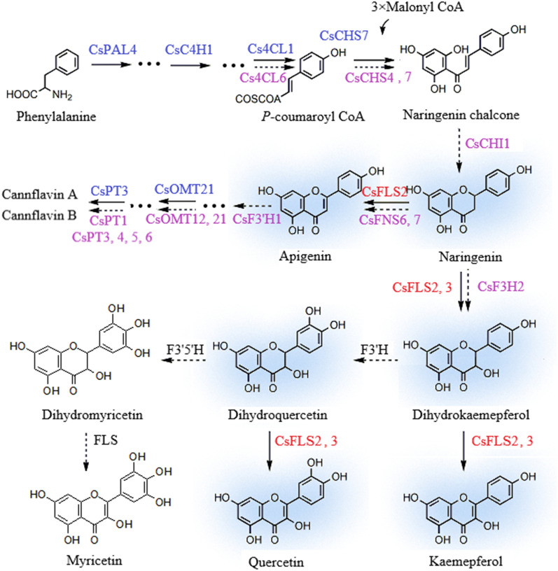
The accumulation of flavonols, such as quercetin and kaempferol, and flavanone varied in tissue specificity and chemovars of C. sativa, suggesting the involvement of CsFLSs. Coincidentally, FLS and other flavonoid-related structural genes responded positively to different environmental variations [44, 45] and developmental growth stages [37]. Hence, demonstrating the flavonoid gene cascade (Fig. 4) and understanding how to manage stress-induced flavonoids will be essential for developing environmentally resilient C. sativa plants and the biosynthesis of a substantial amount of bioactive flavonoids for downstream usage. Additionally, it was reported that flavonoids, synergistic with other non-phytocannabinoids compounds, exerts the entourage effect of boosting the bioactivities of phytocannabinoids [14]. Therefore, elucidating the mechanism of flavonoid production will have significance in the accumulation of phytocannabinoids and other non-phytocannabinoid compounds and the development of therapeutics in C. sativa.
Fig.4.
Proposed model of flavonoid metabolism in C. sativa. Naringenin (Nar) is converted to dihydrokaempferol (DHK) and keamepferol (K) by CsFLS 2 and 3. Meanwhile, Nar is catalyzed to apigenin (Api) by CsFLS2. CsFLS2 and 3 convert dihydroflavonols to flavonols. Genes highlighted in red are verified in this study. Genes in purple mean that these genes are predicted to exert catalytic action during the steps in this study. Genes marked in blue have been identified in previous studies. Dashed lines represent genes have not yet identified via enzymatic reaction
Conclusions
This study proposes step-by-step potential enzymes involved in the flavonoid biosynthetic pathway in C. sativa via a combination of transcriptomics and metabolomics of tissues of different chemovars (Fig. 4). Among these identified genes, CsFLS2 and 3 encoding enzymes were verified to be the key enzymes controlling flavonol flux via the activity analysis of recombinant proteins. Besides the primitive function of FLS converting dihydroflavonol to flavonol, CsFLS2, with versatile properties, can directly orient the production of both flavonol and flavone. Therefore, this study paves the way for reconstructing the entire pathway in heterologous systems or plant culture to yield flavonols and cannflavins in C. sativa. Additionally, this study provides a theoretical foundation for discovering new cannabis-specific flavonoids.
Methods
Plant materials and growth conditions
High-CBD chemovars, Dinamed Kush (DiKu), is a feminized plant, crossing Purple Kush and Dinamed Autoflowering CBD. DiKu and other commercial chemovars, Terra Italia, Swiss Dream, Pain killer, Gorilla Glue, and Red Pure, were grown in controlled growth chambers at 25 ℃ with 16:8 (light: dark) photoperiod in Yunnan Dali, China. The samples used in this study, including flowers, bracts, leaves, stems, and roots, were collected 20 days post-flowering (DPF). Once collected, samples were frozen in liquid nitrogen and stored at − 80 ℃ for further use.
Data sources
The reference genome and annotation files of C. sativa (Accession number: GCA_900626175.1) were obtained from NCBI (National Center of Biotechnology Information) [46]. The transcriptomes of data for six different tissues of DiKu were available at NCBI (Accession No.: bract: SMAN16122880-SAMN16122882; stem: SAMN16122883-SAMN16122885; flower: SAMN16122886-SAMN16122888; leaf: SAMN16122889-SAMN16122891).
Identification and characterization of genes related to the flavonoid metabolic pathway
Protein sequences of PAL, C4H, 4CL, CHS, CHI, FNS, F3′H, OMT, PT, F3H, and FLS from Arabidopsis thaliana were downloaded from the Uniport [47] and homology matching was conducted by using BLASTP search and ‘Blast Several Sequences to a Big Database’ in TBtools with the setting of E-value < 10–5 and removing redundant sequences. Subsequently, the conserved domain (CD) search tool of NCBI was used to screen out those which did not have complete conserved domains to obtain the members of 11 classes in C. sativa. Molecular weights and isoelectric points were predicted using the ExPASy-ProSite website [48]. Subcellular localization predictions were performed using the softberry website [49].
RNA isolation and first-strand cDNA synthesis
Total RNA was extracted from the DiKu tissues of 20 DPF, with three biological replicates using RNAprep Pure (DP441, Tiangen, Beijing) according to the manufacturer’s instruction. First-strand cDNA was subsequently synthesized using StarLighter Script RT all-in-one Mix (FS-P1001, Foreverstar, Beijing, CN).
Transcriptomic data analysis and Gene expression pattern analysis
RNA sample sequencing was conducted via Illumina and PackBio sequencing platforms. After trimming the redundancy reads, clean reads were mapped to the reference C. sativa genome sequence by employing HISAT2 tools, where 31,170 genes referred to 41,553 transcripts were annotated. Gene expression levels were estimated as fragments per kilobase of transcript per million fragments mapped (FPKM), of which log 2 (FPKM + 1) was assessed using TBtools [50] to represent gene expression from different tissues of DiKu (flowers, leaves, bracts, roots, seeds, and stems).
Quantitative real-time PCR validation
qRT–PCR was performed on Rotor-Gene Q (QIAGEN, Germany) using a StarLighter SYBR Green qPCR Mix kit (FS-Q1002, Foreverstar, Beijing, CN). The program was set as 95 °C for five minutes, 95 ℃ for 30 s, 60 ℃ for 20 s, and 72 ℃ for 15 s for 40 cycles. Each sample was repeated at least thrice, and the data were analyzed using the 2−(ΔΔCt) method [51] with EF1α [52] as the reference gene. The relevant primers are shown in Additional file 2: Table S2.
Analysis of flavonoid content in different tissues of C. sativa
The collected samples were lyophilized and then ground into power. Next, 100 mg of the ground sample was exposed to 1 ml of 70% methanol, sonicated at room temperature for 30 min, and placed at 4 °C overnight. The supernatant was retained after a 12,000 rpm centrifugation for 15 min. All samples were extracted twice following the steps above, and the mixed supernatant was filtered through a 0.22-μm organic membrane for total flavonoid content determination and LC–MS/MS analysis.
120 μl of diluted solution (in an appropriate proportion) and 60-ul NaNO3 (5%) were mixed and stayed for six minutes with an addition of 60-μl Al(NO3)3 (10%) with another six minutes stay. Then, 800-μl NaOH (4%) was added and fixed in 5-ml methanol with a thorough blending. After staying at RT for 15 min, the absorbance values were measured at 416 nm, and the standard curve was plotted using rutin standards to calculate the total flavonoid content of each sample. At least three biological replicates were determined.
The Agilent UPLC 1290II-G6400 triple quadrupole mass spectrometer (QQQ; Agilent Technologies, Santa Clara, CA, United States) was employed to determine the relative quantity of synthetic constituents. MS/MS spectra were obtained in negative ionization mode using a C18 column (Eclipse Plus C18, 2.1 × 100 mm, 1.8 μm). The mobile phases were ammonium acetate (A) and acetonitrile (B) solutions with a linear gradient program: 0/5, 2/5, 2.5/18.5, 10.5/41, 11/59, 18/77, 22/95, 24/95, 24.1/5, and 26/5 (min/B%).
Phylogenetic analysis of CsFLS and structural analysis of gene and protein
The CsFLS phylogenetic tree was constructed based on the NJ method by obtaining amino acid sequences from various plant FLSs from the GenBank database. Bootstrap tests with 1000 replicates were performed using the MEGA 6.0 software [53].
TBtools software was used to investigate the chromosomal location of CsFLS genes and their distribution of exons and introns. Motif conserved motifs (motif parameter set to 10) were predicted using the MEME [54].
Molecular docking
Swiss-Model [55] was used to model the homologic 3D structures of CsFLS2 and CsFLS3 protein. CsFLS2 and CsFLS3 were modeled based on the anthocyanidin synthase from A. thaliana (PDB ID: 1GP4) [56] as the template, and the similarities were 44.55% and 78.10%, respectively. Molecular docking of CsFLS2 and CsFLS3 protein models with their three substrates was performed separately using AutodockTools software, with the substrates DHQ (ChEBI: 17948), DHK (ChEBI: 15404), and Nar (ChEBI:50202) data obtained from CHEBI [57]. In this study, flexible docking was used, and the results were analyzed and plotted by using the PyMOL software.
Gene cloning and purification and enzymatic activities of recombinant proteins in CsFLS
CsFLS2 and CsFLS3 were cloned using the cDNA as the template and then constructed into a pET28a (+) expression vector. The primers are shown in Additional file 8: Table S5. The constructed recombinant plasmids were transferred into the E. coli BL21 (DE3) strain and incubated at 37 ℃, 160 rpm for 2–3 h until OD 600 reached 0.6. The final concentration of 0.4-mM isopropylthio-β-galactoside was added and induced at 16 ℃, 130 rpm for 20 h. After sonication and centrifugation, it was purified through a nickel column and eluted with 250-mM imidazole. N/C-terminal fusion proteins with two His-6 tags were obtained from the condensed elution. After protein concentrations were determined using Bradford reagents (DQ101, Transgen, Beijing, CN), SDS–PAGE electrophoresis and subsequent Western blotting probed using mouse monoclonal antibody Anti-His (30401ES10, Yeasen, Shanghai, CN) were performed. Enzyme reactions with recombinant FLS in a 500 μL system contained 20-mM Tris–Hcl (PH7.0), 1-mg ascorbic acid, 0.1-mg/ml bovine serum albumin, 50-μM ferrous sulfate, 1.5-mg/ml 2-ketoglutarate, 20-μg/ml substrate, and 30-μg recombinant protein reaction at 30 ℃ for 20 min followed by twice extraction using 500-μL ethyl acetate and evaporated dry at low temperature. The reactant was dissolved in 500-μL methanol and filtered for further Q-TOF analysis using the mobile phases of ammonium acetate (A) and acetonitrile (B) and a linear gradient program of 0/5, 2/5, 2.5/18.5, 20/30, 20.5/95, 23/95, 23.5/5, and 26/5 (min/B%). An Agilent 6460 (Agilent, USA) triple quadrupole liquid mass spectrometer in the negative ion mode was used to perform the test as per the parameters set as follows: the scan range setting: 100–1000 m/z, atomization pressure: 35 psi, drying gas flow rate: 8 L min−1, protective gas flow rate: 11 L min−1, and protective gas temperature: 350 °C. At least three replicates were performed for each sample.
CsFLS protein activity assays
The substrate concentration was changed to 50 μg/mL, and other conditions were unchanged. The catalytic products were quantified using HPLC with the corresponding standards, and at least three biological replicates were performed.
The optimum pH was determined by conducting the enzymatic reaction at 30 ℃ for 30 min in three buffers (pH 5.0–5.5, 10-mM sodium acetate buffer; pH 6.0–7.5, 10-mM sodium phosphate buffer; pH 8.0–8.5, 10-mM Tris–HCl buffer) at 0.5 intervals. Meanwhile, the optimal temperature was determined by performing the catalytic reaction in pH 7.0 at a 15–50 ℃ gradient range with 5 ℃ interval for every 30 min. The reaction was conducted under the optimum conditions verified above with a substrate concentration range of 0–300 μM. The reactant was detected using HPLC with a linear gradient program of 0/5, 2/5, 2.5/18, 20/30, 20.5/95, 23/95, 23.5/5, and 26/5 (min/B%). The data was stimulated using. The corresponding enzyme kinetic parameters, such as Vmax and Kmax, were calculated by non-linearly fitting the Michaelis–Menten in the Graphpad software. The experiment was repeated at least thrice.
Supplementary Information
Additional file 1: Table S1. Physical characteristics of the major enzyme-encoding genes of flavonoid metabolic pathway in C. sativa.
Additional file 2: Table S2. Quantitative primers of the selected enzyme-encoding genes of flavonoid metabolic pathway in C. sativa.
Additional file 3: Figure S1. Analysis of chromosomal location and gene structure of the CsFLS genes in C. sativa.
Additional file 4:Table S3. Secondary structure predication of the CsFLS proteins in C. sativa.
Additional file 5: Table S4. The list of flavonoid-related genes in the phylogenetic analysis from C. sativa and other plant species.
Additional file 6: Figure S2. Western blotting of recombinant protein of CsFLS2 and CsFLS3.
Additional file 7: Figure S3. Comparsion of CsFLS2 and CsFLS3 with other proteins belonging to DOXC 28/47 subgroup of 2-ODD superfamily.
Additional file 8: Table S5. Cloning primers of CsFLS2 and CsFLS3 in C. sativa.
Additional file 9: Figure S4. Cis-acting elements within the promoters of CsFLS2 and CsFLS3.
Acknowledgements
Not applicable.
Abbreviations
- C. sativa
Cannabis sativa
- PAL
Phenylalanine ammonia lyase
- C4H
Cinnamate 4-hydroxylase
- 4CL
4-coumaric acid: CoA ligase
- CHS
Chalcone synthase
- CHI
Chalcone isomerase
- F3H
Flavanone 3-hydroxylase
- FNS
Flavone synthase
- F3′H
Flavonoid 3′-hydroxylase
- F3′5′H
Flavonoid 3′5′-hydroxylase
- FLS
Flavonol synthase
- OMT
O-methyltransferase
- PT
Prenyltransferase
- K
Kaempferol
- Q
Quercetin
- DHQ
Dihydroquercetin
- DHK
Dihydrokaempferol
- Nar
Naringenin
- Api
Apigenin
- M
Myricetin
- 2-ODD
2-Oxoglutarate and Fe(II)-dependent dioxygenases
- ANS
Anthocyanidin synthase
Author contributions
WS and SC conceived the ideas, designed the skeleton of this study and supervised the whole experiments. XZ and YM performed most of the experiments and prepared the initial draft of the manuscript. XM analyzed the transcriptome data. YZ, HW, WY, and JL prepared the standard substance and established the methods of flavonoid determination. SW and XC maintained the plantation and the sample collection. WS, WC, ZX, and AtW revised the manuscript and provided some constructive advices. All authors read and approved the final manuscript.
Funding
This work was supported by Scientific and technological innovation project of China Academy of Chinese Medical Sciences (C12021A04008) and the National Key R&D Program of China (2021YFE0100900).
Availability of data and materials
The datasets presented in this study can be found in online repositories. The names of repositories and accesion numbers can be found in this manuscript and the additional files.
Declarations
Ethics approval and consent to participate
Not applicable.
Consent for publication
Not applicable.
Competing interests
The authors declare that they have no competing interests.
Footnotes
Publisher's Note
Springer Nature remains neutral with regard to jurisdictional claims in published maps and institutional affiliations.
Xuewen Zhu and Yaolei Mi contributed equally to the work and share first authorship
Contributor Information
Yaolei Mi, Email: xiaomi20063@sina.com.
Shilin Chen, Email: slchen@icmm.ac.cn.
Wei Sun, Email: wsun@icmm.ac.cn.
References
- 1.Treutter D. Significance of flavonoids in plant resistance: a review. Environ Chem Lett. 2006;4:147–157. doi: 10.1007/s10311-006-0068-8. [DOI] [Google Scholar]
- 2.Peer WA, Murphy AS. Flavonoids and auxin transport: modulators or regulators? Trends Plant Sci. 2007;12:556–563. doi: 10.1016/j.tplants.2007.10.003. [DOI] [PubMed] [Google Scholar]
- 3.Williams RJ, Spencer JP, Rice-Evans C. Flavonoids: antioxidants or signalling molecules? Free Radic Biol Med. 2004;36:838–849. doi: 10.1016/j.freeradbiomed.2004.01.001. [DOI] [PubMed] [Google Scholar]
- 4.Mandal SM, Chakraborty D, Dey S. Phenolic acids act as signaling molecules in plant-microbe symbioses. Plant Signal Behav. 2010;5:359–368. doi: 10.4161/psb.5.4.10871. [DOI] [PMC free article] [PubMed] [Google Scholar]
- 5.Hollman PCH, Katan MB. Dietary flavonoids: intake, health effects and bioavailability. Food Chem Toxicol. 1999;37:937–942. doi: 10.1016/S0278-6915(99)00079-4. [DOI] [PubMed] [Google Scholar]
- 6.Hanasaki Y, Ogawa S, Fukui S. The correlation between active oxygens scavenging and antioxidative effects of flavonoids. Free Radical Biol Med. 1994;16:845–850. doi: 10.1016/0891-5849(94)90202-X. [DOI] [PubMed] [Google Scholar]
- 7.Kim HP, Son KH, Chang HW, Kang SS. Anti-inflammatory plant flavonoids and cellular action mechanisms. J Pharmacol Sci. 2004 doi: 10.1254/jphs.crj04003x. [DOI] [PubMed] [Google Scholar]
- 8.Kopustinskiene DM, Jakstas V, Savickas A, Bernatoniene J. Flavonoids as anticancer agents. Nutrients. 2020;12:457. doi: 10.3390/nu12020457. [DOI] [PMC free article] [PubMed] [Google Scholar]
- 9.Vinson JA, Dabbagh YA, Serry MM, Jang J. Plant flavonoids, especially tea flavonols, are powerful antioxidants using an in vitro oxidation model for heart disease. J Agric Food Chem. 1995;43:2800–2802. doi: 10.1021/jf00059a005. [DOI] [Google Scholar]
- 10.Cook NC, Samman S. Flavonoids—chemistry, metabolism, cardioprotective effects, and dietary sources. J Nutr Biochem. 1996;7:66–76. doi: 10.1016/0955-2863(95)00168-9. [DOI] [Google Scholar]
- 11.Nam TG, Lee SM, Park JH, Kim DO, Baek NI, Eom SH. Flavonoid analysis of buckwheat sprouts. Food Chem. 2015;170:97–101. doi: 10.1016/j.foodchem.2014.08.067. [DOI] [PubMed] [Google Scholar]
- 12.Zhou X, Wang F, Zhou R, Song X, Xie M. Apigenin: a current review on its beneficial biological activities. J Food Biochem. 2017;41:e12376. doi: 10.1111/jfbc.12376. [DOI] [Google Scholar]
- 13.Zhang B, Xing J, Lang Y, Liu H. Synthesis of amino-silane modified magnetic silica adsorbents and application for adsorption of flavonoids from Glycyrrhiza uralensis Fisch. Sci China Ser B Chem. 2008;51:145–151. doi: 10.1007/s11426-007-0104-y. [DOI] [Google Scholar]
- 14.Bautista JL, Yu S, Tian L. Flavonoids in Cannabis sativa: biosynthesis, bioactivities, and biotechnology. ACS Omega. 2021;6:5119–5123. doi: 10.1021/acsomega.1c00318. [DOI] [PMC free article] [PubMed] [Google Scholar]
- 15.Hillig KW, Mahlberg PG. A chemotaxonomic analysis of cannabinoid variation in Cannabis (Cannabaceae) Am J Bot. 2004;91:966–975. doi: 10.3732/ajb.91.6.966. [DOI] [PubMed] [Google Scholar]
- 16.Liu FH, Hu HR, Du GH, Deng G, Yang Y. Ethnobotanical research on origin, cultivation, distribution and utilization of hemp (Cannabis sativa L.) in China. 2017.
- 17.Pollastro F, Minassi A, Fresu LG. Cannabis phenolics and their bioactivities. Curr Med Chem. 2018;25:1160–1185. doi: 10.2174/0929867324666170810164636. [DOI] [PubMed] [Google Scholar]
- 18.Nallathambi R, Mazuz M, Namdar D, Shik M, Namintzer D, Vinayaka AC. Identification of synergistic interaction between Cannabis-derived compounds for cytotoxic activity in colorectal cancer cell lines and colon polyps that induces apoptosis-related cell death and distinct gene expression. Cannabis Cannabinoid Res. 2018;3:120–135. doi: 10.1089/can.2018.0010. [DOI] [PMC free article] [PubMed] [Google Scholar]
- 19.Flores-Sanchez IJ, Verpoorte R. Secondary metabolism in cannabis. Phytochem Rev. 2008;7:615–639. doi: 10.1007/s11101-008-9094-4. [DOI] [Google Scholar]
- 20.Hollman PCH, Arts ICW. Flavonols, flavones and flavanols–nature, occurrence and dietary burden. J Sci Food Agric. 2000;80:1081–1093. doi: 10.1002/(SICI)1097-0010(20000515)80:7<1081::AID-JSFA566>3.0.CO;2-G. [DOI] [Google Scholar]
- 21.Rea KA, Casaretto JA, Al-Abdul-Wahid MS, Sukumaran A, Geddes-McAlister J, Rothstein SJ, Akhtar TA. Biosynthesis of cannflavins A and B from Cannabis sativa L. Phytochemistry. 2019;164:162–171. doi: 10.1016/j.phytochem.2019.05.009. [DOI] [PubMed] [Google Scholar]
- 22.Barrett M, Gordon D, Evans F. Isolation from Cannabis sativa L. of cannflavin—a novel inhibitor of prostaglandin production. Biochem Pharmacol. 1985;34:2019–2024. doi: 10.1016/0006-2952(85)90325-9. [DOI] [PubMed] [Google Scholar]
- 23.Werz O, Seegers J, Schaible AM, Weinigel C, Barz D, Koeberle A, Allegrone G, Pollastro F, Zampieri L, Grassi G. Cannflavins from hemp sprouts, a novel cannabinoid-free hemp food product, target microsomal prostaglandin E2 synthase-1 and 5-lipoxygenase. PharmaNutrition. 2014;2:53–60. doi: 10.1016/j.phanu.2014.05.001. [DOI] [Google Scholar]
- 24.Zhao Q, Zhang Y, Wang G, Hill L, Weng J-K, Chen X-Y, Xue H, Martin C. A specialized flavone biosynthetic pathway has evolved in the medicinal plant Scutellaria baicalensis. Sci Adv. 2016;2:e1501780. doi: 10.1126/sciadv.1501780. [DOI] [PMC free article] [PubMed] [Google Scholar]
- 25.Zhao Q, Cui M-Y, Levsh O, Yang D, Liu J, Li J, Hill L, Yang L, Hu Y, Weng J-K. Two CYP82D enzymes function as flavone hydroxylases in the biosynthesis of root-specific 4′-deoxyflavones in Scutellaria baicalensis. Mol Plant. 2018;11:135–148. doi: 10.1016/j.molp.2017.08.009. [DOI] [PMC free article] [PubMed] [Google Scholar]
- 26.Docimo T, Consonni R, Coraggio I, Mattana M. Early phenylpropanoid biosynthetic steps in Cannabis sativa: link between genes and metabolites. Int J Mol Sci. 2013;14:13626–13644. doi: 10.3390/ijms140713626. [DOI] [PMC free article] [PubMed] [Google Scholar]
- 27.Wang Y, Shi Y, Li K, Yang D, Liu N, Zhang L, Zhao L, Zhang X, Liu Y, Gao L. Roles of the 2-oxoglutarate-dependent dioxygenase superfamily in the flavonoid pathway: a review of the functional diversity of F3H, FNS I, FLS, and LDOX/ANS. Molecules. 2021;26:6745. doi: 10.3390/molecules26216745. [DOI] [PMC free article] [PubMed] [Google Scholar]
- 28.Cheng A-X, Han X-J, Wu Y-F, Lou H-X. The function and catalysis of 2-oxoglutarate-dependent oxygenases involved in plant flavonoid biosynthesis. Int J Mol Sci. 2014;15:1080–1095. doi: 10.3390/ijms15011080. [DOI] [PMC free article] [PubMed] [Google Scholar]
- 29.Araújo WL, Martins AO, Fernie AR, Tohge T. 2-Oxoglutarate: linking TCA cycle function with amino acid, glucosinolate, flavonoid, alkaloid, and gibberellin biosynthesis. Front Plant Sci. 2014;5:552. doi: 10.3389/fpls.2014.00552. [DOI] [PMC free article] [PubMed] [Google Scholar]
- 30.Farrow SC, Facchini PJ. Functional diversity of 2-oxoglutarate/Fe (II)-dependent dioxygenases in plant metabolism. Front Plant Sci. 2014;5:524. doi: 10.3389/fpls.2014.00524. [DOI] [PMC free article] [PubMed] [Google Scholar]
- 31.Kawai Y, Ono E, Mizutani M. Evolution and diversity of the 2–oxoglutarate-dependent dioxygenase superfamily in plants. Plant J. 2014;78:328–343. doi: 10.1111/tpj.12479. [DOI] [PubMed] [Google Scholar]
- 32.Wang H, Liu S, Wang T, Liu H, Xu X, Chen K, Zhang P. The moss flavone synthase I positively regulates the tolerance of plants to drought stress and UV-B radiation. Plant Sci. 2020;298:110591. doi: 10.1016/j.plantsci.2020.110591. [DOI] [PubMed] [Google Scholar]
- 33.Nam H, Lewis NE, Lerman JA, Lee D-H, Chang RL, Kim D, Palsson BO. Network context and selection in the evolution to enzyme specificity. Science. 2012;337:1101–1104. doi: 10.1126/science.1216861. [DOI] [PMC free article] [PubMed] [Google Scholar]
- 34.Johnson KA, Goody RS. The original Michaelis constant: translation of the 1913 Michaelis-Menten paper. Biochemistry. 2011;50:8264–8269. doi: 10.1021/bi201284u. [DOI] [PMC free article] [PubMed] [Google Scholar]
- 35.Lv Z-Y, Sun W-J, Jiang R, Chen J-F, Ying X, Zhang L, Chen W-S. Phytohormones jasmonic acid, salicylic acid, gibberellins, and abscisic acid are key mediators of plant secondary metabolites. World J Tradit Chin Med. 2021;7:307–325. doi: 10.4103/wjtcm.wjtcm_20_21. [DOI] [Google Scholar]
- 36.Flores-Sanchez IJ, Verpoorte R. PKS activities and biosynthesis of cannabinoids and flavonoids in Cannabis sativa L. plants. Plant Cell Physiol. 2008;49:1767–1782. doi: 10.1093/pcp/pcn150. [DOI] [PubMed] [Google Scholar]
- 37.Vu TT, Jeong CY, Nguyen HN, Lee D, Lee SA, Kim JH, Hong S-W, Lee H. Characterization of Brassica napus flavonol synthase involved in flavonol biosynthesis in Brassica napus L. J Agric Food Chem. 2015;63:7819–7829. doi: 10.1021/acs.jafc.5b02994. [DOI] [PubMed] [Google Scholar]
- 38.Peer WA, Brown DE, Tague BW, Muday GK, Taiz L, Murphy AS. Flavonoid accumulation patterns of transparent testa mutants of Arabidopsis. Plant Physiol. 2001;126:536–548. doi: 10.1104/pp.126.2.536. [DOI] [PMC free article] [PubMed] [Google Scholar]
- 39.Xu F, Li L, Zhang W, Cheng H, Sun N, Cheng S, Wang Y. Isolation, characterization, and function analysis of a flavonol synthase gene from Ginkgo biloba. Mol Biol Rep. 2012;39:2285–2296. doi: 10.1007/s11033-011-0978-9. [DOI] [PubMed] [Google Scholar]
- 40.Wan Q, Bai T, Liu M, Liu Y, Xie Y, Zhang T, Huang M, Zhang J. Comparative analysis of the chalcone-flavanone isomerase genes in six citrus species and their expression analysis in sweet orange (Citrus sinensis) Front Genetics. 2022 doi: 10.3389/fgene.2022.848141. [DOI] [PMC free article] [PubMed] [Google Scholar]
- 41.Chua CS, Biermann D, Goo KS, Sim T-S. Elucidation of active site residues of Arabidopsis thaliana flavonol synthase provides a molecular platform for engineering flavonols. Phytochemistry. 2008;69:66–75. doi: 10.1016/j.phytochem.2007.07.006. [DOI] [PubMed] [Google Scholar]
- 42.Welford RW, Turnbull JJ, Claridge TD, Prescott AG, Schofield CJ. Evidence for oxidation at C-3 of the flavonoid C-ring during anthocyanin biosynthesis. Chem Commun. 2001;18:1828–1829. doi: 10.1039/b105576n. [DOI] [PubMed] [Google Scholar]
- 43.Thomazella DP, Seong K, Mackelprang R, Dahlbeck D, Geng Y, Gill US, Qi T, Pham J, Giuseppe P, Lee CY. Loss of function of a DMR6 ortholog in tomato confers broad-spectrum disease resistance. Proc Natl Acad Sci. 2021;118:e2026152118. doi: 10.1073/pnas.2026152118. [DOI] [PMC free article] [PubMed] [Google Scholar]
- 44.Weisshaar B, Jenkins GI. Phenylpropanoid biosynthesis and its regulation. Curr Opin Plant Biol. 1998;1:251–257. doi: 10.1016/S1369-5266(98)80113-1. [DOI] [PubMed] [Google Scholar]
- 45.Dixon RA, Paiva NL. Stress-induced phenylpropanoid metabolism. Plant Cell. 1995;7:1085. doi: 10.2307/3870059. [DOI] [PMC free article] [PubMed] [Google Scholar]
- 46.dbSNP: the NCBI database of genetic variation. 2022. https://www.ncbi.nlm.nih.gov. Accessed 1 Apr 2022. [DOI] [PMC free article] [PubMed]
- 47.Marger MD, Saier MH., Jr A major superfamily of transmembrane facilitators that catalyse uniport, symport and antiport. Trends Biochem Sci. 1993;18:13–20. doi: 10.1016/0968-0004(93)90081-W. [DOI] [PubMed] [Google Scholar]
- 48.Protein identification and analysis tools on the ExPASy server. 2022. http://web.expasy.org/protparam/. Accessed 3 Apr 2022. [DOI] [PubMed]
- 49.PlantProm: a database of plant promoter sequences. http://www.softberry.com/cgi-bin/programs/proloc/protcomppl.pl. 2022. Accessed 8 Apr 2022.
- 50.Chen C, Chen H, Zhang Y, Thomas HR, Frank MH, He Y, Xia R. TBtools: an integrative toolkit developed for interactive analyses of big biological data. Mol Plant. 2020;13:1194–1202. doi: 10.1016/j.molp.2020.06.009. [DOI] [PubMed] [Google Scholar]
- 51.Livak KJ, Schmittgen TD. Analysis of relative gene expression data using real-time quantitative PCR and the 2− ΔΔCT method. Methods. 2001;25:402–408. doi: 10.1006/meth.2001.1262. [DOI] [PubMed] [Google Scholar]
- 52.Guo R, Guo H, Zhang Q, Guo M, Xu Y, Zeng M, Lv P, Chen X, Yang M. Evaluation of reference genes for RT-qPCR analysis in wild and cultivated Cannabis. Biosci Biotechnol Biochem. 2018;82:1902–1910. doi: 10.1080/09168451.2018.1506253. [DOI] [PubMed] [Google Scholar]
- 53.Kumar S, Tamura K, Nei M. MEGA: molecular evolutionary genetics analysis software for microcomputers. Bioinformatics. 1994;10:189–191. doi: 10.1093/bioinformatics/10.2.189. [DOI] [PubMed] [Google Scholar]
- 54.The meme machine. 2022. http://meme-suite.org/tools/meme. Accessed 9 Apr 2022.
- 55.SWISS‐MODEL and the Swiss‐Pdb Viewer: an environment for comparative protein modeling. 2022. https://swissmodel.expasy.org/. Accessed 12 Apr 2022.
- 56.Sun X, Zhou D, Kandavelu P, Zhang H, Yuan Q, Wang B-C, Rose J, Yan Y. Structural insights into substrate specificity of feruloyl-CoA 6’-hydroxylase from Arabidopsis thaliana. Sci Rep. 2015;5:1–10. doi: 10.1038/srep10355. [DOI] [PMC free article] [PubMed] [Google Scholar]
- 57.ChEBI: a database and ontology for chemical entities of biological interest. 2022. http://www.ebi.ac.uk/chebi/init.do. Accessed 15 May 2022. [DOI] [PMC free article] [PubMed]
Associated Data
This section collects any data citations, data availability statements, or supplementary materials included in this article.
Supplementary Materials
Additional file 1: Table S1. Physical characteristics of the major enzyme-encoding genes of flavonoid metabolic pathway in C. sativa.
Additional file 2: Table S2. Quantitative primers of the selected enzyme-encoding genes of flavonoid metabolic pathway in C. sativa.
Additional file 3: Figure S1. Analysis of chromosomal location and gene structure of the CsFLS genes in C. sativa.
Additional file 4:Table S3. Secondary structure predication of the CsFLS proteins in C. sativa.
Additional file 5: Table S4. The list of flavonoid-related genes in the phylogenetic analysis from C. sativa and other plant species.
Additional file 6: Figure S2. Western blotting of recombinant protein of CsFLS2 and CsFLS3.
Additional file 7: Figure S3. Comparsion of CsFLS2 and CsFLS3 with other proteins belonging to DOXC 28/47 subgroup of 2-ODD superfamily.
Additional file 8: Table S5. Cloning primers of CsFLS2 and CsFLS3 in C. sativa.
Additional file 9: Figure S4. Cis-acting elements within the promoters of CsFLS2 and CsFLS3.
Data Availability Statement
The datasets presented in this study can be found in online repositories. The names of repositories and accesion numbers can be found in this manuscript and the additional files.


