Abstract
Extracts from Drosera rotundifolia are traditionally used to treat cough symptoms during a common cold. The present study aimed to investigate the impact of extracts from D. rotundifolia and active compounds on the respiratory tract. Tracheal slices of C57BL/6N mice were used ex vivo to examine effects on airway smooth muscle (ASM) and ciliary beat frequency (CBF). Phosphodiesterase (PDE) inhibition assays were carried out to test whether PDE1 or PDE4 are targeted by the active compounds. An ethanol–water extract, as well as an aqueous fraction of this extract, exerted antispasmodic properties against acetylcholine-induced contractions. In addition, contractions induced by 60 mM K+ were abrogated by the aqueous fraction. Effects on ASM could be attributed to the flavonoids quercetin, 2″-O-galloylhyperoside and hyperoside. Moreover, the Drosera extract and the aqueous fraction increased the CBF of murine tracheal slices. Quercetin and 2″-O-galloylhyperoside were identified as active compounds involved in the elevation of CBF. Both compounds inhibited PDE1A and PDE4D. The elevation of CBF was mimicked by the subtype-selective PDE inhibitor rolipram (PDE4) and by 8-methoxymethyl-IBMX. In summary, our study shows, for the first time, that a Drosera extract and its flavonoid compounds increase the CBF of murine airways while antispasmodic effects were transferred to ASM.
Keywords: Drosera rotundifolia, antispasmodic, trachea, ciliary beat frequency, phosphodiesterase
1. Introduction
Drosera rotundifolia L. is a carnivorous plant that natively grows in peatland or other wet and oligotrophic regions of the world [1]. The medicinal use to treat symptoms of a common cold, especially to treat a dry cough, is due to tradition and belongs to the area of complementary medicine. The extraction process of the fresh undried herbal drug is defined by the European Pharmacopoeia leading to the mother tincture, which serves as the raw material for medicinal drugs available in Germany [2,3]. In addition to D. rotundifolia, D. anglica and D. intermedia are of pharmaceutical relevance [3].
The phytochemical composition of D. rotundifolia is well characterized and consists of a complex mix of flavonoids, mainly flavonols and their glycosides (e.g., quercetin, isoquercitrin, hyperoside and 2″-O-galloylhyperoside) [4,5]. 2″-O-galloylhyperoside is one flavonoid of high relevance because of the infrequent occurrence in the plant kingdom compared to quercetin or isoquercitrin. 1,4-Naphtoquinones described for D. rotundifolia are particularly the main naphthoquinone 7-methyljuglone as well as plumbagin, hydroxydroserone, ramentone and their glycosides [6,7]. Other compounds reported are ellagic acid and dimethyl ellagic acid, as well as anthocyanins [4,6].
From the pharmacological point of view, studies focused on ex vivo experiments investigating antispasmodic effects and anti-inflammatory and antibacterial properties [8,9,10,11,12,13]. In addition to functional aspects, recent studies also provide evidence for the influence of Drosera on gene expression [14]. Despite the fact that the indication area of extracts from Drosera spp. addresses the respiratory tract, many ex vivo studies were performed using intestine smooth muscle [8,9,10]. Until now, effects on the lower respiratory tract have been insufficiently studied. Of note, a phytochemically uncharacterized ethanolic extract of Drosera was reported to have only a slightly antispasmodic effect on guinea pig tracheal smooth muscle, as not more than 15% of a carbachol-induced contraction of the trachea could be abrogated [10]. The same extract exerted much stronger effects on intestine muscles, inhibiting almost 50% of acetylcholine-induced contractions [10]. In the same line, an ethanolic extract of D. madagascariensis was ineffective against PGF2-induced contractions on guinea-pig tracheal slices [8].
Considerably better understood are the anti-inflammatory properties of Drosera, as extracts of D. rotundifolia and D. madagascariensis and their natural compounds (quercetin, isoquercitrin and hyperoside) turned out to be inhibitors of the neutrophil elastase [8,9]. In addition, extracts of D. rotundifolia and D. madagascariensis and their flavonoids were potent anti-inflammatory agents in a HET-CAM assay [11], and fractions of D. rotundifolia and D. tokaiensis were reported to suppress the activation of human mast cells [12]. Antibacterial activity against oral bacteria was reported for extracts of D. peltata, pointing out that plumbagin is the active component [13].
To the best of our knowledge, no experiments have been performed examining the influence of extracts from Drosera on ciliary beat frequency (CBF), ciliary beating amplitude (CBA) or mucociliary clearance until now. The mucociliary clearance is a major host defense mechanism of the upper and lower airways, which is responsible for eliminating inhaled particles of dust or pathogens [15,16,17] and thereby represents a powerful target to assess. Parameters with an impact on mucociliary clearance are particularly the CBF and CBA [15,16,18]. Certain spasmolytics used for the treatment of chronic obstructive pulmonary disease (COPD) and bronchial asthma were reported to increase the CBF of airway epithelium cells. For instance, salbutamol, a β2 adrenergic agonist, increases ciliary beating by rising cAMP levels [16,19]. Furthermore, the phosphodiesterase 4 (PDE 4) inhibitor roflumilast-N-oxide, which is the active metabolite of roflumilast, was reported to increase CBF of cultured bronchial epithelium cells via a rise in cAMP [17,20].
The present study provides a detailed investigation of the effects of extracts and natural compounds from D. rotundifolia on airway smooth muscle (ASM) and CBF. Characterization of the ethanolic extract was performed by HPLC-MS. Murine tracheal slices were used to examine the influence on smooth muscle contractility and ciliary beating of ciliated tracheal epithelium cells. Further experiments were carried out with single compounds to test for an influence on PDE1A and PDE4D.
2. Results
2.1. Phytochemical Characterization of Extracts and Quantification of Flavonoids
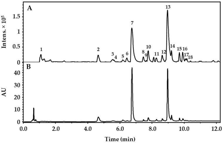
The fresh undried herbal material of Drosera rotundifolia was extracted with ethanol:water (9:1, v/v), as described in detail in the methods for the preparation of homeopathic stocks and potentisation (method 1.1.3) of the European Pharmacopoeia [2]. After the removal of ethanol in vacuo, the remaining aqueous extract was lyophilized, yielding the Drosera dry extract. For the experiments, the lyophilisate was either fully solved (“Drosera extract”, DE) or an aqueous fraction (“Drosera fraction, aqueous”, DFA) was generated by suspending the lyophilisate in Krebs–Henseleit buffer and removing insoluble components (see methods, 4.2, for details). DE was characterized by UHPLC-(+)-ESI-qTOF-MS, and peaks were related to known natural compounds described for Drosera spp. (Table 1 and Figure 1). The amount of the flavonoids quercetin, hyperoside and 2″-O-galloylhyperoside was determined by UHPLC-PDA (Table 2) for DE (acetonitrile/water, 1:1, v/v) and for DFA.
Table 1.
LC-qTOF-MS peak characteristics of Drosera dry extract (DE). Peaks were assigned to known constituents of the genus Drosera as denominated in the references. Assignments without references are based on the de novo interpretation of MS, MS2 and UV-spectra.
| Peak No. | tR (min) | λmax (nm) | [M+H]+ (m/z) |
MS2 Fragments (m/z) |
Ion Formula |
Error (mDa) | mSigma | Tentative Compound Identity |
Ref. |
|---|---|---|---|---|---|---|---|---|---|
| 1 | 2.377 | - | 221.0676 | 203.0558, 175.0605, 157.0409, 129.0178, 115.0398 |
C8H13O7 | −2.0 | 54.2 | 1,5-Dimethylcitrate | |
| 2 | 4.654 | 220, 272 |
199.0608 | 171.0270, 153.0166 | C9H11O5 | −0.6 | 1.6 | Ethyl gallate | |
| 3 | 5.557 | 216, 270, 357 |
633.1103 | 319.0437, 315.0719, 153.0167, 127.0427 |
C28H25O17 | −1.6 | 4.3 | Myricetin-3-O-(6″-O-galloyl)-hexoside | [21] |
| 4 | 5.641 | 212, 269, 360 |
779.1669 | 617.1122, 477.1330, 315.0704, 303.0497, 153.0156 |
C34H35O21 | −0.3 | 10.5 | tentatively identified Quercetin- (galloylhexosyl)-hexoside |
|
| 5 | 6.186 | 214, 252, 363 |
303.0142 | 285.0069, 275.0145, 257.0081, 247.0264, 229.0135, 219.0262, 201.0202, 173.0215, |
C14H7O8 | 0.6 | 18.8 | Ellagic acid | [21] |
| 6 | 6.435 | 212, 259, 356 |
465.1036 | 303.0496 | C21H21O12 | 0.9 | 7.5 | Hyperoside | [4] |
| 7 | 6.736 | 216, 264, 356 |
617.1173 | 303.0527, 233.0455, 153.0181 |
C28H25O16 | 0.6 | 106.1 | 2″-O-galloylhyperoside | [4] |
| 8 | 7.455 | 216, 268, 354 |
601.1183 | 315.0710, 287.0545, 233.0410, 205.0485, 153.0160 |
C28H25O15 | 0.5 | 16.6 | Kaempferol-3-O-(2-O-galloyl)-β- galactopyranoside |
[4,5] |
| 9 | 7.6480 | 216, 262, 360 |
601.1191 | 465.1033, 303.0501, 299.0769, 137.0214 |
C28H25O15 | 0.3 | 10.4 | tentatively identified Quercetin- (dihydroxybenzoyl-)hexoside |
|
| 10 | 7.759 | 212, 256, 368 |
319.0441 | 273.0387, 245.0431, 217.0476, 179.0311, 165.0150, 153.0150 |
C15H11O8 | −0.8 | 1.8 | Myricetin | |
| 11 | 5.741 | 206, 262, 364 |
585.1246 | 303.0509, 283.0835, 265.0676, 121.0268 |
C28H25O14 | −0.7 | 8.8 | tentatively identified Quercetin- (hydroxybenzoyl-)hexoside |
|
| 12 | 8.846 | 212, 256, 368 |
611.1389 | 309.0973, 303.0471, 147.0428 |
C30H27O14 | 0.7 | 8.3 | tentatively identified Quercetin- (cumaroyl-)hexoside |
|
| 13 | 8.956 | 220, 256, 368 |
303.0497 | 229.0487, 153.0161, 137.0223 |
C15H11O7 | 0.7 | 37.6 | Quercetin | [4] |
| 14 | 9.222 | 220, 243, 371 |
331.0463 | 316.0227, 300.9989, 271.0255, 253.0097 |
C16H11O8 | 0.8 | 3.8 | 3,3′-Di-O-methylellagic acid | [4] |
| 15 | 9.699 | 220, 269, 356 |
569.1301 | 303.0504, 267.0856, 123.0420, 105.0330 |
C28H25O13 | −1.2 | 6.3 | Quercetin-(benzoyl-)hexoside | [21] |
| 16 | 9.910 | 220, 265, 362 |
287.0558 | 213.0574, 165.0160, 153.0168, 121.0290 |
C15H11O6 | −0.1 | 35.0 | Kaempferol | [4] |
| 17 | 10.075 | 220, 268, 355 |
553.1359 | 287.0558, 267.0863, 123.0409, 105.0338 |
C28H25O12 | −1.8 | 11.4 | tentatively identified Kaempferol-(benzoyl-)hexoside | |
| 18 | 10.157 | 220, 268, 355 |
553.1374 | 287.0546, 267.0870, 123.0462, 105.0334 |
C28H25O12 | −3.3 | 55.3 | tentatively identified Kaempferol-(benzoyl-)hexoside |
Figure 1.
Base peak chromatogram (A) and UV chromatogram at λ = 280 nm (B) of the Drosera dry extract (DE). Identified compounds are represented by numbers as classified in Table 1.
Table 2.
Quantification of the flavonoids 2″-O-galloylhyperoside (GH), quercetin (Q) and hyperoside (H) in DE and DFA at a concentration of 0.5 mg/mL by UHPLC-PDA.
| Sample | GH (µM) | Q (µM) | H (µM) |
|---|---|---|---|
| DE | 88 ± 3 | 71 ± 30 | 1.8 ± 0.2 |
| DFA | 67 ± 5 | 9 ± 3 | 2.3 ± 0.3 |
2.2. Effects of the Drosera Extract, the Aqueous Drosera Fraction and Isolated Flavonoids on ASM Contractions
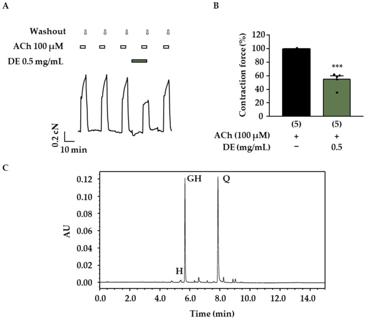
In order to systematically investigate the antispasmodic activity of D. rotundifolia on the trachea, first, the influence of DE and DFA on tracheal contractions induced by stimulation of muscarinic receptors with acetylcholine (ACh, 100 µM) was addressed. DE and DFA were added 10 min before and during the application of the cholinergic stimulus in these experiments. Both preparations showed antispasmodic effects (Figure 2A,B and Figure 3A,B). The contraction maximum evoked by 100 µM ACh was reduced to 55 ± 11% (p < 0.001, Figure 2B) by 0.5 mg/mL DE (Krebs–Henseleit buffer, 1% DMSO).
Figure 2.
Influence of DE on the ACh-induced contraction of mouse tracheal slices. (A) Representative measurement examining the antispasmodic activity of DE on the pharmacomechanical coupling. At least 3 ACh-induced control contractions (100 µM ACh) were recorded before DE (0.5 mg/mL) was added to the bath solution for a 10-minute pretreatment period. Thereafter, another contraction was induced by ACh. In the presence of DE, the contraction force was significantly reduced. Reversibility was proven by a washout period followed by another ACh bolus. (B) The scatterplot illustrates the mean contraction force in % ± SD (related to the control contraction) and single data points of 5 independent mouse preparations. (C) UV chromatogram of DE at λ = 360 nm. 2″-O-galloylhyperoside (GH), quercetin (Q) and hyperoside (H) are labeled as the characteristic flavonoids contained in the extract. (***: p < 0.001 vs. control).
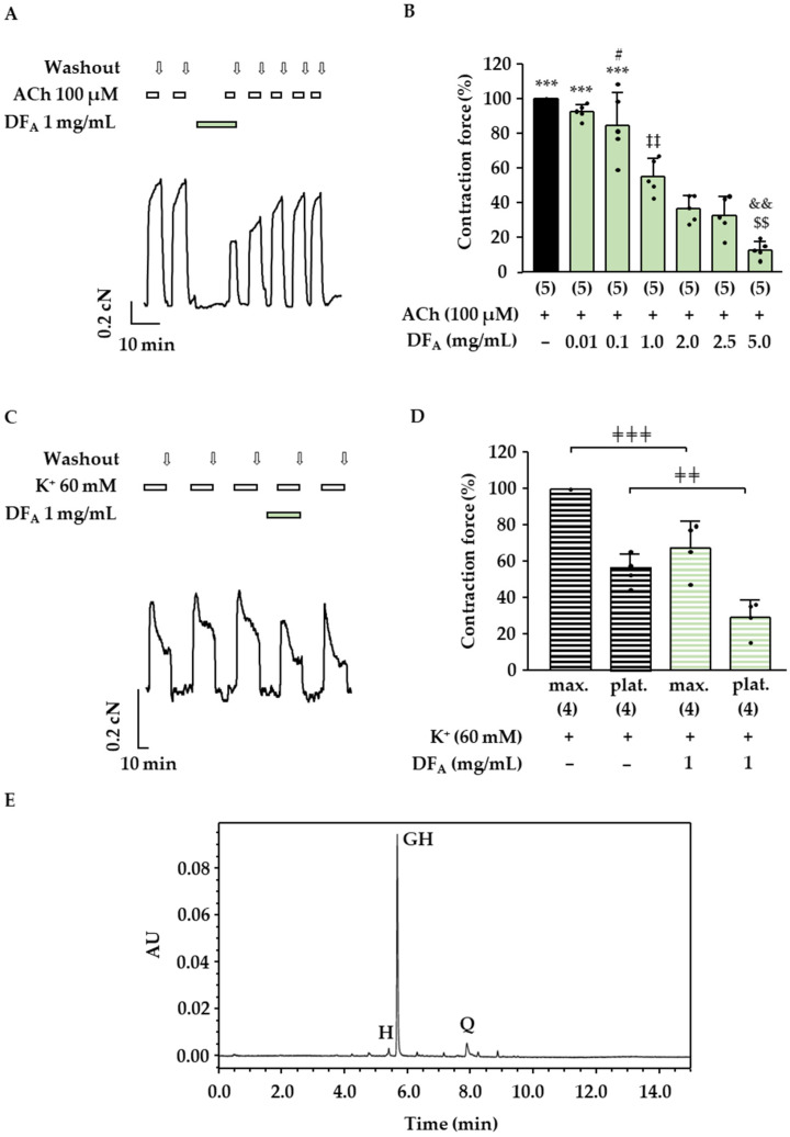
Figure 3.
Influence of DFA on the ACh-induced contraction of mouse tracheal slices. (A) Representative measurement investigating the effect on the ACh-induced contraction of ASM. First, ACh-induced control contractions (100 µM ACh) were induced. Afterward, DFA (1 mg/mL) was added to the bath solution for a pretreatment time of 10 min, and another contraction was induced by ACh in the presence of DFA. (B) The scatterplot represents the decrease in the mean contraction force in % ± SD in response to treatment with DFA (5 independent mouse preparations). (C) Representative measurement to investigate the influence on the 60 mM K+-induced contractions of ASM. At least 3 constant contractions evoked by K+ were recorded. Afterward, DFA was added for 10 min, and K+ was applied again. This maneuver was followed by a washout phase to check for reversibility. (D) The scatterplots illustrate the mean contraction maximum (max.) and the plateau (plat.) in % ± SD (related to the respective control condition) and the single data points of four independent mouse preparations. (E) UV chromatogram at λ = 360 nm of DFA at a concentration of 0.5 mg/mL. 2″-O-galloylhyperoside (GH), quercetin (Q) and hyperoside (H) are labeled as characteristic flavonoids contained in the extract. (#: p ≤ 0.05 vs. control contraction, ***: p < 0.001 vs. DFA ≥ 1.0 mg/mL, ‡‡: p < 0.01 vs. ≥ DFA 1 mg/mL, &&: p < 0.01 vs. DFA 2 mg/mL, $$: p < 0.01 vs. DFA 2.5 mg/mL, ╪╪╪: p < 0.001, ╪╪: p < 0.01).
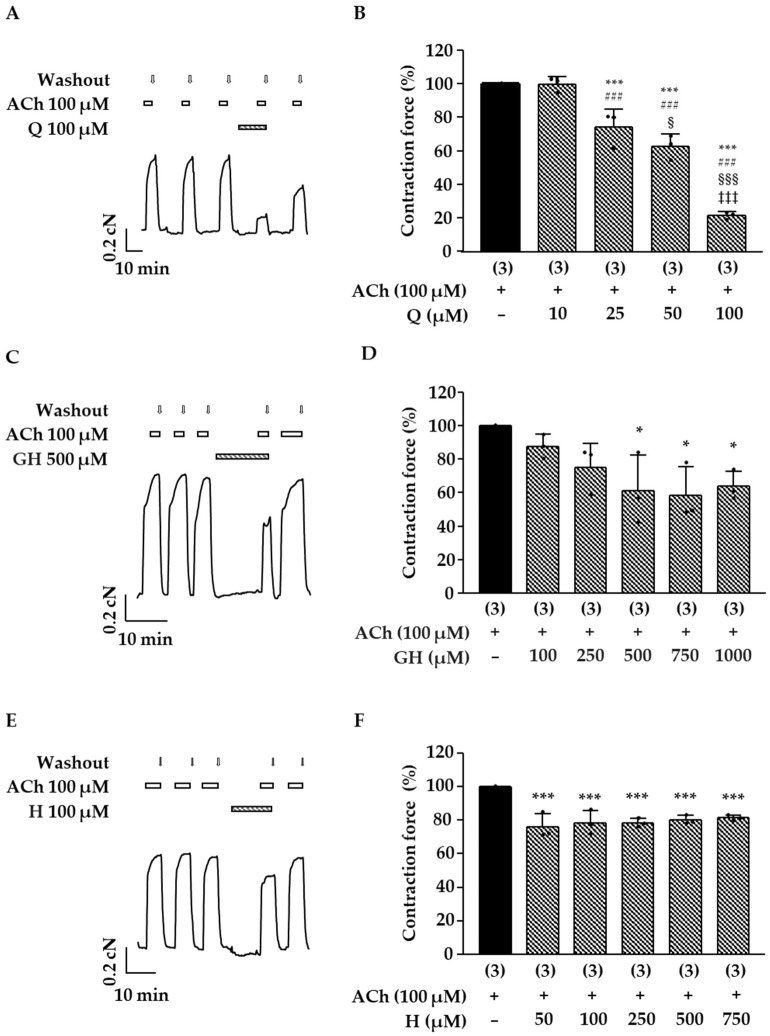
Similar results were obtained with the double dose of the more hydrophilic fraction DFA (Figure 3A,B: contraction force DFA 1.0 mg/mL + ACh 55 ± 9%, related to the control with ACh, which was set to 100%; p < 0.001). Further experiments revealed a clear concentration–response correlation for DFA and an IC50 value of 1.1 mg/mL (CI: 0.8–1.4 mg/mL) was calculated. These data show that the compounds solubilized in DE and DFA affect the pharmacomechanical coupling of mouse trachea. Next, the influence of DFA on the electromechanical coupling was investigated. After a pretreatment period of 10 min, the contraction force induced by 60 mM K+ was significantly decreased in the presence of 1 mg/mL DFA (Figure 3C,D). In this series of experiments, the initial peak and the plateau phase of the K+-mediated contractions were evaluated, revealing that both parameters were affected by DFA. The initial K+-induced peak was reduced to 68 ± 15% of the control contraction maximum without DFA (p < 0.01). The contraction plateau amounted to 30 ± 10% after treatment with DFA (p ≤ 0.05 vs. control contraction plateau). The chromatograms presented in Figure 2C and Figure 3E show that 2″-O-galloylhyperoside is a major flavonoid in both preparations, whereas quercetin is an additional main component of DE. In order to assess the relevance of these compounds for the effect of DE and DFA, they were tested on ACh-induced contractions of the mouse trachea. Hyperoside, a flavonoid contained in the dry extract of D. rotundifolia (Table 1) in low amounts, is structurally closely related to quercetin and 2″-O-galloylhyperoside and was investigated in the same manner. Quercetin exerted a dose-dependent antispasmodic effect (Figure 4A,B) and decreased the effect of 100 µM ACh to 74 ± 11% (25 µM quercetin, p < 0.001 vs. ACh alone), 63 ± 7% (50 µM quercetin, p < 0.001) and 21 ± 2% (100 µM quercetin, p < 0.001). In the case of 2″-O-galloylhyperoside and hyperoside, no dose-dependency was observed. The ACh-induced contractions were significantly inhibited by 500 µM 2″-O-galloylhyperoside and reduced to 61 ± 21% of control (p ≤ 0.05 vs. ACh alone), while higher concentrations (750 and 1000 µM) did not significantly differ from the aforementioned effect (Figure 4C,D). Hyperoside (50 to 750 µM) had the smallest effect of the three flavonoids and decreased ACh-mediated contraction force to approximately 80% of control at all concentrations tested (Figure 4E,F).
Figure 4.
Influence of flavonoids contained in D. rotundifolia on ACh-induced contractions of mouse tracheal slices. Experiments were performed as described in Figure 2A. (A,C,E) Representative recordings are shown. Quercetin (Q, 100 µM), 2″-O-galloylhyperoside (GH, 500 µM) and hyperoside (H, 100 µM) were added to the bath solution for a period of 10 min preceding the next bolus of ACh (100 µM). (B,D,F) The scatterplots represent the single data points and the mean contraction force in % ± SD of 3 independent mouse preparations. (*: p ≤ 0.05 and ***: p < 0.001 vs. control contraction, ###: p < 0.001 vs. Q 10 µM, §: p < 0.05 and §§§: p < 0.001 vs. Q 25 µM, ‡‡‡: p < 0.001 vs. Q 50 µM).
2.3. Influence of DE and Flavonoids on CBF
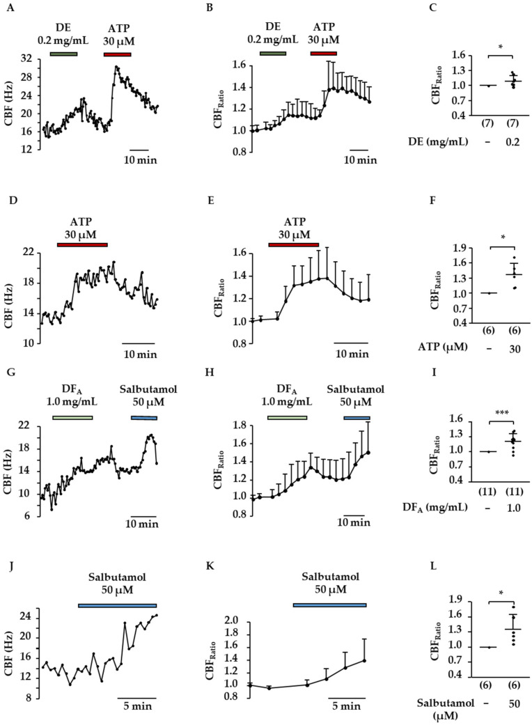
The influence of DE and DFA on CBF was measured using murine tracheal slices cultured for 1 to 7 days. Test solutions and compounds were added during constant perfusion (1 mL/min). ATP (30 µM) or salbutamol (50 µM) were applied at the end of each experiment as positive controls and both substances increased the CBF as expected (Figure 5F: CBFRatio-ATP = 1.4 ± 0.2, p ≤ 0.05 compared to CBFBasal; Figure 5L: CBFRatio-Salbutamol = 1.4 ± 0.3, p ≤ 0.05 compared to CBFBasal). Both Drosera preparations significantly increased the CBF of ciliated tracheal epithelial cells (Figure 5A–C,G–I). The more lipophilic DE elevated the CBF by approximately 10% already at a concentration of 0.2 mg/mL (Krebs–Henseleit buffer, 0.3% DMSO) (Figure 5C: CBFRatio = 1.1 ± 0.1, p ≤ 0.05 vs. CBFBasal). DFA was tested at a concentration of 1 mg/mL and induced a rise in CBF of 20% on average (Figure 5I: CBFRatio = 1.2 ± 0.2, p < 0.001 vs. CBFBasal).
Figure 5.
Influence of DE and DFA on the CBF of ciliated tracheal epithelium cells. In (A,D,G,J), representative measurements are shown. After determination of the basal CBF test solutions containing DE (0.2 mg/mL), DFA (1.0 mg/mL), ATP (30 µM) or salbutamol (50 µM) were added for 15, or in case of salbutamol for 10 min. The sequence of treatments and washout periods was performed as indicated in the exemplary recordings. Graphs (B,E,H,K) represent the averaged time-dependent change in CBF normalized to the basal CBF for each experiment. The scatterplots (C,F,I,L) illustrate the mean CBFRatio ± SD, related to CBFBasal. Here the CBFRatio represents the averaged values in minutes 10 to 15 and, in the case of salbutamol, in minutes 5 to 10 after changing to the aforementioned test solutions. The number of experiments is shown in the brackets. (***: p < 0.001, *: p ≤ 0.05).
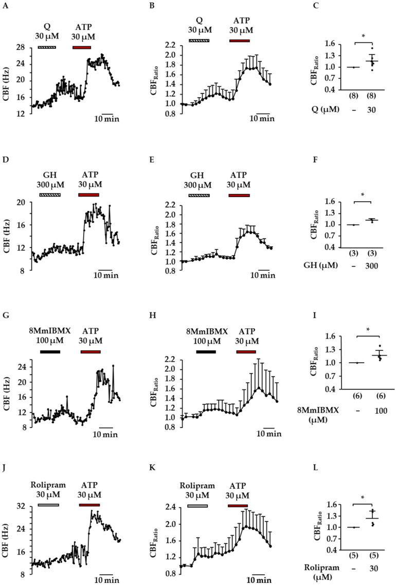
Effects in the same order of magnitude were observed by the flavonoids quercetin (30 µM, Figure 6A–C) and 2″-O-galloylhyperoside (300 µM, Figure 6D–F). In the presence of quercetin, the CBFRatio amounted to 1.2 ± 0.2 (p ≤ 0.05 vs. control), and a tenfold higher concentration of 2″-O-galloylhyperoside increased the CBFRatio to 1.11 ± 0.04 (p ≤ 0.05 vs. control). It is known that cAMP and cGMP play an important role in the regulation of CBF. Due to the fact that quercetin was reported to inhibit PDE 1–5, two PDE inhibitors were used to elucidate whether the subtypes PDE1A and PDE4 could mimic the effects observed for flavonoids and preparations of Drosera dry extract. Both the PDE1A selective inhibitor 8-methoxymethyl-(8Mm)IBMX (Figure 6G–I) and the PDE4 selective inhibitor rolipram (Figure 6J–L) significantly increased the CBF by 20% on average (CBFRatio (8MmIBMX, 100 µM): 1.2 ± 0.1, CBFRatio (rolipram, 30 µM): 1.2 ± 0.2, p ≤ 0.05 compared to CBFBasal for both inhibitors).
Figure 6.
Influence of flavonoids from D. rotundifolia on the CBF of ciliated tracheal epithelial cells. In (A,D,G,J), representative example measurements are illustrated. In analogy to the other measurements, the basal CBF was recorded before changing to test solutions. Quercetin (Q), 2″-O-galloylhyperoside (GH), 8MmIBMX and rolipram were added for 15 min. Afterward, test substances were removed, and the positive control ATP (30 µM) was added, followed by another washout period. In (B,E,H,K), the time-dependent change in CBF normalized to CBFBasal is illustrated in 2.5 min steps. The scatterplots (C,F,I,L) illustrate the mean CBFRatio ± SD related to CBFBasal. The CBFRatio represents the averaged values in minutes 10 to 15 after changing to the aforementioned test solutions. The number of experiments is represented in the brackets. (*: p ≤ 0.05).
2.4. Inhibition of PDE1A and PDE4D by Flavonoids Present in D. rotundifolia
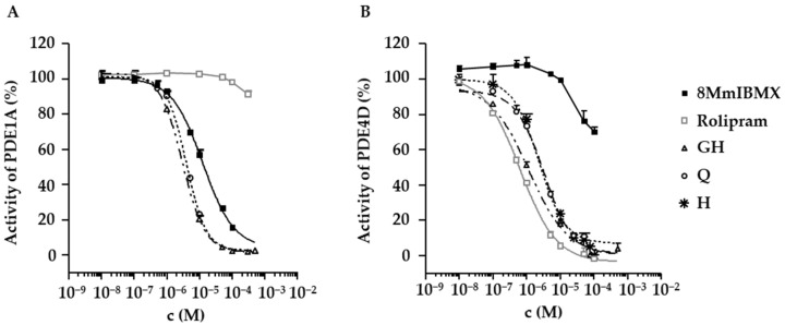
In order to verify the proposed targets of the natural compounds contained in the DE, two PDE inhibitor assays were performed. As expected, quercetin reduced the in vitro activity of the PDE1A and PDE4D (Table 3 and Figure 7). The 2″-O-galloylhyperoside and hyperoside, which to our knowledge have not been described as inhibitors of the PDE until now, turned out to inhibit the PDE4D with IC50 values in the low µM range (Table 3 and Figure 7B). The 2″-O-galloylhyperoside was also tested for its inhibitory potency on the PDE1A (Table 3 and Figure 7A) and was shown to be very effective on this PDE as well. Rolipram did not have an influence on the activity of PDE1A up to a concentration of 100 µM (PDE1A activity: 98.2 ± 0.6%) and only slightly inhibited PDE1A at 300 µM (PDE1A activity: 92 ± 2%). Regarding PDE4D, rolipram inhibited this PDE in a dose-dependent manner as expected with an IC50 below 1 µM. Interestingly, 8MmIBMX lost its selectivity for PDE1A and influenced PDE4D already at a concentration of 50 µM (PDE4D activity with 50 µM 8MmIBMX: 77 ± 6%, with 100 µM 8MmIBMX: 70 ± 2%).
Table 3.
Inhibition of the PDE1A and PDE4D by the flavonoids 2″-O-galloylhyperoside (GH), quercetin (Q) and hyperoside (H) and the respective controls.
| GH IC50 (µM) |
Q IC50 (µM) |
H IC50 (µM) |
8MmIBMX IC50 (µM) |
Rolipram IC50 (µM) |
|
|---|---|---|---|---|---|
| PDE1A | 3.0 (CI: 2.8–3.3) | 4 (CI: 3–5) | not tested | 12 (CI: 9–18) | − |
| PDE4D | 1.1 (CI: 0.7–1.5) | 2 (CI: 2–3) | 3 (CI: 3–4) | − | 0.6 (CI: 0.5–0.7) |
Figure 7.
Influence of flavonoids from D. rotundifolia, 8MmIBMX and rolipram on the activity of the PDE1A (A) and PDE4D (B). 2″-O-galloylhyperoside (GH), quercetin (Q), hyperoside (H) and the respective positive controls 8MmIBMX and rolipram were tested on the activity of the PDEs in a cell-free assay. Samples were measured as duplicates. Enzyme activity is given in % ± SD and refers to the control without inhibitor.
3. Discussion
Drosera extracts are traditionally used to treat common cold symptoms, especially due to proposed antispasmodic activities. The inhibition of PDEs, especially the inhibition of PDE3 and PDE4, are known targets for airway dilatation by rising cAMP and cGMP levels [22,23,24]. Thus, it is likely, that the effects observed are due to an inhibition of the PDEs by the flavonoids. Data supporting the latter are given by the PDE inhibitor assays. It has to be taken into account that there is a different response to sub-selective PDE inhibitors in different organ tissues [23,25], exemplifying the limits of using intestine smooth muscle as a model for drugs addressing the lower respiratory tract. In previous experiments, Drosera extracts were reported to exhibit rather weak relaxing effects on ASM using guinea-pig tracheal slices [8,10], while current results imply strong effects, as ACh- and K+-induced contractions were significantly abrogated.
Interestingly, in the present study, quercetin failed to abrogate ACh-induced contraction on ASM at a concentration of 10 µM (Figure 4), while this concentration was reported to inhibit carbachol-induced contractions on guinea-pig ileum by more than 50% underlining the aforementioned tissue-specific differences [26]. These divergences in antispasmodic potency on different organ tissues might explain weak effects on guinea-pig ASM and the strong effects on guinea-pig ileum reported for Drosera extracts in the past. The tissue specificity of plant extracts and natural compounds are indeed of high relevance. For instance, a tissue specific antispasmodic activity on uterine and intestine smooth muscle has been reported for an extract of Marrubium vulgare, whereas it was absent in ASM [27]. Regarding the active compounds, current results coincide with the literature, as in earlier studies on intestine smooth muscle, antispasmodic effects were related to the flavonoids quercetin and isoquercitrin [9,26]. In line with these previous findings, the dry extract used within this study was pharmacologically active despite the absence of naphthoquinones [9], which have formerly often been regarded as the active principle in Drosera extracts and considered as marker compounds for quality control [28]. These findings are certainly favorable with respect to the general toxicity of this compound class [29,30].
Our study showed that quercetin and its glycosides were responsible for the antispasmodic activities on ASM, with quercetin being the most potent substance (Figure 4). Effects of quercetin on murine tracheal slices and the potent inhibition of PDE4D concur with a previous study, which demonstrated that its antispasmodic property is mediated via dual inhibition of PLCβ and PDE4 [31]. For 2″-O-galloylhyperoside, a flavonoid that is described to exert anti-inflammatory properties [32], it is the first time that an antispasmodic activity on ASM could be demonstrated. Hyperoside, which only appears in very low amounts, turned out to be less potent on the maximal effect against ACh-induced contractions on ASM and reduced the contraction force by maximally 20–25% (Figure 4). In the literature, only antispasmodic effects on KCl-induced (80 mM) contractions have been reported thus far [33]. Nevertheless, in synergy with the other flavonoids, it might contribute to the antispasmodic effects under cholinergic stimulation. As shown in earlier studies, the glycosides were less potent than the respective aglycon, which might be explained by their hydrophilicity resulting in a decreased ability to penetrate cell membranes [34,35]. Differences in PDE selectivity, as were reported for luteolin and luteolin-7-O-glycoside [34], are also possible.
In addition to the antispasmodic properties of Drosera extracts and flavonoids, their effects on mucociliary clearance are of high relevance. Previous studies demonstrated that increasing the CBF has a crucial influence on mucociliary clearance; for instance, an increase in CBF by 16% results in an increase in mucociliary clearance by even 56% [18]. Our data show that the CBF of ciliated tracheal epithelium cells was significantly increased by low amounts of DE (0.2 mg/mL) and by the aqueous Drosera fraction (1.0 mg/mL). Quercetin and 2″-O-galloylhyperoside were identified as active compounds. Their effects could be mimicked by the PDE inhibitors rolipram and 8MmIBMX, which are already known to increase ciliary beating [36,37]. PDE1A was shown to be located directly in the cilia [36]. The PDE inhibition assays demonstrate that, besides quercetin, which was already characterized as an inhibitor of PDE 1–5 [34], 2″-O-galloylhyperoside effectively targets PDE1A and 4D. Certainly, it cannot be excluded that inhibition of other PDEs is involved as well. While an increase in the ciliary beating of both tracheal epithelium and lung epithelium cells was described for PDE4 inhibitors by various groups, the role of PDE1A was only addressed in lung epithelium cells [36,37] but not in the trachea. Our experiments with 8MmIBMX indicate that PDE1A also regulates the activity of tracheal cilia. Of note, Figure 7 illustrates that 8MmIBMX is not fully selective. In consequence, acceleration of CBF by 8MmIBMX cannot be definitely assigned to PDE1A inhibition. Nevertheless, for the two flavonoids tested, the effects on ciliated epithelium cells of the lower respiratory tract are shown for the first time. Quercetin was reported to increase cystic fibrosis transmembrane conductance regulator (CFTR)-mediated chloride transport and ciliary beating of ciliated sinonasal epithelium cells, which could support this idea [38]. As for quercetin and some other flavonoids, an inhibition of PDEs was reported [31,34], it is likely that the observed effects are due to this mode of action. Since flavonoids are known to be poorly bioavailable after an oral intake [39], drug administration through inhalation of lyophilized and pulverized Drosera extracts should be taken into consideration. Of note, in in vivo experiments, a reduction in methacholine-induced airway resistance by nebulized quercetin was demonstrated [31], indicating that beneficial effects on ciliary beating might be possible by this way of administration. Standardized extracts containing defined amounts of quercetin and 2″-O-galloylhyperoside should be used for further pharmacological experiments since Drosera spp. differ in flavonoid composition with respect to quality and quantity [4].
In conclusion, our study shows that extracts, fractions and pure compounds from Drosera rotundifolia exert pharmacological effects on the musculature of the lower respiratory tract and ciliary beating. It is likely that extracts provide beneficial effects during a common cold by reducing spasms of ASM. Furthermore, the accelerated CBF identified as another mode of action for quercetin and 2″-O-galloylhyperoside is expected to improve mucociliary clearance, supporting an important innate defense mechanism of the lower airways.
4. Materials and Methods
4.1. Chemicals and Reagents
Solvents were of analytical quality and obtained from VWR International (Darmstadt, Germany). Acetylcholine chloride (purity >99%), quercetin (purity >95%) and 8MmIBMX (purity >98%) were purchased from Sigma-Aldrich (Steinheim, Germany). The 2″-O-galloylhyperoside used for contraction experiments and measurements of ciliary beating (purity >98%) was isolated from D. rotundifolia as described below. The 2″-O-galloylhyperoside (purity >98%) tested in the PDE assays and used for quantification was purchased from Biopurify Phytochemicals (Chengdu, China). Hyperoside (purity >95%) was purchased from Carl Roth (Karlsruhe, Germany). Rolipram (purity >98%) was obtained from Biotrend (Cologne, Germany).
4.2. Plant Material and Extraction
Drosera herbal plant material was collected in Rastede (Germany, 53°15′59.9″ N 8°15′01.3″ E) and identified as Drosera rotundifolia L. by Dr. J. Schulz. The extraction process was executed according to method 1.1.3 of the European Pharmacopoeia [2], which is used for mother tinctures of D. rotundifolia. The amount of ethanol 50% (v/v) was calculated to receive 1.2% of dry residue, as required in the monograph of the German Homeopathic Pharmacopoeia [3]. For better reproducibility, handling, stability and in deviation from the extraction process described in the European Pharmacopoeia, the liquid was removed using a rotary evaporator to obtain a dry extract. The lyophilisate was solved in acetonitrile/water (1:1, v/v) for phytochemical characterization of the dry extract. For pharmacological investigations, Krebs–Henseleit buffer (in mM: NaCl 118.1, KCl 4.7, CaCl2 2.5, MgSO4 1.2, KH2PO4 1.2, NaHCO3 25, glucose 5.6, pH of 7.4) supplemented with 0.3 or 1% DMSO depending on the final concentration of the extract was used. Preparations containing organic solvents are referred to as “DE″. For the preparation of an aqueous fraction (DFA), the dry extract was solved in DMSO-free Krebs–Henseleit buffer and centrifuged before use to remove insoluble compounds (1000 rpm, 10 min, 22 °C).
4.3. Phytochemical Characterization of the Drosera Dry Extract
UHPLC-ESI-qTOF-MS analysis was performed using a Dionex Ultimate 3000 RS Liquid Chromatography System with an AcclaimTM RSLC 120 C18 (2.1 × 100 mm, 2.2 μm) column (ThermoFisher Scientific, Waltham, MA, USA) at 40 °C. Injection volume: 2 µL. Flow rate: 0.5 mL/min. Samples were dissolved in acetonitrile/water 1:1 (v/v) at a concentration of 12.34 mg/mL. The binary gradient comprised of (A) water with 0.1% formic acid and (B) acetonitrile with 0.1% formic acid. Gradient: 0.00 to 1.12 min isocratic at 10% B; 1.12 to 4.12 min linear to 20% B; 4.12 to 10.12 min linear to 55% B; 10.12 to 11.62 min linear to 100% B; 11.62 to 15.00 min isocratic at 100% B; 15.00 to 15.10 min linear to 10% B; 15.10 to 20.00 min isocratic at 10% B. Eluted compounds were detected using a Dionex Ultimate DAD-3000 RS (λ = 200 to 800 nm) and a Bruker Daltonics micrOTOF-QII time-of-flight mass spectrometer with an Apollo electrospray ionization source in positive mode at 3 Hz over a mass range of m/z 50–1500. The UV chromatogram at λ = 280 nm and the base peak chromatogram (representing the most intense peaks at any time) [20] of the DE are displayed in Figure 1. The major peaks of the base peak chromatogram were identified by comparison of the m/z values from the [M + H]+ ion and fragments in the respective MS2 spectra with the respective datasets from the literature (Table 1).
4.4. Isolation and Identification of 2″-O-galloylhyperoside
An amount of 230 g of fresh plant material was pulverized with liquid nitrogen. Ethanol 90% (v/v) was added, and the plant was extracted with an Ultra-Turrax rotor stator system under ice cooling (9500 rpm, 15 min). Then, the liquid extract was filtered, concentrated and lyophilized. Fast centrifugal partition chromatography FCPC (Gilson, Middleton, WI, USA) was used to fractionate the ethanol 90% extract from D. rotundifolia. For each fractionation procedure, 0.5 g of the dry extract was dissolved in 5 mL ethyl acetate/water 1:1 (v/v). Mobile phase: Ethyl acetate (saturated with water), stationary phase: water (saturated with ethyl acetate), ascending mode: 195 × g; 10 mL/min, fraction size: 5 mL. Selected fractions were purified via column chromatography on Sephadex® LH-20 (column size 130 × 29 mm i.d.) by isocratic elution with ethanol/water 1:1 (v/v) to isolate 2″-O-galloylhyperoside (yield: 51.4 mg).
Identity was determined by mass spectrometry, followed by 1H and 13C NMR. NMR spectra were recorded on an Agilent DD2 spectrometer (Agilent Technologies, Santa Clara, USA) at 600 MHz (1H) or 150 MHz (13C). The sample was dissolved in methanol-d4, and chemical shifts were referenced to the respective residual solvent signals (3.31; 49.00 ppm).
Measured data were in accordance with those reported in the literature [4]: ESI-qTOF-MS m/z 617.1160 [M + H]+ (calculated for C28H25O16, 617.1137). 1H NMR (CD3OD, 600 MHz) δ 7.64 (1H, d, J = 2.3 Hz; H-2′), 7.50 (1H, dd, J = 8.5, 2.2 Hz; H-6′), 7.13 (2H, s; H-6′″/2′″), 6.78 (1H, d, J = 8.5 Hz; H-5′), 6.33 (1H, d, J = 2.2 Hz; H-8), 6.16 (1H, d, J = 2.1 Hz; H-6), 5.68 (1H, d, J = 7.9 Hz; H-1′’), 5.45 (1H, dd, J = 10.0, 7.9 Hz; H-2′’), 3.93 (1H, d, J = 3.4 Hz; H-4′’), 3.82 (1H, dd, J = 9.9, 3.4 Hz; H-3′’), 3.69 (2H, m; H-6′’), 3.59 (1H, t, J = 6.2 Hz; H-5′’). 13C NMR (CD3OD, 150 MHz) δ 179.1 (C-4), 168.2 (C-7′″), 166.1 (C-7), 163.1 (C-5), 158.3 (C-8a), 158.0 (C-2), 149.8 (C-4′), 146.3 (C-3′″/5′″), 145.9 (C-3′), 139.9 (C-4′″), 135.0 (C-3), 123.1, 123.0 (C-1′), 121.6 (C-1′″), 117.1 (C-2′), 116.2 (C-5′), 110.6 (C-6′″/2′″), 105.7 (C-4a), 101.2 (C-1′’), 99.8 (C-8), 94.6 (C-6), 77.5 (C-5′’), 74.5 (C-2′’), 73.5 (C-3′’), 70.6 (C-4′’), 62.0 (C-6′’).
4.5. UHPLC-PDA Analysis: Quantification of 2″-O-galloylhyperoside, Quercetin and Hyperoside
Drosera dry extract was dissolved in acetonitrile/H2O, 1:1, v/v, (DE) or Krebs–Henseleit buffer (DFA) and analyzed at a concentration of 0.5 mg/mL via Waters Acquity UPLC system on an Acquity UPLC HSS T3 (1.8 µm, 2.1 × 100 mm) stationary phase (Waters, Milford, U.S.A.). Mobile phase: (A) water with formic acid 0.1%; (B) acetonitrile with 0.1% formic acid; gradient: 0 to 2 min: isocratic at 10% B; 2 to 5 min: linear to 20% B; 5 to 11 min: linear to 45% B; 11 to 12.5 min: linear to 100% B; 12.5 to 14.5 min: isocratic at 100% B; 14.5 to 15 min: linear to 10% B. Column temperature: 40 °C. Flow rate: 0.5 mL/min. Injection volume: 2 µL. Compounds were detected using a Waters Acquity PDA eλ (λ = 200 to 800 nm) and a Waters Acquity QDa detector in positive ionization mode (cone voltage of 15 V, capillary voltage of 0.8 V). For quantification, the respective UV maxima were used (2″-O-galloylhyperoside: λ = 359 nm, hyperoside: λ = 355 nm, quercetin: λ = 370 nm). For determination of the amount of 2″-O-galloylhyperosid, a four-point calibration, and for quercetin and hyperoside, a five-point calibration curve with standards (double injection) was used. Samples of DE and DFA were measured as independent triplicates (n = 3). Representative UV chromatograms of DE and DFA at λ = 360 nm are displayed in Figure 2C and Figure 3D.
4.6. Animals and Dissection of Mouse Tracheal Slices
Male and female C57BL/6N mice (Charles River, Sulzfeld, Germany, and own breeding at the Institute of Pharmaceutical and Medicinal Chemistry, University of Münster, Germany) were housed under standard conditions. Animal care followed the rules of German laws (Az. 53.5.32.7.1/MS-12668, Health and Veterinary Office Münster, Germany). The mice were sacrificed using CO2.
4.7. Tissue Bath Experiments
A Mayflower horizontal tissue bath (Hugo Sachs Elektronik, Germany) was used to measure contractions. Murine tracheal slices of 3 mm were placed in the 5 mL tissue bath at a preload of 0.2 cN. Krebs–Henseleit buffer (37 °C) was continuously gassed (O2:CO2 95:5). Drosera dry extract (0.5 mg/mL) was solved in Krebs–Henseleit buffer containing 1 % DMSO (DE). DFA was obtained as described above. Hyperoside, 2″-O-galloylhyperoside and quercetin were dissolved in Krebs–Henseleit buffer; for quercetin, 0.5% DMSO was added to ensure solubility up to the highest concentration (100 µM). Control solutions contained equal amounts of DMSO depending on the respective test compound. Experiments were started after an equilibration time of 60 min. Tracheal tension was measured isometrically (force transducer F10 Type 375, amplifier TAM-A Type 705/1, evaluation software HSE ACAD 2.0, Harvard Apparatus GmbH, March-Hugstetten, Germany). The last ACh-induced contraction (100 µM) before adding test compounds was set as 100%. Test solutions were added 10 min before the next ACh bolus. When using a high potassium solution (60 mM K+), NaCl concentration was reduced to maintain osmolarity. At least 3 K+-induced contractions (each 20 min) were averaged and taken as control contraction. The maximal contraction represents the highest value, while the plateau (plat.) was calculated in minutes 19–20. IC50 values were calculated by the fitting of sigmoidal dose–response curves with a variable slope using GraphPad Prism version 3.00 for Windows (GraphPad, San Diego, CA, USA).
4.8. Measurements of the Ciliary Beat Frequency (CBF)
Murine tracheal explants were placed in LHC-9 medium (Invitrogen, CA, USA), cut into 0.5 mm slices and stored at 37 °C and an atmosphere of 5% CO2. The medium was changed every 3 days. CBF was measured up to 7 days after dissection. Slices were placed in a 0.7 mL perfusion chamber at 37 °C and perfused with Krebs–Henseleit buffer (1 mL/min). The tissue bath was located at an inverted Nikon Eclipse Ti2 microscope (Nikon GmbH, Düsseldorf, Germany) with a 100× oil objective. Drosera dry extract (0.2 mg/mL) was solved in Krebs–Henseleit buffer containing 0.3% DMSO (DE). The amount of DMSO was not exceeding 0.3%; as for this concentration, no effects on CBF were reported [40]. DFA was prepared as described above. Quercetin, 8MmIBMX, and rolipram were solved in Krebs–Henseleit buffer containing 0.1% DMSO. 2″-O-Galloylhyperoside, ATP and salbutamol were solved without DMSO. In case DMSO was added, all test solutions of one experiment contained the same amount of DMSO. For calculation of the CBF, videos were recorded with a high-speed (200 frames per second) Basler aceA1300–200um camera (BaslerVision Technologies, Ahrensburg, Germany) after an equilibration period of 20 min. Videos were captured and analyzed by the SAVA software (Ammons Engineering, Clio, MI, USA) [41]. Ciliated cells exerted baseline frequencies in a range of 5 to 20 Hz, in agreement with the literature [42]. Data were normalized to values of the last 5 min before changing to a test solution (CBFbasal), and effects on ciliary beating were expressed as a ratio (CBFRatio).
4.9. Phosphodiesterase Inhibitor Assays
The activity of PDE4D and PDE1A was measured with assay kits #60345 and #60310 from Biomol (Hamburg, Germany). Assays were based on fluorescence polarization using fluorescein amidites-labeled cAMP and were performed according to the manufacturer’s instructions. Samples were recorded as duplicates in a 96-well plate containing equal amounts of DMSO (1%). The fluorescence polarization was measured at excitation wavelengths between 480 and 516 nm and emission wavelengths between 530 and 540 nm by using the plate reader CLARIOstar (BMG Labtech, Ortenberg, Germany). The IC50 values were calculated by a fitting of sigmoidal dose–response curves (variable slope) with GraphPad Prism version 3.00 for Windows (GraphPad, San Diego, CA, USA).
4.10. Statistics
Tissue bath experiments were performed using tracheal slices of 3 to 5 different mouse preparations. Measurements of CBF were carried out by using tracheal slices of at least 3 different preparations, while a single measurement represents an independent experiment. All results are expressed as the means ± SD. Different conditions were compared by a two-tailed paired Student’s t-test, while ANOVA (analysis of variance) followed by Student–Newman–Keul’s post hoc test was used for multiple comparisons. The null hypothesis of single experiments was that extracts or test solutions do not differ in the respective parameter. Statistical significance is considered if p-values are ≤0.05. Confidence intervals (CI) were used when IC50 values were calculated.
Acknowledgments
We thank Christian Nauert and the Klosterfrau Vertriebsgesellschaft mbH for supporting our research. We are also grateful to Jens Köhler and Claudia Thier for NMR measurements and to Jandirk Sendker for recording mass spectra.
Author Contributions
The manuscript consists of contributions of all the aforementioned authors. The full phytochemical characterization of the extract was carried out by A.H. (Alexander Hake) and V.S. Pharmacological experiments, phytochemical analysis and fractionation were performed by A.H. (Alexander Hake). Analysis and interpretation of the data were performed by A.H. (Alexander Hake), F.B., V.S., N.S., A.H. (Andreas Hensel) and M.D.; Statistical analysis was performed by A.H. (Alexander Hake). All authors contributed to the conceptualization of the project and methodology. The authors involved in the drafting of the manuscript were A.H. (Alexander Hake), F.B., V.S., N.S., A.H. (Andreas Hensel) and M.D. The critical revision of the manuscript was performed by F.B., A.H. (Andreas Hensel) and M.D. All authors have read and agreed to the published version of the manuscript.
Institutional Review Board Statement
Animal care followed the rules of German laws (Az. 53.5.32.7.1/MS-12668, Health and Veterinary Office Münster, Germany).
Informed Consent Statement
Not applicable.
Data Availability Statement
Raw data are available at the Institute of Pharmaceutical and Medicinal Chemistry (University of Münster, Germany).
Conflicts of Interest
The research was financially supported by Klosterfrau Vertriebsgesellschaft mbH.
Sample Availability
A voucher specimen (DE 001) of the Drosera dry extract is deposited at the archives of the Institute of Pharmaceutical and Medicinal Chemistry (Münster, Germany).
Funding Statement
The research was funded by Klosterfrau Vertriebsgesellschaft mbH, Germany (Funding No. 3550084100).
Footnotes
Publisher’s Note: MDPI stays neutral with regard to jurisdictional claims in published maps and institutional affiliations.
References
- 1.Baranyai B., Joosten H. Biology, ecology, use, conservation and cultivation of round-leaved sundew (Drosera rotundifolia L.): A review. Mires Peat. 2016;18:1–28. [Google Scholar]
- 2.Council of Europe . Methods of Preparation of Homeopathic Stocks and Potentisation. Council of Europe; Strasbourg, France: Jan, 2020. European Pharmacopoiea 10th Ed. [Google Scholar]
- 3.Homöopathisches Arzneibuch: Monographie Drosera. Deutscher Apotheker Verlag; Stuttgart, Germany: 2011. [Google Scholar]
- 4.Zehl M., Braunberger C., Conrad J., Crnogorac M., Krasteva S., Vogler B., Beifuss U., Krenn L. Identification and quantification of flavonoids and ellagic acid derivatives in therapeutically important Drosera species by LC-DAD, LC-NMR, NMR, and LC-MS. Anal. Bioanal. Chem. 2011;400:2565–2576. doi: 10.1007/s00216-011-4690-3. [DOI] [PubMed] [Google Scholar]
- 5.Braunberger C., Zehl M., Conrad J., Wawrosch C., Strohbach J., Beifuss U., Krenn L. Flavonoids as chemotaxonomic markers in the genus Drosera. Phytochemistry. 2015;118:74–82. doi: 10.1016/j.phytochem.2015.08.017. [DOI] [PubMed] [Google Scholar]
- 6.Egan P.A., van der Koo F. Phytochemistry of the carnivorous sundew genus Drosera (Droseraceae)—Future perspectives and ethnopharmacological relevance. Chem. Biodivers. 2013;10:1774–1790. doi: 10.1002/cbdv.201200359. [DOI] [PubMed] [Google Scholar]
- 7.Tienaho J., Reshamwala D., Karonen M., Silvan N., Korpela L., Marjomäki V., Sarjala T. Field-Grown in vitro propagated round-leaved Sundew (Drosera rotundifolia L.) show differences in metabolic profiles and biological activities. Molecules. 2021;26:3581. doi: 10.3390/molecules26123581. [DOI] [PMC free article] [PubMed] [Google Scholar]
- 8.Melzig M.F., Pertz H.H., Krenn L. Anti-inflammatory and spasmolytic activity of extracts from Droserae herba. Phytomedicine. 2001;8:225–229. doi: 10.1078/0944-7113-00031. [DOI] [PubMed] [Google Scholar]
- 9.Krenn L., Beyer G., Pertz H.H., Karall E., Kremser M., Galambosi B., Melzig M.F. In vitro antispasmodic and anti-inflammatory effects of Drosera rotundifolia. Arzneimittelforschung. 2004;54:402–405. doi: 10.1055/s-0031-1296991. [DOI] [PubMed] [Google Scholar]
- 10.van den Broucke C.O., Lemli J.A. Pharmacological and chemical investigation of thyme liquid extracts. Planta Med. 1981;41:129–135. doi: 10.1055/s-2007-971689. [DOI] [PubMed] [Google Scholar]
- 11.Paper D.H., Karall E., Kremser M., Krenn L. Comparison of the antiinflammatory effects of Drosera rotundifolia and Drosera madagascariensis in the HET-CAM assay. Phytother. Res. 2005;19:323–326. doi: 10.1002/ptr.1666. [DOI] [PubMed] [Google Scholar]
- 12.Fukushima K., Nagai K., Hoshi Y., Masumoto S., Mikami I., Takahashi Y., Oike H., Kobori M. Drosera rotundifolia and Drosera tokaiensis suppress the activation of HMC-1 human mast cells. J. Ethnopharmacol. 2009;125:90–96. doi: 10.1016/j.jep.2009.06.009. [DOI] [PubMed] [Google Scholar]
- 13.Didry N., Dubreuil L., Trotin F., Pinkas M. Antimicrobial activity of aerial parts of Drosera peltata Smith on oral bacteria. J. Ethnopharmacol. 1998;60:91–96. doi: 10.1016/S0378-8741(97)00129-3. [DOI] [PubMed] [Google Scholar]
- 14.Arruda-Silva F., Bellavite P., Marzotto M. Low-dose Drosera rotundifolia induces gene expression changes in 16HBE human bronchial epithelial cells. Sci. Rep. 2021;11:2356. doi: 10.1038/s41598-021-81843-y. [DOI] [PMC free article] [PubMed] [Google Scholar]
- 15.Bustamante-Marin X.M., Ostrowski L.E. Cilia and mucociliary clearance. Cold Spring Harb. Perspect. Biol. 2017;9:a028241. doi: 10.1101/cshperspect.a028241. [DOI] [PMC free article] [PubMed] [Google Scholar]
- 16.Komatani-Tamiya N., Daikokua E., Yoshizumi T., Shimamoto C., Nakano T., Iwasakia Y., Kohda Y., Matsumura H., Marunaka Y., Nakahari T. Procaterol-stimulated increases in ciliary bend amplitude and ciliary beat frequency in mouse bronchioles. J. Cell. Physiol. Biochem. 2012;29:511–522. doi: 10.1159/000338505. [DOI] [PubMed] [Google Scholar]
- 17.Joskova M., Mokry J., Franova S. Respiratory cilia as a therapeutic target of phosphodiesterase inhibitors. Front. Pharmacol. 2020;11:609. doi: 10.3389/fphar.2020.00609. [DOI] [PMC free article] [PubMed] [Google Scholar]
- 18.Seybold Z.V., Mariassy A.T., Stroh D., Kim C.S., Gazeroglu H., Wanner A. Mucociliary interaction in vitro: Effects of physiological and inflammatory stimuli. J. Appl. Physiol. 1990;68:1421–1426. doi: 10.1152/jappl.1990.68.4.1421. [DOI] [PubMed] [Google Scholar]
- 19.Begrow F., Böckenholt C., Ehmen M., Wittig T., Verspohl E.J. Effect of myrtol standardized and other substances on the respiratory tract: Ciliary beat frequency and mucociliary clearance as parameters. Adv. Ther. 2012;29:350–358. doi: 10.1007/s12325-012-0014-z. [DOI] [PubMed] [Google Scholar]
- 20.Milara J., Armengot M., Bañuls P., Tenor H., Beume R., Artigues E., Cortijo J. Roflumilast N-oxide, a PDE4 inhibitor, improves cilia motility and ciliated human bronchial epithelial cells compromised by cigarette smoke in vitro. Br. J. Pharmacol. 2012;166:2243–2262. doi: 10.1111/j.1476-5381.2012.01929.x. [DOI] [PMC free article] [PubMed] [Google Scholar]
- 21.Bienenfeld W., Katzlmeier H. Flavonoide aus Drosera rotundifolia L. Arch. Pharm. 1966;299:598–602. doi: 10.1002/ardp.19662990705. [DOI] [PubMed] [Google Scholar]
- 22.de Boer J., Philpott A.J., van Amsterdam R.G.M., Shahid M., Zaagsma J., Nicholson C.D. Human bronchial cyclic nucleotide phosphodiesterase isoenzymes: Biochemical and pharmacological analysis using selective inhibitors. Br. J. Pharmacol. 1992;106:1028–1034. doi: 10.1111/j.1476-5381.1992.tb14451.x. [DOI] [PMC free article] [PubMed] [Google Scholar]
- 23.Bernareggi M.M., Belvisi M.G., Patel H., Barnes P.J., Giembycz M.A. Anti-spasmogenic activity of isoenzyme-selective phosphodiesterase inhibitors in guinea-pig trachealis. Br. J. Pharmacol. 1999;128:327–336. doi: 10.1038/sj.bjp.0702779. [DOI] [PMC free article] [PubMed] [Google Scholar]
- 24.Méhats C., Jin S.-L.C., Wahlstrom J., Law E., Umetsu D.T., Conti M. PDE4D plays a critical role in the control of airway smooth muscle contraction. FASEB J. 2003;17:1831–1841. doi: 10.1096/fj.03-0274com. [DOI] [PubMed] [Google Scholar]
- 25.Tomkinson A., Raeburn D. The effect of isoenzyme-selective PDE inhibitors on methacholine-induced contraction of guineapig and rat ileum. Br. J. Pharmacol. 1996;118:2131–2139. doi: 10.1111/j.1476-5381.1996.tb15653.x. [DOI] [PMC free article] [PubMed] [Google Scholar]
- 26.Koloziej H., Pertz H.H., Humke A. Main constituents of a commercial Drosera fluid extract and their antagonist activity at muscarinic M3 receptors in guinea-pig ileum. Pharmazie. 2002;57:201–203. [PubMed] [Google Scholar]
- 27.Schlemper V., Ribas A., Nicolau M., Cechinel Filho V. Antispasmodic effects of hydroalcoholic extract of Marrubium vulgare on isolated tissues. Phytomedicine. 1996;3:211–216. doi: 10.1016/S0944-7113(96)80038-9. [DOI] [PubMed] [Google Scholar]
- 28.Krenn L., Blaeser U., Hausknost-Chenicek N. Determination of Naphthoquinones in Droserae herba by Reversed-Phase High Performance Liquid Chromatography. J. Liq. Chromatogr. Relat. Technol. 1998;21:3149–3160. doi: 10.1080/10826079808001264. [DOI] [Google Scholar]
- 29.Babula P., Adam V., Havel L., Kizek R. Noteworthy Secondary Metabolites Naphthoquinones—Their Occurrence, Pharmacological Properties and Analysis. CPA. 2009;5:47–68. doi: 10.2174/157341209787314936. [DOI] [Google Scholar]
- 30.Tikkanen L., Matsushima T., Natori S., Yoshihira K. Mutagenicity of natural naphthoquinones and benzoquinones in the Salmonella/microsome test. Mutat. Res. Genet. Toxicol. 1983;124:25–34. doi: 10.1016/0165-1218(83)90182-9. [DOI] [PubMed] [Google Scholar]
- 31.Townsend E.A., Emala C.W. Quercetin acutely relaxes airway smooth muscle and potentiates β-agonist-induced relaxation via dual phosphodiesterase inhibition of PLCβ and PDE4. Am. J. Physiol. Lung Cell. Mol. Physiol. 2013;305:396–403. doi: 10.1152/ajplung.00125.2013. [DOI] [PMC free article] [PubMed] [Google Scholar]
- 32.Zhang S.-D., Wang P., Zhang J., Wang W., Yao L.-P., Gu C.-B., Efferth T., Fu Y.-J. 2′O-galloylhyperin attenuates LPS-induced acute lung injury via up-regulation antioxidation and inhibition of inflammatory responses in vivo. Chem. Biol. Interact. 2019;304:20–27. doi: 10.1016/j.cbi.2019.02.029. [DOI] [PubMed] [Google Scholar]
- 33.Khan A., Gilani A.-H., Najeeb-ur-Rehman Pharmacological studies on Hypericum perforatum fractions and constituents. Pharm. Biol. 2011;49:46–56. doi: 10.3109/13880209.2010.494307. [DOI] [PubMed] [Google Scholar]
- 34.Ko W.-C., Shih C.-M., Lai Y.-H., Chen J.-H., Huang H.-L. Inhibitory effects of flavonoids on phosphodiesterase isozymes from guinea pig and their structure-activity relationships. Biochem. Pharmacol. 2004;68:2087–2094. doi: 10.1016/j.bcp.2004.06.030. [DOI] [PubMed] [Google Scholar]
- 35.Nmngoong Y., Son K.H., Chang H.W., Kang S.S., Kim H.P. Effects of naturally occuring flavonoids on mitogen-induced lymphocyte proliferation and mixed lymphocyte culture. Life Sci. 1993;54:313–320. doi: 10.1016/0024-3205(94)00787-X. [DOI] [PubMed] [Google Scholar]
- 36.Kogiso H., Hosogi S., Ikeuchi Y., Tanaka S., Shimamoto C., Matsumura H., Nakano T., Sano K.-I., Inui T., Marunaka Y., et al. A low Ca2+i -induced enhancement of cAMP-activated ciliary beating by PDE1A inhibition in mouse airway cilia. Pflügers Arch. 2017;469:1215–1227. doi: 10.1007/s00424-017-1988-9. [DOI] [PubMed] [Google Scholar]
- 37.Kogiso H., Hosogi S., Ikeuchi Y., Tanaka S., Marunaka Y., Nakahari T. Ca2+i modulation of cAMP-stimulated ciliary beat frequency via PDE1 in airway ciliary cells of mice. Exp. Physiol. 2018;103:381–390. doi: 10.1113/EP086681. [DOI] [PubMed] [Google Scholar]
- 38.Zhang S., Smith N., Schuster D., Azbell C., Sorscher E.J., Rowe S.M., Woodworth B.A. Quercetin increases cystic fibrosis transmembrane conductance regulator–mediated chloride transport and ciliary beat frequency: Therapeutic implications for chronic rhinosinusitis. Am. J. Rhinol. Allergy. 2011;25:307–311. doi: 10.2500/ajra.2011.25.3643. [DOI] [PMC free article] [PubMed] [Google Scholar]
- 39.Reinboth M., Wolffram S., Abraham G., Ungemach F.R., Cermak R. Oral bioavailability of quercetin from different quercetin glycosides in dogs. Br. J. Nutr. 2010;104:198–203. doi: 10.1017/S000711451000053X. [DOI] [PubMed] [Google Scholar]
- 40.Boeckenholt C., Begrow F., Verspohl E.J. Effect of silymarin and harpagoside on inflammation reaction of BEAS-2B cells, on ciliary beat frequency (CBF) of trachea explants and on mucociliary clearance (MCC) Planta Med. 2012;78:761–766. doi: 10.1055/s-0031-1298374. [DOI] [PubMed] [Google Scholar]
- 41.Sisson J.H., Stoner J.A., Ammons B.A. All-digital image capture and whole-field analysis of ciliary beat frequency. J. Microsc. 2003;211:103–111. doi: 10.1046/j.1365-2818.2003.01209.x. [DOI] [PubMed] [Google Scholar]
- 42.Delmotte P., Sanderson M.J. Ciliary beat frequency is maintained at a maximal rate in the small airways of mouse lung slices. Am. J. Respir. Cell Mol. Biol. 2006;35:110–117. doi: 10.1165/rcmb.2005-0417OC. [DOI] [PMC free article] [PubMed] [Google Scholar]
Associated Data
This section collects any data citations, data availability statements, or supplementary materials included in this article.
Data Availability Statement
Raw data are available at the Institute of Pharmaceutical and Medicinal Chemistry (University of Münster, Germany).


