Abstract
Background: Virtually all patients with heart failure with reduced ejection fraction have a reduction of myocardial mechano-energetic efficiency (MEE). Cardiac contractility modulation (CCM) is a novel therapy for the treatment of patients with HFrEF, in whom it improves the quality of life and functional capacity, reduces hospitalizations, and induces biventricular reverse remodeling. However, the effects of CCM on MEE and global longitudinal strain (GLS) are still unknown; therefore, this study aims to evaluate whether CCM therapy can improve the MEE of patients with HFrEF. Methods: We enrolled 25 patients with HFrEF who received an Optimizer Smart implant (the device that develops CCM therapy) between January 2018 and January 2021. Clinical and echocardiographic evaluations were performed in all patients 24 h before and six months after CCM therapy. Results: At six months, follow-up patients who underwent CCM therapy showed an increase of left ventricular ejection fraction (30.8 ± 7.1 vs. 36.1 ± 6.9%; p = 0.032) as well a rise of GLS 10.3 ± 2.7 vs. −12.9 ± 4.2; p = 0.018), of MEE (32.2 ± 10.1 vs. 38.6 ± 7.6 mL/s; p = 0.013) and of MEE index (18.4 ± 6.3 vs. 24.3 ± 6.7 mL/s/g; p = 0.022). Conclusions: CCM therapy increased left ventricular performance, improving left ventricular ejection fraction, GLS, as well as MEE and MEEi.
Keywords: cardiac contractility modulation, heart failure with reduced ejection fraction, global longitudinal strain, myocardial mechano-energetics efficiency
1. Introduction
Myocardial mechano-energetic efficiency (MEE) expresses the heart’s ability to convert adenosine triphosphate (ATP), obtained from aerobic metabolism, into mechanical work [1]. Increased energy dissipation is a pathophysiologic hallmark of heart failure (HF) with reduced ejection fraction (HFrEF), in which MEE is reduced [2]. Although the gold standard for quantification of MEE is cardiac catheterization (bilateral and of the coronary sinus) [3], recently, an echocardiographic approach has been proposed, enabling more extensive clinical applications [4,5]. Cardiac contractility modulation (CCM) is an innovative therapy for the treatment of patients with HF [6] that through delivery, via an implantable device (Optimizer Smart®, Impulse Dynamics, Marlton, NJ, USA), of high-energy biphasic non-excitatory impulses during the absolute refractory period of the cardiomyocytes results in improved calcium handling [7], reverses titin downregulation and fetal gene expression [8,9] and reduces adrenergic tone and myocardial fibrosis [10,11]. These effects on failing myocardium biology result in an improvement of quality of life and functional capacity [12], reduction of hospitalizations [13], and a biventricular reverse remodeling [14,15] in patients with HFrEF. However, the effects of CCM on the MEE of patients with HFrEF are still unknown; therefore, in this study, we evaluate whether CCM therapy can improve the MEE of patients with HFrEF.
2. Materials and Methods
2.1. Study Design
We evaluated for inclusion in the study all patients who underwent an Optimizer Smart implant between January 2018 and January 2021 at the Heart Failure Unit of Monaldi Hospital.
The following inclusion criteria were used:
-
(1)
left ventricular ejection fraction ≤ 40%,
-
(2)
New York Heart Association Class (NYHA) II-IV,
-
(3)
Persistence of HF-related symptoms and/or >2 unplanned HF-related visits or hospitalization in the last 12 months despite optimal medical therapy (OMT),
-
(4)
QRS duration < 120 ms.
The following exclusion criteria were used:
-
(1)
acute coronary syndrome in the previous three months,
-
(2)
cardiac resynchronization therapy device implantation in the previous 12 months,
-
(3)
absence of aortic stenosis or left ventricular outflow tract (LVOT) obstruction,
-
(4)
non-target dose of OMT for HFrEF,
-
(5)
end-stage kidney disease required renal replacement therapy.
During the study period, 27 patients underwent an Optimizer Smart® implant, however, 2 patients died before the six-months follow-up, so the final enrolled population consisted of 25 patients.
Study data were obtained from all patients 24 h before and six months after CCM therapy. In addition, all patients signed informed consent, the recommendations of the Helsinki Declaration were followed, and the ethics committee of the AORN dei Colli-Monaldi Hospital approved the study (resolution No. 903/2020).
2.2. Echocardiography
Standard transthoracic echocardiography and Doppler assessment were performed with Vivid E9 (GE Healthcare, Chicago, IL, USA) as recommended elsewhere [16,17,18]. Three cardiologists with expertise in echocardiography, blinded to this study, acquired and analyzed all echocardiographic images.
An average of 3 cardiac cycles in patients with sinus rhythm and 5 cardiac cycles in patients with atrial fibrillation was used for the individual measures. According to common practice [19], stroke volume (SV) was calculated as:
| SV = Left ventricular outflow tract (LVOT) radius2 × time velocity integral (TVI) of LVOT. |
The global longitudinal strain (GLS) of the left ventricle was measured using the Q-Analysis software package (EchoPAC BT2.02; GE Vingmed, Horten, Norway).
After manually identifying the end-systolic endocardial boundary of the left ventricle by locating three points, a region of interest (ROI) was automatically generated. Next, the ROI was adjusted by the operator in order to include the entire left ventricular walls. Finally, according to international recommendations, we calculated the GLS value as the average of the values obtained from the four chambers, two chambers, and three chambers’ views. The echocardiographic evaluations were performed 24 h before and six months after CCM therapy.
2.3. MEE Evaluation
The MEE of a system is the ratio of the work produced to the amount of energy required to produce that work [20]. The MEE of the left ventricle is determined by the ratio of systolic work (SW) to myocardial volume oxygen (MVO2), which expresses the amount of oxygen used by the cardiomyocytes [21].
The following formula were used for calculations:
| SW = systolic blood pressure (SBP) × stroke volume (SV), |
| MVO2 = SBP × heart rate (HR), |
| MEE = SV/HR (where HR is expressed in second, HR/60), |
| MEEi = MEE/body surface area (BSA). |
2.4. Statistical Analysis
Prism 9 statistical software (GraphPad Software, San Diego, CA, USA) was used to do all statistical analyses. Clinical and population variables are shown as mean ± standard deviation, and categorical variables are expressed as numbers and percentages. Variations between variables at baseline and follow-up were compared using the Wilcoxon test for variables with nonnormal distribution and the t-test for variables with normal distribution. All p values were two-sided; statistical significance was considered for p values < 0.05.
3. Results
The final study population consisted of 25 patients, whose clinical and echocardiographic characteristics are shown in Table 1.
Table 1.
Clinical and echocardiographic patients’ characteristics at baseline.
| Variable | Overall Population (25) |
|---|---|
| Age (mean ± SD) | 62.8 ± 9.7 years |
| Female sex (n,%) | 3 (12%) |
| Ischemic etiology (n%) | 13 (52%) |
| Hypertension (n, %) | 12 (48%) |
| Diabetes (n,%) | 9 (36%) |
| COPD (n,%) | 7 (28%) |
| NYHA class II (n,%) | 4 (16%) |
| NYHA class III (n,%) | 13 (52%) |
| NYHA class IV (n, %) | 8 (32%) |
| ICD-DR (n,%) | 16 (64%) |
| S-ICD | 2 (8%) |
| CRT-D | 7 (28%) |
| SBP (mean ± SD) | 101 ± 11 mmHg |
| DBP (mean ± SD) | 72 ± 6 mmHg |
| NT-pro BNP (mean ± SD) | 2185 ± 1738 pg/mL |
| e-GFR (CKD-EPI) | 62.3 ± 12 ml/min/1.73 m2 |
| BUN/Creatinine | 18.4 ± 9.7 mg/dL |
| Atrial fibrillation | 9 (36%) |
| LVEDV (mean ± SD) | 208.2 ± 73.2 mL |
| LVESV (mean ± SD) | 125.3 ± 43.5 mL |
| LVEF (mean ± SD) | 32.8 ± 7.1% |
| LAVi | 41.9 ± 4.3 mL/m2 |
| E/e’ ratio | 16.3 ± 7.5 cm/sec |
| Loop diuretic (n,%) | 16 (64%) |
| Beta-Blockers (n,%) | 25 (100%) |
| ARNI (n%) | 25 (100%) |
| MRA (n,%) | 18 (72%) |
COPD: chronic obstructive pulmonary disease; NYHA: New York Heart Association; ICD-DR: dual chamber implantable cardioverter defibrillator; S-ICD: subcutaneous implantable cardioverter defibrillator; CRT-D: cardiac resynchronization therapy with defibrillator back-up SBP: systolic blood pressure; DBP: diastolic blood pressure; NT-pro BNP: N terminal-pro brain natriuretic peptide; e-GFR: estimated glomerular filtration rate; CKD-EPI: chronic kidney disease epidemiology collaboration; BUN: blood urea nitrogen; LVEDV: left ventricular end-diastolic volume; LVESV: left ventricular end-systolic volume; LVEF: left ventricular ejection fraction; LAVi: left atrium volume index; E/e’ ratio: Ratio of mitral peak velocity of early filling to early diastolic mitral annular velocity ARNI: angiotensin receptor-neprilysin inhibitor; MRA: mineral receptor antagonist.
Most of the patients were male (22; 88%), 13 patients (52%) had an ischemic etiology, and 9 patients (36%) had atrial fibrillation. Additionally, all patients have a previous implantable cardioverter defibrillator, and 7 patients (28%) have a device for cardiac resynchronization therapy.
3.1. Effects of CCM Therapy on Left Ventricular Function
The echocardiographic index of left ventricular systolic function improved at the six-months follow-up (Table 2).
Table 2.
Echocardiographic index of left ventricular systolic function of the study population.
| Variable | Baseline | 6 Months Follow-Up | p-Value |
|---|---|---|---|
| LVEDV (mL) | 211.8 ± 45.8 | 188.3 ± 38.5 | 0.041 |
| LVESV (mL) | 141.8 ± 51.5 | 119.6 ± 49.7 | 0.024 |
| LVEF (%) | 32.8 ± 7.1 | 36.1 ± 6.9 | 0.032 |
| GLS (%) | −10.3 ± −2.7 | −12.9 ± −4.2 | 0.018 |
LVEDV: left ventricular end-diastolic volume; LVESV: left ventricular end-systolic volume; LVEF: left ventricular ejection fraction; GLS: global longitudinal strain.
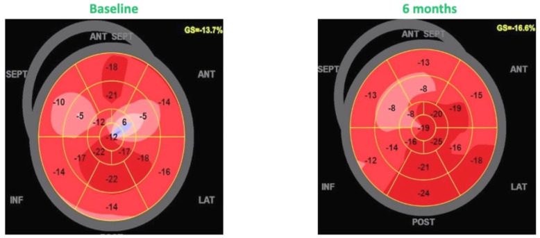
There was a significant left ventricular reverse remodeling with a reduction of end-diastolic (211.8 ± 45.8 vs. 88.3 ± 38.5 mL; p = 0.041) and end-systolic volumes (141.8 ± 51.5 vs. 119.6 ± 49.7 mL; p = 0.024), with a consequent improvement of left ventricular ejection fraction (30.8 ± 71 vs. 36.1 ± 6.9%; p = 0.032). In addition, there was a significant increase in the most specific and reproducible echocardiographic index of left ventricular function, the GLS (−10.3 ± −2.7 vs. −12.9 ± −4.2%; p = 0.018; Figure 1). In addition, diastolic function indices also improved, particularly the E/e’ ratio was significantly reduced at six-month follow-up (16.3 ± 7.5 vs. 10.8 ± 4.2; p = 0.041).
Figure 1.
Effects of CCM on global longitudinal strain.
3.2. Effects of CCM Therapy on Natriuretic Peptides, NYHA Class, and Quality of Life
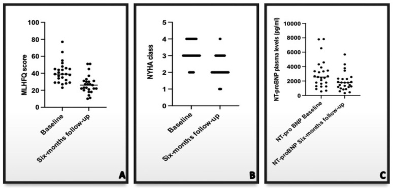
As shown in Figure 2 (panel A) at the six months follow-up, a significant reduction of plasma levels of N-terminal Brian Natriuretic Peptide (NT-proBNP) was observed in the enrolled patients (2975 ± 1988 vs. 1911 ± 1268 pg/mL; p = 0.029).
Figure 2.
Effects of CCM therapy on NT-proBNP plasma levels (panel (A)), NYHA class (panel (B)), and MLHFQ score (panel (C)). NT-proBNP: N terminal-pro brain natriuretic peptide; NYHA: New York Heart Association; MLHFQ: Minnesota Living with Heart Failure Questionnaire.
Simultaneously with the reduction of natriuretic peptides plasma levels, an improvement in the symptom reported by the patients occurred; in fact, at follow-up, a statistical reduction in both NYHA class (3.1 ± 0.62 vs. 2.3 ± 0.56; p = 0.0001; Figure 2B) and of the Minnesota Living with Heart Failure score occurred (40.08 ± 12.31 vs. 26.9 ± 10.8; p = 0.0001—Figure 2C).
3.3. Effects of CCM on MEE
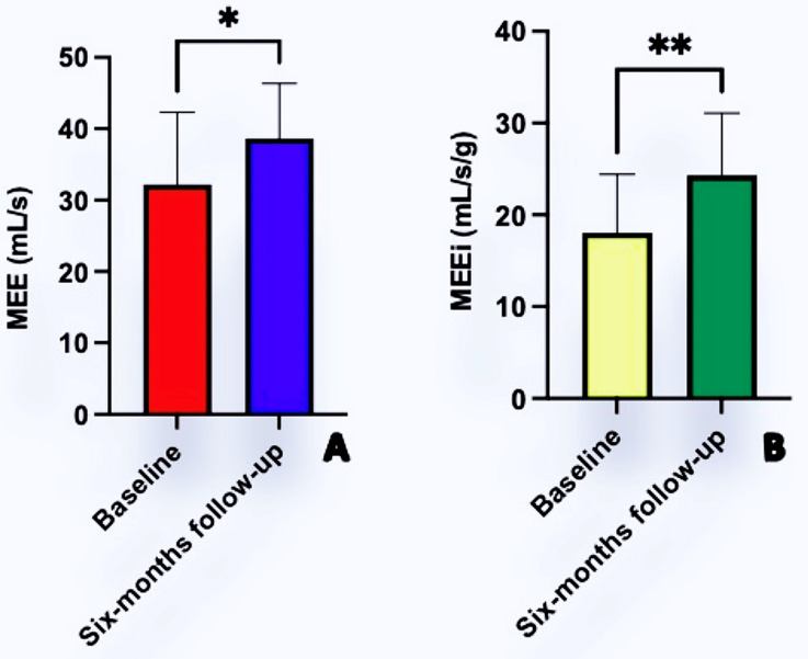
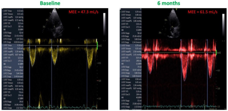
As showed in Figure 3, both MEE (32.2 ± 10.1 vs. 38.6 ± 7.6; mL/s p = 0.013) and MEEi (18.4 ± 6.3 vs. 24.3 ± 6.7 mL/s/g; p = 0.022) increased after six months of CCM therapy. The improvement of these indexes was due essentially due to the increase of SV without a concomitant increase in HR (Figure 4). From a pathophysiological point of view, this indicates an increase in cardiac contractility in the absence of a corresponding increase in myocardial oxygen consumption, thus leading to an improved mechano-energetic coupling of the heart.
Figure 3.
Improvements of Myocardial Mechano-Energetic Efficiency (MEE; Panel (A)) and Mechano-Energetic Efficiency index (MEEi; Panel (B)) after six months of CCM therapy. * = p < 0.05; ** = p < 0.001.
Figure 4.
Effects of CCM therapy on MME. Note the increase in stroke volume without an increase in heart rate.
4. Discussion
In this study, for the first time, we demonstrate that left ventricular GLS and MEE increased after 6 months of CCM therapy in patients with HFrEF. Longitudinal deformation of the left ventricle is due to the contraction of subendocardial fibers, which are the most susceptible to altered calcium handling [22], increased myocardial stiffness [23], and myocardial fibrosis [24], typical features of the failing heart.
Therefore, longitudinal left ventricular dysfunction and consequentially reduced GLS values develop early in patients with HFrEF [25]. In ex vivo intact hearts, CCM therapy improves calcium handling through several mechanisms, such as rapid normalization of phospholamban phosphorylation [26], upregulation of L-type calcium channels, and increased calcium uptake into the sarcoplasmic reticulum [27]. The latter mechanism results in a rise of extracellular calcium flux during the subsequent cardiac cycle and increased calcium release from the SR itself (the so-called “calcium-induced calcium release”) mechanism [28].
Animal models have demonstrated benefits of CCM therapy. In a canine HFrEF model, CCM therapy reduced left ventricular filling pressure due to the improvement of ventricular compliance and relaxation and improved diastolic Ca++ physiology [29]. In a rabbit HFrEF model, CCM therapy reduced cardiac expression of connective tissue growth factor and galectin-3 (a pro-fibrotic marker involved in myocardial structural remodeling) with a reduction of myocardial fibrosis [11]. These effects of CCM therapy observed in animal models may explain the improvement in diastolic function and GLS observed in this study, as well as a reduction of the E/e’ ratio and of the NT-proBNP plasma levels both expression of left ventricular filling pressure.
The improvement in diastolic function justifies the improvement in NYHA class and quality of life observed in patients enrolled in the study. In fact, diastolic function is the main determinant of functional capacity and quality of life in patients with HF [30,31,32], and therefore its improvement is associated with an improvement in these parameters [33].CCM has also been shown to increase stroke volume in a canine HFrEF model [34]; in our study, we documented for the first time that CCM therapy results in an increase in SV at 6 months, even in a population of patients with HFrEF in optimal medical treatment.
Notably, the improvement in MME observed in our study was caused by an increase in SV without a rise in HR and, consequently, of MVO2. This confirms the findings of a prior study in which CCM increased dP/dt (an index of myocardial contractility) without an increase of MVO2 in nine patients with HFrEF [35].
In conclusion, CCM induces an increase of SV and consequently of cardiac output without a concomitant increase in myocardial oxygen demand acting as a smart inotropic therapy.
5. Study Limitations
The relatively small number of patients as well as the single-center, observational design of the study with the lack of a control group may influence our results. In addition, although the echocardiographic evaluations were performed in stable patients, the assessments of SV and GLS may be influenced by loading conditions. Seven patients have a CRT-D implanted 12 months before the inclusion in the study; for these patients, late response to this therapy cannot be excluded.
6. Conclusions
At six months of follow-up, CCM therapy increased left ventricular performance, improving left ventricular ejection fraction, E/e’ ratio, GLS, as well as MEE and MEEi in patients with HFrEF on optimal medical therapy.
These echocardiographic improvements are associated with a clear clinical benefit documented by reduction of NT-pro BNP plasma levels NYHA class and MLHFQ score.
Additional larger studies are needed to provide a greater understanding of the long-term impact of CCM on left ventricular function, as well as the prognostic significance of these observations.
Author Contributions
Conceptualization, D.M. and M.M.K.; methodology, C.C., S.D.V., M.L.M., A.D., E.A. and G.N.; data curation, D.M., M.L.M. and V.E.; writing—original draft preparation, D.M.; writing—review and editing, M.M.K., S.D.V., A.D., E.A., G.N. and G.P. All authors have read and agreed to the published version of the manuscript.
Institutional Review Board Statement
The study was conducted in accordance with the Declaration of Helsinki and approved by the ethics committee of the AORN dei Colli-Monaldi Hospital (resolution No. 903/2020).
Informed Consent Statement
Informed consent was obtained from all subjects involved in the study.
Data Availability Statement
The data presented in this study are available on request from the corresponding author.
Conflicts of Interest
The authors declare no conflict of interest.
Funding Statement
This research received no external funding.
Footnotes
Publisher’s Note: MDPI stays neutral with regard to jurisdictional claims in published maps and institutional affiliations.
References
- 1.Ferrara F., Capone V., Cademartiri F., Vriz O., Cocchia R., Ranieri B., Franzese M., Castaldo R., D’Andrea A., Citro R., et al. Physiologic Range of Myocardial Mechano-Energetic Efficiency among Healthy Subjects: Impact of Gender and Age. J. Pers. Med. 2022;12:996. doi: 10.3390/jpm12060996. [DOI] [PMC free article] [PubMed] [Google Scholar]
- 2.Kim I.S., Izawa H., Sobue T., Ishihara H., Somura F., Nishizawa T., Nagata K., Iwase M., Yokota M. Prognostic value of mechanical efficiency in ambulatory patients with idiopathic dilated cardiomyopathy in sinus rhythm. J. Am. Coll. Cardiol. 2002;39:1264–1268. doi: 10.1016/S0735-1097(02)01775-8. [DOI] [PubMed] [Google Scholar]
- 3.Knaapen P., Germans T., Knuuti J., Paulus W.J., Dijkmans P.A., Allaart C.P., Lammertsma A.A., Visser F.C. Myocardial energetics and efficiency: Current status of the noninvasive approach. Circulation. 2007;115:918–927. doi: 10.1161/CIRCULATIONAHA.106.660639. [DOI] [PubMed] [Google Scholar]
- 4.Losi M.A., Izzo R., Mancusi C., Wang W., Roman M.J., Lee E.T., Howard B.V., Devereux R.B., de Simone G. Depressed Myocardial Energetic Efficiency Increases Risk of Incident Heart Failure: The Strong Heart Study. J. Clin. Med. 2019;8:1044. doi: 10.3390/jcm8071044. [DOI] [PMC free article] [PubMed] [Google Scholar]
- 5.Manzi M.V., Mancusi C., Lembo M., Esposito G., Rao M.A., de Simone G., Morisco C., Trimarco V., Izzo R., Trimarco B. Low mechano-energetic efficiency is associated with future left ventricular systolic dysfunction in hypertensives. ESC Heart Fail. 2022;9:2291–2300. doi: 10.1002/ehf2.13908. [DOI] [PMC free article] [PubMed] [Google Scholar]
- 6.Patel P.A., Nadarajah R., Ali N., Gierula J., Witte K.K. Cardiac contractility modulation for the treatment of heart failure with reduced ejection fraction. Heart Fail Rev. 2021;26:217–226. doi: 10.1007/s10741-020-10017-1. [DOI] [PubMed] [Google Scholar]
- 7.Gupta R.C., Mishra S., Rastogi S., Wang M., Rousso B., Mika Y., Remppis A., Sabbah H.N. Ca(2+)-binding proteins in dogs with heart failure: Effects of cardiac contractility modulation electrical signals. Clin. Transl. Sci. 2009;2:211–215. doi: 10.1111/j.1752-8062.2009.00097.x. [DOI] [PMC free article] [PubMed] [Google Scholar]
- 8.Rastogi S., Mishra S., Zacà V., Mika Y., Rousso B., Sabbah H.N. Effects of chronic therapy with cardiac contractility modulation electrical signals on cytoskeletal proteins and matrix metalloproteinases in dogs with heart failure. Cardiology. 2008;110:230–237. doi: 10.1159/000112405. [DOI] [PubMed] [Google Scholar]
- 9.Butter C., Rastogi S., Minden H.H., Meyhöfer J., Burkhoff D., Sabbah H.N. Cardiac contractility modulation electrical signals improve myocardial gene expression in patients with heart failure. J. Am. Coll Cardiol. 2008;51:1784–1789. doi: 10.1016/j.jacc.2008.01.036. [DOI] [PubMed] [Google Scholar]
- 10.Tschöpe C., Kherad B., Klein O., Lipp A., Blaschke F., Gutterman D., Burkhoff D., Hamdani N., Spillmann F., Van Linthout S. Cardiac contractility modulation: Mechanisms of action in heart failure with reduced ejection fraction and beyond. Eur. J. Heart Fail. 2019;21:14–22. doi: 10.1002/ejhf.1349. [DOI] [PMC free article] [PubMed] [Google Scholar]
- 11.Ning B., Zhang F., Song X., Hao Q., Li Y., Li R., Dang Y. Cardiac contractility modulation attenuates structural and electrical remodeling in a chronic heart failure rabbit model. J. Int. Med. Res. 2020;48:300060520962910. doi: 10.1177/0300060520962910. [DOI] [PMC free article] [PubMed] [Google Scholar]
- 12.Giallauria F., Cuomo G., Parlato A., Raval N.Y., Kuschyk J., Stewart Coats A.J. A comprehensive individual patient data meta-analysis of the effects of cardiac contractility modulation on functional capacity and heart failure-related quality of life. ESC Heart Fail. 2020;7:2922–2932. doi: 10.1002/ehf2.12902. [DOI] [PMC free article] [PubMed] [Google Scholar]
- 13.Anker S.D., Borggrefe M., Neuser H., Ohlow M.-A., Röger S., Goette A., Remppis B.A., Kuck K., Najarian K.B., Gutterman D.D., et al. Cardiac contractility modulation improves long-term survival and hospitalizations in heart failure with reduced ejection fraction. Eur. J. Heart Fail. 2019;21:1103–1111. doi: 10.1002/ejhf.1374. [DOI] [PubMed] [Google Scholar]
- 14.Yücel G., Fastner C., Hetjens S., Toepel M., Schmiel G., Yazdani B., Husain-Syed F., Liebe V., Rudic B., Akin I., et al. Impact of baseline left ventricular ejection fraction on long-term outcomes in cardiac contractility modulation therapy. Pacing Clin. Electrophysiol. 2022;45:639–648. doi: 10.1111/pace.14478. [DOI] [PubMed] [Google Scholar]
- 15.Contaldi C., De Vivo S., Martucci M.L., D’Onofrio A., Ammendola E., Nigro G., Errigo V., Pacileo G., Masarone D. Effects of Cardiac Contractility Modulation Therapy on Right Ventricular Function: An Echocardiographic Study. Appl. Sci. 2022;2:7917. doi: 10.3390/app12157917. [DOI] [Google Scholar]
- 16.Lang R.M., Badano L.P., Mor-Avi V., Afilalo J., Armstrong A., Ernande L., Flachskampf F.A., Foster E., Goldstein S.A., Kuznetsova T., et al. Recommendations for cardiac chamber quantification by echocardiography in adults: An update from the American Society of Echocardiography and the European Association of Cardiovascular Imaging. J. Am. Soc. Echocardiogr. 2015;28:1–39.e14. doi: 10.1016/j.echo.2014.10.003. [DOI] [PubMed] [Google Scholar]
- 17.Quiñones M.A., Otto C.M., Stoddard M., Waggoner A., Zoghbi W.A. Doppler Quantification Task Force of the Nomenclature and Standards Committee of the American Society of Echocardiography. Recommendations for quantification of Doppler echocardiography: A report from the Doppler Quantification Task Force of the Nomenclature and Standards Committee of the American Society of Echocardiography. J. Am. Soc Echocardiogr. 2002;15:167–184. doi: 10.1067/mje.2002.120202. [DOI] [PubMed] [Google Scholar]
- 18.Mitchell C., Rahko P.S., Blauwet L.A., Canaday B., Finstuen J.A., Foster M.C., Horton K., Ogunyankin K.O., Palma R.A., Velazquez E.J. Guidelines for Performing a Comprehensive Transthoracic Echocardiographic Examination in Adults: Recommendations from the American Society of Echocardiography. J. Am. Soc. Echocardiogr. 2019;32:1–64. doi: 10.1016/j.echo.2018.06.004. [DOI] [PubMed] [Google Scholar]
- 19.Sattin M., Burhani Z., Jaidka A., Millington S.J., Arntfield R.T. Stroke Volume Determination by Echocardiography. Chest. 2022;161:1598–1605. doi: 10.1016/j.chest.2022.01.022. [DOI] [PubMed] [Google Scholar]
- 20.Tran P., Maddock H., Banerjee P. Myocardial Fatigue: A Mechano-energetic Concept in Heart Failure. Curr. Cardiol. Rep. 2022;24:711–730. doi: 10.1007/s11886-022-01689-2. [DOI] [PubMed] [Google Scholar]
- 21.Juszczyk A., Jankowska K., Zawiślak B., Surdacki A., Chyrchel B. Depressed Cardiac Mechanical Energetic Efficiency: A Contributor to Cardiovascular Risk in Common Metabolic Diseases-From Mechanisms to Clinical Applications. J. Clin. Med. 2020;9:2681. doi: 10.3390/jcm9092681. [DOI] [PMC free article] [PubMed] [Google Scholar]
- 22.Ong G., Yan A.T., Connelly K.A. Clinical application of echocardiographic-derived myocardial strain imaging in subclinical disease: A primer for cardiologists. Curr. Opin. Cardiol. 2019;34:147–155. doi: 10.1097/HCO.0000000000000592. [DOI] [PubMed] [Google Scholar]
- 23.Haji K., Marwick T.H. Clinical Utility of Echocardiographic Strain and Strain Rate Measurements. Curr. Cardiol. Rep. 2021;23:18–25. doi: 10.1007/s11886-021-01444-z. [DOI] [PubMed] [Google Scholar]
- 24.Tops L.F., Delgado V., Marsan N.A., Bax J.J. Myocardial strain to detect subtle left ventricular systolic dysfunction. Eur. J. Heart Fail. 2017;19:307–313. doi: 10.1002/ejhf.694. [DOI] [PubMed] [Google Scholar]
- 25.Russo C., Jin Z., Elkind M.S., Rundek T., Homma S., Sacco R.L., Di Tullio M.R. Prevalence and prognostic value of subclinical left ventricular systolic dysfunction by global longitudinal strain in a community-based cohort. Eur. J. Heart Fail. 2014;16:1301–1309. doi: 10.1002/ejhf.154. [DOI] [PMC free article] [PubMed] [Google Scholar]
- 26.Brunckhorst C.B., Shemer I., Mika Y., Ben-Haim S.A., Burkhoff D. Cardiac contractility modulation by non-excitatory currents: Studies in isolated cardiac muscle. Eur. J. Heart Fail. 2006;8:7–15. doi: 10.1016/j.ejheart.2005.05.011. [DOI] [PubMed] [Google Scholar]
- 27.Burkhoff D., Shemer I., Felzen B., Shimizu J., Mika Y., Dickstein M., Prutchi D., Darvish N., Ben-Haim S.A. Electric currents applied during the refractory period can modulate cardiac contractility in vitro and in vivo. Heart Fail. Rev. 2001;6:27–34. doi: 10.1023/A:1009851107189. [DOI] [PubMed] [Google Scholar]
- 28.Endo M. Calcium-induced calcium release in skeletal muscle. Physiol. Rev. 2009;89:1153–1176. doi: 10.1152/physrev.00040.2008. [DOI] [PubMed] [Google Scholar]
- 29.Imai M., Rastogi S., Gupta R.C., Mishra S., Sharov V.G., Stanley W.C., Mika Y., Rousso B., Burkhoff D., Ben-Haim S., et al. Therapy with cardiac contractility modulation electrical signals improves left ventricular function and remodeling in dogs with chronic heart failure. J. Am. Coll. Cardiol. 2007;49:2120–2128. doi: 10.1016/j.jacc.2006.10.082. [DOI] [PubMed] [Google Scholar]
- 30.Smart N., Haluska B., Leano R., Case C., Mottram P.M., Marwick T.H. Determinants of functional capacity in patients with chronic heart failure: Role of filling pressure and systolic and diastolic function. Am. Heart J. 2005;149:152–158. doi: 10.1016/j.ahj.2004.06.017. [DOI] [PubMed] [Google Scholar]
- 31.Terzi S., Sayar N., Bilsel T., Enc Y., Yildirim A., Ciloğlu F., Yesilcimen K. Tissue Doppler imaging adds incremental value in predicting exercise capacity in patients with congestive heart failure. Heart Vessel. 2007;22:237–244. doi: 10.1007/s00380-006-0961-x. [DOI] [PubMed] [Google Scholar]
- 32.Daullxhiu I., Haliti E., Poniku A., Ahmeti A., Hyseni V., Olloni R., Vela Z., Elezi S., Bajraktari G., Daullxhiu T., et al. Predictors of exercise capacity in patients with chronic heart failure. J. Cardiovasc. Med. 2011;12:223–225. doi: 10.2459/JCM.0b013e328343e950. [DOI] [PubMed] [Google Scholar]
- 33.Bussoni M.F., Guirado G.N., Roscani M.G., Polegato B.F., Matsubara L.S., Bazan S.G., Matsubara B. Diastolic function is associated with quality of life and exercise capacity in stable heart failure patients with reduced ejection fraction. Braz. J. Med. Biol. Res. 2013;46:803–838. doi: 10.1590/1414-431X20132902. [DOI] [PMC free article] [PubMed] [Google Scholar]
- 34.Mohri S., He K.L., Dickstein M., Mika Y., Shimizu J., Shemer I., Yi G.H., Wang J., Ben-Haim S., Burkhoff D. Cardiac contractility modulation by electric currents applied during the refractory period. Am. J. Physiol. Heart Circ. Physiol. 2002;282:1642–1647. doi: 10.1152/ajpheart.00959.2001. [DOI] [PubMed] [Google Scholar]
- 35.Butter C., Wellnhofer E., Schlegl M., Winbeck G., Fleck E., Sabbah H.N. Enhanced inotropic state of the failing left ventricle by cardiac contractility modulation electrical signals is not associated with increased myocardial oxygen consumption. J. Card. Fail. 2007;13:137–142. doi: 10.1016/j.cardfail.2006.11.004. [DOI] [PubMed] [Google Scholar]
Associated Data
This section collects any data citations, data availability statements, or supplementary materials included in this article.
Data Availability Statement
The data presented in this study are available on request from the corresponding author.


