

Abstract
Background
Land‐based exercise therapy is recommended in clinical guidelines for hip or knee osteoarthritis. Adjunctive non‐pharmacological therapies are commonly used alongside exercise in hip or knee osteoarthritis management, but cumulative evidence for adjuncts to land‐based exercise therapy is lacking.
Objectives
To evaluate the benefits and harms of adjunctive therapies used in addition to land‐based exercise therapy compared with placebo adjunctive therapy added to land‐based exercise therapy, or land‐based exercise therapy only for people with hip or knee osteoarthritis.
Search methods
We searched CENTRAL, MEDLINE, PsycINFO, EMBASE, CINAHL, Physiotherapy Evidence Database (PEDro) and clinical trials registries up to 10 June 2021.
Selection criteria
We included randomised controlled trials (RCTs) or quasi‐RCTs of people with hip or knee osteoarthritis comparing adjunctive therapies alongside land‐based exercise therapy (experimental group) versus placebo adjunctive therapies alongside land‐based exercise therapy, or land‐based exercise therapy (control groups). Exercise had to be identical in both groups. Major outcomes were pain, physical function, participant‐reported global assessment, quality of life (QOL), radiographic joint structural changes, adverse events and withdrawals due to adverse events. We evaluated short‐term (6 months), medium‐term (6 to 12 months) and long‐term (12 months onwards) effects.
Data collection and analysis
Two review authors independently assessed study eligibility, extracted data, and assessed risk of bias and certainty of evidence for major outcomes using GRADE.
Main results
We included 62 trials (60 RCTs and 2 quasi‐RCTs) totalling 6508 participants. One trial included people with hip osteoarthritis, one hip or knee osteoarthritis and 60 included people with knee osteoarthritis only. Thirty‐six trials evaluated electrophysical agents, seven manual therapies, four acupuncture or dry needling, or taping, three psychological therapies, dietary interventions or whole body vibration, two spa or peloid therapy and one foot insoles. Twenty‐two trials included a placebo adjunctive therapy. We presented the effects stratified by different adjunctive therapies along with the overall results. We judged most trials to be at risk of bias, including 55% at risk of selection bias, 74% at risk of performance bias and 79% at risk of detection bias. Adverse events were reported in 11 (18%) trials.
Comparing adjunctive therapies plus land‐based exercise therapy against placebo therapies plus exercise up to six months (short‐term), we found low‐certainty evidence for reduced pain and function, which did not meet our prespecified threshold for a clinically important difference. Mean pain intensity was 5.4 in the placebo group on a 0 to 10 numerical pain rating scale (NPRS) (lower scores represent less pain), and 0.77 points lower (0.48 points better to 1.16 points better) in the adjunctive therapy and exercise therapy group; relative improvement 10% (6% to 15% better) (22 studies; 1428 participants). Mean physical function on the Western Ontario and McMaster (WOMAC) 0 to 68 physical function (lower scores represent better function) subscale was 32.5 points in the placebo group and reduced by 5.03 points (2.57 points better to 7.61 points better) in the adjunctive therapy and exercise therapy group; relative improvement 12% (6% better to 18% better) (20 studies; 1361 participants). Moderate‐certainty evidence indicates that adjunctive therapies did not improve QOL (SF‐36 0 to 100 scale, higher scores represent better QOL). Placebo group mean QOL was 81.8 points, and 0.75 points worse (4.80 points worse to 3.39 points better) in the placebo adjunctive therapy group; relative improvement 1% (7% worse to 5% better) (two trials; 82 participants). Low‐certainty evidence (two trials; 340 participants) indicates adjunctive therapies plus exercise may not increase adverse events compared to placebo therapies plus exercise (31% versus 13%; risk ratio (RR) 2.41, 95% confidence interval (CI) 0.27 to 21.90). Participant‐reported global assessment was not measured in any studies.
Compared with land‐based exercise therapy, low‐certainty evidence indicates that adjunctive electrophysical agents alongside exercise produced short‐term (0 to 6 months) pain reduction of 0.41 points (0.17 points better to 0.63 points better); mean pain in the exercise‐only group was 3.8 points and 0.41 points better in the adjunctive therapy plus exercise group (0 to 10 NPRS); relative improvement 7% (3% better to 11% better) (41 studies; 3322 participants). Mean physical function (0 to 68 WOMAC subscale) was 18.2 points in the exercise group and 2.83 points better (1.62 points better to 4.04 points better) in the adjunctive therapy plus exercise group; relative improvement 9% (5% better to 13% better) (41 studies; 3323 participants). These results are not clinically important. Mean QOL in the exercise group was 56.1 points and 1.04 points worse in the adjunctive therapies plus exercise therapy group (1.04 points worse to 3.12 points better); relative improvement 2% (2% worse to 5% better) (11 studies; 1483 participants), indicating no benefit (low‐certainty evidence). Moderate‐certainty evidence indicates that adjunctive therapies plus exercise probably result in a slight increase in participant‐reported global assessment (short‐term), with success reported by 45% in the exercise therapy group and 17% more individuals receiving adjunctive therapies and exercise (RR 1.37, 95% CI 1.15 to 1.62) (5 studies; 840 participants). One study (156 participants) showed little difference in radiographic joint structural changes (0.25 mm less, 95% CI ‐0.32 to ‐0.18 mm); 12% relative improvement (6% better to 18% better). Low‐certainty evidence (8 trials; 1542 participants) indicates that adjunctive therapies plus exercise may not increase adverse events compared with exercise only (8.6% versus 6.5%; RR 1.33, 95% CI 0.78 to 2.27).
Authors' conclusions
Moderate‐ to low‐certainty evidence showed no difference in pain, physical function or QOL between adjunctive therapies and placebo adjunctive therapies, or in pain, physical function, QOL or joint structural changes, compared to exercise only. Participant‐reported global assessment was not reported for placebo comparisons, but there is probably a slight clinical benefit for adjunctive therapies plus exercise compared with exercise, based on a small number of studies. This may be explained by additional constructs captured in global measures compared with specific measures. Although results indicate no increased adverse events for adjunctive therapies used with exercise, these were poorly reported. Most studies evaluated short‐term effects, with limited medium‐ or long‐term evaluation. Due to a preponderance of knee osteoarthritis trials, we urge caution in extrapolating the findings to populations with hip osteoarthritis.
Keywords: Humans; Exercise Therapy; Osteoarthritis, Hip; Osteoarthritis, Hip/therapy; Osteoarthritis, Knee; Osteoarthritis, Knee/therapy; Pain; Pain Measurement; Randomized Controlled Trials as Topic
Plain language summary
Additional therapies used with exercise therapy for hip or knee osteoarthritis
What was the aim of this review?
Osteoarthritis, a chronic degenerative condition that commonly affects hip and knee joints, causes pain and difficulty with everyday activities such as walking. Land‐based exercise therapy refers to exercise conducted on land (as opposed to exercise in the water) and is a first‐line treatment. This review aimed to find out if adding additional therapies to land‐based exercise therapy improved pain, function, quality of life, participant‐reported overall change or X‐ray changes in people with hip or knee osteoarthritis. Additional therapies include manual (hands‐on) therapy, psychological or dietary therapies, electrophysical agents (such as heat, cold, nerve stimulation, ultrasound or laser therapy) or acupuncture. We included studies comparing additional therapies plus land‐based exercise therapy to either 1) sham (or dummy) therapy plus land‐based exercise therapy or 2) land‐based exercise therapy only.
Search date
This systematic review is up‐to‐date to 10 June 2021.
What did we find?
We found 62 randomised controlled trials with 6508 participants, mostly women, from 24 countries. The average age was between 52 and 83 years, with symptoms present from 9 months to 12 years. Sixty studies enrolled people with knee osteoarthritis, one enrolled people with hip osteoarthritis and one enrolled people with knee or hip osteoarthritis. Twenty‐two trials compared additional therapies plus exercise therapy to sham additional therapies plus exercise therapy, and 41 compared to exercise therapy. Thirty‐eight trials studied electrophysical agents, seven studied manual therapies, four studied acupuncture/dry needling or use of tape, three studied psychological or dietary interventions, whole body vibration (this involves standing on a vibration platform), or spa/mud therapy, and one studied foot orthotics (shoe insoles).
Funding source
Thirty‐eight studies were funded, four received no funding and funding support was not reported in 20.
Main results
Eleven trials (18%) measured adverse (unwanted harmful) events, which included both non‐serious and serious adverse events. The most common were increased pain, stiffness or swelling. There was no difference in adverse events between additional therapies used with exercise and sham therapies with exercise.
Additional therapies plus exercise therapy compared with sham additional therapies plus exercise therapy (22 studies)
Compared with sham additional therapies used with land‐based exercise therapy, additional therapies such as electrophysical agents, acupuncture, dry needling or taping, used with exercise therapy, may not be more effective in improving pain, physical function or quality of life up to six months after treatment.
Pain (lower scores mean less pain)
Improved by 10% or 0.77 points on a zero to 10‐point scale.
Physical function scores (lower scores mean better physical function)
Improved by 12% or 5.03 points on a zero to 68‐point scale.
Quality of life (higher scores mean better quality of life)
Worse by 1% or 0.75 points worse on a zero to 100‐point scale.
Adverse events
Although not commonly reported in studies, there was no difference in adverse events between additional therapies used with exercise and sham therapies with exercise.
Additional therapies plus exercise therapy compared with exercise therapy (41 studies)
Compared with land‐based exercise therapy, additional therapies (manual therapies, electrotherapy, dietary interventions, psychological therapies, whole body vibration, acupuncture, dry needling, taping, spa/mud therapy or foot orthotics) plus exercise therapy, may not be more effective in improving pain, physical function, quality of life or joint changes measured with X‐rays up to six months after treatment.
Pain (lower scores mean less pain)
Improved by 7% or 0.41 points on a zero to 10‐point scale.
Physical function scores (lower scores mean better physical function)
Improved by 9% or 2.83 points on a zero to 68‐point scale.
Quality of life (higher scores mean better quality of life)
Worse by 2%, or 1.04 points worse on a zero to 100‐point scale.
Patient‐reported overall change
17% more people rated their treatment a success.
X‐ray changes
Improved by 12% (based on one study)
Adverse effects
Although not commonly reported in studies, risks appear no greater for additional therapies used with exercise compared to exercise only.
Fewer studies assessed outcomes six or 12 months after treatment. Additional therapies plus land‐based exercise therapy may be no better in reducing pain or improving physical function or quality of life than exercise therapy at 6 or 12 months. In patient‐reported overall assessment, 31% reported improvement at 6 months, and 42% reported improvement at 12 months.
Conclusions and certainty of evidence
Additional therapies plus exercise therapy do not appear to offer meaningful improvements in pain, function, quality of life or overall change for people with hip or knee osteoarthritis compared with sham additional therapies plus land‐based exercise therapy; or in pain, function, quality of life or changes on X‐rays when compared with exercise therapy only. Compared with exercise therapy there is probably a clinical benefit in patient‐reported overall change for additional therapies plus exercise therapy, based on a small number of studies. Our confidence in the evidence varies between moderate to little or no confidence for different outcomes. Although results indicate no increased adverse events from additional therapies used with exercise therapy, this was poorly reported. Most studies evaluated short‐term effects, with limited medium‐ or long‐term evaluation.
Summary of findings
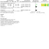
Summary of findings 1. Summary of findings table ‐ Adjunctive therapies in addition to land‐based exercise therapy compared to placebo adjunctive therapies in addition to land‐based exercise therapy for osteoarthritis of the hip or knee.
| Adjunctive therapies in addition to land‐based exercise therapy compared to placebo adjunctive therapies in addition to land‐based exercise therapy for osteoarthritis of the hip or knee | ||||||
| Patient or population: osteoarthritis of the hip or knee Setting: outpatient clinics, inpatient units, hospitals, specialist rehabilitation units or the community Intervention: adjunctive therapies in addition to land‐based exercise therapy Comparison: placebo adjunctive therapies in addition to land‐based exercise therapy | ||||||
| Outcomes | Anticipated absolute effects* (95% CI) | Relative effect (95% CI) | № of participants (studies) | Certainty of the evidence (GRADE) | Comments | |
| Risk with placebo adjunctive therapies in addition to land‐based exercise therapy | Risk with adjunctive therapies in addition to land‐based exercise therapy | |||||
| Pain assessed with: numerical pain rating scale (lower scores represent less pain) Scale from: 0 to 10 | The mean pain was 5.4 | MD 0.77 lower (1.16 lower to 0.48 lower) | ‐ | 1428 (22 RCTs) | ⊕⊕⊝⊝ Lowa,b | The evidence suggests that adjunctive therapies in addition to land‐based exercise result in little to no difference in participant‐reported pain. Absolute difference: 0.77 points better (0.48 points better to 1.16 points better). Relative difference: 10% better (6% better to 15% better). Clinically unimportant change. 1,c |
| Physical function assessed with: Western Ontario McMaster Osteoarthritis Index (WOMAC) (lower scores represent better physical function) Scale from: 0 to 68 | The mean physical function was 32.5 | MD 5.03 lower (7.61 lower to 2.57 lower) | ‐ | 1361 (20 RCTs) | ⊕⊕⊝⊝ Lowa,b | The evidence suggests that adjunctive therapies in addition to land‐based exercise therapy result in little to no difference in participant‐reported physical function. Absolute difference: 5.03 points better (2.57 points better to 7.61 points better). Relative difference: 12% better (6% better to 18% better). Clinically unimportant change.1,d |
| Quality of life assessed with: Short‐Form 36 (SF‐36) (higher scores represent better quality of life) Scale from: 0 to 100 | The mean quality of life was 81.8 | MD 0.75 higher (3.39 lower to 4.8 higher) | ‐ | 82 (2 RCTs) | ⊕⊕⊕⊝ Moderatee,f | The evidence suggests that adjunctive therapies used in addition to land‐based exercise therapy likely do not improve quality of life. Absolute difference: 0.75 points (4.80 points worse to 3.39 points better). Relative difference: 1% (7% worse to 5% better). 2,g |
| Participant‐reported global assessment | 0 per 1000 | 0 per 1000 (0 to 0) | Not estimable | (0 studies) | ‐ | Not measured in included studies |
| Radiographic joint structure changes | 0 per 1000 | 0 per 1000 (0 to 0) | Not estimable | (0 studies) | ‐ | Not measured in included studies |
| Adverse events | 127 per 1000 | 306 per 1000 (34 to 1000) | RR 2.41 (0.27 to 21.90) | 340 (2 RCTs) | ⊕⊕⊝⊝ Lowa,e,f | The evidence suggests that adjunctive therapies used in addition to land‐based exercise do not increase adverse events compared with placebo adjunctive therapies used in addition to land‐based exercise. Relative difference: 141% more (73% fewer to 2090% more). |
| Withdrawals due to adverse events | 0 per 1000 | 0 per 1000 (0 to 0) | Not estimable | (0 studies) | ‐ | No studies reported withdrawals due to adverse events. |
| *The risk in the intervention group (and its 95% confidence interval) is based on the assumed risk in the comparison group and the relative effect of the intervention (and its 95% CI). CI: confidence interval; MD: mean difference; RR: risk ratio | ||||||
| GRADE Working Group grades of evidence High certainty: we are very confident that the true effect lies close to that of the estimate of the effect. Moderate certainty: we are moderately confident in the effect estimate: the true effect is likely to be close to the estimate of the effect, but there is a possibility that it is substantially different. Low certainty: our confidence in the effect estimate is limited: the true effect may be substantially different from the estimate of the effect. Very low certainty: we have very little confidence in the effect estimate: the true effect is likely to be substantially different from the estimate of effect. | ||||||
| See interactive version of this table: https://gdt.gradepro.org/presentations/#/isof/isof_question_revman_web_429355504372610521. | ||||||
a Downgraded one level due to inconsistency (I2 > 50%). b Downgraded one level due to publication bias, based on inspection of funnel plot. c We calculated the relative change based on a 0 to 10 VAS using the baseline SD (1.85) from the trial by Atamaz 2012. d We calculated the relative change based on a 0 to 68 WOMAC physical function subscale using the baseline SD (11.7) from the trial by Atamaz 2012. e Downgraded one level due to wide confidence intervals of the pooled estimate. f Publication bias was not assessed due to the small number of studies (< 10 studies). g We calculated the relative change based on a 0 to 100 SF‐36 quality of life scale using the baseline SD (9.42) from the trial by Sardim 2020. 1 Atamaz FC, Durmaz B,Baydar M,Demircioglu OY,Iyiyapici A,Kuran B,Oncel S,Sendur OF. Comparison of the efficacy of transcutaneous electrical nerve stimulation, interferential currents, and shortwave diathermy in knee osteoarthritis: a double‐blind, randomized, controlled, multicenter study. Archives of Physical Medicine And Rehabilitation; 2012. 2 Sardim AC, Prado RP,Pinfildi CE. Effect of photobiomodulation associated withexercise on pain and functionality of patients with knee osteoarthritis: a pilot study. Fisioterapia and Pesquisa (Physical Therapy and Research); 2020.
Summary of findings 2. Summary of findings table ‐ Adjunctive therapies in addition to land‐based exercise therapy compared to land‐based exercise therapy for osteoarthritis of the hip or knee.
| Adjunctive therapies in addition to land‐based exercise therapy compared to land‐based exercise therapy for osteoarthritis of the hip or knee | ||||||
| Patient or population: osteoarthritis of the hip or knee Setting: outpatient clinics, inpatient units, hospitals, specialist rehabilitation units or the community Intervention: adjunctive therapies in addition to land‐based exercise therapy Comparison: land‐based exercise therapy | ||||||
| Outcomes | Anticipated absolute effects* (95% CI) | Relative effect (95% CI) | № of participants (studies) | Certainty of the evidence (GRADE) | Comments | |
| Risk with land‐based exercise therapy | Risk with adjunctive therapies in addition to land‐based exercise therapy | |||||
| Pain ‐ total assessed with: numerical pain rating scale (lower scores represent less pain) Scale from: 0 to 10 | The mean pain ‐ total was 3.8 | MD 0.41 lower (0.63 lower to 0.17 lower) | ‐ | 3322 (41 RCTs) | ⊕⊕⊝⊝ Lowa,b | The evidence suggests that adjunctive therapies used in addition to land‐based exercise therapy result in little to no difference in pain. Absolute difference: 0.41 points better (0.17 points better to 0.63 points better). Relative difference: 7% better (3% better to 11% better). The difference did not meet the predefined clinically relevant change.1,c,d |
| Physical function ‐ total assessed with: Western Ontario McMaster Osteoarthritis Index (WOMAC) (lower scores represent better physical function) Scale from: 0 to 68 | The mean physical function ‐ total was 18.2 | MD 2.83 lower (4.04 lower to 1.62 lower) | ‐ | 3323 (41 RCTs) | ⊕⊝⊝⊝ Very lowa,b,e | We are uncertain whether adjunctive therapies have an effect on physical function. Absolute difference: 2.83 points better (1.62 points better to 4.04 points better). Relative difference: 9% better (5% better to 13% better). The difference did not meet the predefined clinically relevant change.1,e,f |
| Quality of life ‐ total assessed with: Short‐Form 36 (SF‐36) (higher scores represent better quality of life) Scale from: 0 to 100 | The mean quality of life ‐ total was 56.1 | MD 1.04 lower (3.12 lower to 1.04 higher) | ‐ | 1483 (10 RCTs) | ⊕⊝⊝⊝ Very lowb,e,g | Adjunctive therapies may have little to no effect on quality of life but the evidence is very uncertain. Absolute difference: 1.04 poorer quality of life (3.12 points worse to 1.04 points better). Relative difference: 2% worse (5% worse to 2% better). 2,d |
| Participant‐reported global assessment‐ total assessed with: Likert scales | 449 per 1000 | 615 per 1000 (516 to 727) | RR 1.37 (1.15 to 1.62) | 840 (5 RCTs) | ⊕⊕⊕⊝ Moderateh,i | Adjunctive therapies likely result in a slight increase in participant‐reported global assessment. Absolute difference: 17% reported more success (7% to 28% more). Relative difference: 37% more reported success (15% more to 62% more). |
| Radiographic joint structure change (lower scores represent less joint structure change) | The mean radiographic joint structure change (lower scores represent less joint structure change) was 3.18 mm | MD 0.25 mm lower (0.32 lower to 0.18 lower) | ‐ | 156 (1 RCT) | ⊕⊕⊝⊝ Lowh,i,j | The evidence suggests that adjunctive therapies result in little to no difference in radiographic joint structure changes. Absolute difference: 0.25 mm lower (lower means less joint space narrowing) (0.18 mm lower to 0.32 mm lower). Relative difference: 12% (6% lower to 18% lower). |
| Adverse events | 65 per 1000 | 86 per 1000 (51 to 148) | RR 1.33 (0.78 to 2.27) | 1542 (8 RCTs) | ⊕⊕⊝⊝ Lowh,i,j | The evidence suggests that adjunctive therapies used in addition to land‐based exercise therapy do not increase adverse events compared to land‐based exercise therapy only. Absolute difference: 50% more events (33% fewer to 392% more). Relative difference: 33% more events (22% fewer to 127% more). |
| Withdrawals due to adverse events | 0 per 1000 | 0 per 1000 (0 to 0) | Not estimable | ( studies) | ‐ | No studies reported withdrawals due to adverse events. |
| *The risk in the intervention group (and its 95% confidence interval) is based on the assumed risk in the comparison group and the relative effect of the intervention (and its 95% CI). CI: confidence interval; MD: mean difference; RR: risk ratio | ||||||
| GRADE Working Group grades of evidence High certainty: we are very confident that the true effect lies close to that of the estimate of the effect. Moderate certainty: we are moderately confident in the effect estimate: the true effect is likely to be close to the estimate of the effect, but there is a possibility that it is substantially different. Low certainty: our confidence in the effect estimate is limited: the true effect may be substantially different from the estimate of the effect. Very low certainty: we have very little confidence in the effect estimate: the true effect is likely to be substantially different from the estimate of effect. | ||||||
| See interactive version of this table: https://gdt.gradepro.org/presentations/#/isof/isof_question_revman_web_429356297214037644. | ||||||
a Downgraded one level due to possible risk of selection, performance and reporting bias. b Downgraded one level due to statistical heterogeneity (> 50%). c We calculated the relative change based on a 0 to 10 numerical pain rating scale using the baseline SD (1.5) from the trial by Bennell 2017. d We calculated the relative change based on a 0 to 100 SF‐36 quality of life scale using the baseline SD (8.68) from the trial by Messier 2013. e Downgraded one level due to possible publication bias, as assessed by funnel plots. f We calculated the relative change based on a 0 to 68 WOMAC physical function subscale using the baseline SD (10.1) from the trial by Bennell 2017. g Downgraded one level due to possible risk of selection and performance bias. h Downgraded one level due to possible performance bias. i Publication bias not assessed due to the small number of studies (< 10 studies). j Downgraded one level due to imprecision associated with wide confidence intervals. 1 Bennell KL, Campbell PK Egerton T Metcalf B Kasza J Forbes A Bills C Gale J Harris A Kolt GS Bunker SJ Hunter DJ Brand CA Hinman RS. Telephone coaching to enhance a home‐based physical activity program for knee osteoarthritis: a randomised clinical trial. Arthritis Care & Research; 2017. 2 Messier SP, Mihalko SL,Legault C,Miller GD,Nicklas BJ,de Vita P,Beavers DP,Hunter DJ,Lyles MF,Eckstein F,Williamson JD,Carr JJ,Guermazi A,Loeser RF. Effects of intensive diet and exercise on knee joint loads, inflammation, and clinical outcomes among overweight and obese adults with knee osteoarthritis: the IDEA randomized clinical trial. JAMA; 2013.
Background
Description of the condition
Osteoarthritis is a chronic degenerative condition that involves the entire joint, including bone, synovium and capsule, but the most critical changes occur in the articular cartilage (L'Hermette 2006), which undergoes progressive degeneration. Osteoarthritis is characterised by pain, loss of joint range of movement, functional limitation and social participation (Fautrel 2005; Hinton 2002). The hip and knee are the most common large joints affected. Prevalence is associated with increasing age and is higher in women than men, particularly over the age of 50 years (Felson 2006). The World Health Organization (WHO) estimated that 10% of the world’s population aged 60 years or more have significant clinical problems associated with osteoarthritis (Woolf 2003). Although prevalence estimates vary depending on whether osteoarthritis is defined by radiographic criteria and the presence or absence of symptoms (Pereira 2011), higher prevalence rates of knee osteoarthritis are consistently found across studies compared with hip osteoarthritis (Grotle 2008; Pereira 2011; Tukker 2009). Overall prevalence of up to 27% for knee osteoarthritis has been reported compared with 12% for hip osteoarthritis (Pereira 2011).
Description of the intervention
A range of conservative adjunctive therapies are available for the management of hip or knee osteoarthritis. International clinical guidelines agree that exercise is a core conservative management approach (Bannaru 2019; Kolasinski 2020; NICE 2014). Exercise can incorporate aerobic exercise, specific muscle strengthening, balance and flexibility exercises. It can be prescribed and delivered by different healthcare professionals such as physiotherapists or doctors and can be delivered in a supervised or unsupervised format, either individually or in groups (Dziedzic 2008). A range of other conservative therapies may be used in combination with exercise therapy. These can include physical therapies, weight loss interventions and psychological therapies. Evidence on their role in the management of osteoarthritis is unclear. Previous Cochrane Reviews have evaluated the effectiveness of some of these therapies as stand‐alone treatments (Cameron 2013; Duivenvoorden 2015; Kroon 2014; Li 2013; Manheimer 2010; Manheimer 2018; Rutjes 2010; Verhagen 2007), but to date no Cochrane Review has appraised their effectiveness when used in combination with land‐based exercise therapy. Land‐based exercise refers to exercise conducted on land (as opposed to exercise conducted in the water). For the purpose of this review, an adjunctive therapy is defined as a non‐surgical and non‐pharmacological intervention used in combination with exercise therapy in the management of hip or knee osteoarthritis.
How the intervention might work
Exercise therapy is associated with different mechanisms of effect in osteoarthritis including improved strength, proprioception and aerobic fitness (Beckwee 2013), weight loss (Shaw 2006), and positive effects on co‐morbidities such as cardiovascular disease and diabetes (Davies 2010; Thomas 2006). Animal studies have shown a protective effect of load‐bearing exercise on cartilage degeneration (Galois 2003), whilst aerobic and strengthening exercise in humans at high risk of osteoarthritis has shown increased knee cartilage glycosaminoglycan content (Roos 2005). Psychosocial benefits include improved mood, well‐being and increased self‐efficacy (Beckwee 2013). Any number of these effects can result in reduced pain and improved physical function. Increased pain associated with exercise appears to be rare in osteoarthritis (Fransen 2014).
Adjunctive therapies may have a variety of effects. Pain can be modulated with electrophysical agents such as transcutaneous electrical nerve stimulation (TENS), cryotherapy, thermotherapy and manual therapies such as massage and joint mobilisation via a cascade of neurophysiological responses from the peripheral and central nervous system (Bialosky 2009; Kalra 2001). Manual therapies such as joint mobilisations are purported to also exert a mechanical effect by the physical loading and unloading of cartilage, which facilitates synovial fluid flow (Hoving 2005). Cold and laser therapy plays a significant role in reducing inflammation by vasoconstriction and reduction of inflammatory mediators (Aimbire 2006; Wojtecka‐Lukasik 2010). Experimental studies have shown that ultrasound, laser and pulsed electromagnetic energy may upregulate cellular activity to increase chondrocyte activity and fibroblast proliferation (Aimbire 2006; Brighton 2008), stimulate proteoglycan synthesis (Kopakkala‐Tani 2006), and increase blood flow (Barzelai 2006).
It is well established that chronic pain in conditions such as osteoarthritis is maintained and influenced by maladaptive emotional, cognitive and behavioural factors (Somers 2009a; Somers 2009b). People with osteoarthritis can also report symptoms associated with depression and anxiety (Sale 2008). Psychological therapies can incorporate behavioural strategies such as relaxation training, biofeedback and goal‐setting and cognitive strategies including stress management, guided imagery and cognitive coping skills. These aim to address negative beliefs and behaviours and enhance self‐efficacy.
Weight loss is recommended as a core management strategy for osteoarthritis in a number of clinical guidelines (Bannaru 2019; Kolasinski 2020; NICE 2014). However, Bannaru 2019 does not recommend dietary weight management for individuals with hip osteoarthritis due to a lack of direct evidence for its effectiveness on hip osteoarthritis symptoms. A dose‐response effect is noted in the American College of Rheumatology (ACR) guidelines, where a loss of ≥ 5% of body weight can improve clinical and mechanistic outcomes, with greater clinical benefits occurring with higher percentages of body weight loss (Kolasinski 2020). Weight loss strategies can comprise dietary interventions, surgery or exercise. As exercise is the core intervention of interest in this review, weight loss strategies considered to be adjunctive therapies may include, but are not limited to, dietary and behavioural strategies to address barriers to diet and physical activity.
Footwear, orthoses and walking aids may also be considered as adjunctive therapies and their proposed mechanism of efficacy is biomechanical through the reduction of joint load (Brouwer 2005; Reeves 2011). Complementary and alternative therapies are also available in the management of osteoarthritis. Complementary and alternative therapies can include, but are not exclusive to, herbal medicine, acupuncture, magnet therapy and homeopathy. Evidence from laboratory studies has demonstrated that acupuncture can have an analgesic effect and suppress inflammation (Han 2003; Hui 2005; Li 2008). Sham‐controlled studies have demonstrated small short‐ and long‐term beneficial effects on pain and function (Li 2008).
Why it is important to do this review
Osteoarthritis is the most common cause of disability in those over the age of 65 and is likely to increase in prevalence due to increased life expectancy (Croft 2005). The effectiveness of exercise in the management of hip or knee osteoarthritis has already been evaluated in previous Cochrane Reviews (Fransen 2014; Fransen 2015). High‐quality evidence from 32 trials with 3616 participants confirmed a small therapeutic effect for pain and physical function in people with knee osteoarthritis. There was variability across the studies in symptom duration, exercise interventions and study methodology (Fransen 2015). With regards to hip osteoarthritis, high‐quality evidence from nine trials and up to 549 participants indicated that exercise had a small beneficial effect on pain and physical function immediately after treatment. Reduced pain and improved function were maintained up to three to six months after the intervention. Only five of these studies recruited people exclusively with hip osteoarthritis (Fransen 2014).
To date, the majority of Cochrane Reviews have assessed the effectiveness of exercise (Fransen 2014; Fransen 2015), or other conservative therapies such as electrophysical agents (Brosseau 2003a; Brosseau 2003b; Li 2013; Rutjes 2009; Rutjes 2010), alternative therapies (Cameron 2013; Cameron 2014; Manheimer 2018), and braces or orthoses as stand‐alone interventions for hip or knee osteoarthritis (Duivenvoorden 2015). In clinical practice, the combination of exercise with these therapies, which can be considered to be adjunctive therapies, is more likely (Cowan 2010; French 2007). Clinical guidelines recommend that many of these therapies be used alongside, or as an adjunct, to core management strategies such as exercise and education (Bannaru 2019; NICE 2014). The National Institute for Health and Care Excellence (NICE) guidelines on the care of osteoarthritis also suggest that future research should evaluate combinations of therapies (NICE 2014).
Whilst various non‐Cochrane systematic reviews have included adjunctive therapies delivered in conjunction with exercise therapy for hip or knee osteoarthritis (Hall 2019; Huang 2015; Li 2019; Stausholm 2019; Wang 2015b; Wang 2017; Zafar 2015), none have specifically focussed on placebo adjunctive therapies used in conjunction with exercise therapy, or exercise therapy only as the comparator intervention. Including only placebo adjunctive therapies used with land‐based exercise therapy, or land‐based exercise therapy only, as the comparators in this review allows the evidence for using adjunctive therapies with land‐based exercise over and above land‐based exercise only to be ascertained, which is an important and relevant clinical question.
Objectives
The purpose of this review was to evaluate the benefits and harms of adjunctive therapies used in addition to land‐based exercise therapy compared with placebo adjunctive therapy added to land‐based exercise therapy, or land‐based exercise therapy only for people with hip or knee osteoarthritis.
Methods
Criteria for considering studies for this review
Types of studies
Randomised controlled trials (RCTs), cluster‐RCTs, controlled clinical trials or trials using quasi‐randomised methods of allocating participants, such as alternation or date of birth. We included those reported as full‐text, those published as abstract only and unpublished data. There was no language restriction.
Types of participants
We included adults aged 18 years or older with a clinical or radiographic diagnosis of hip or knee osteoarthritis as defined in the included trials. Where mixed populations were present, at least 75% of trial participants must have had hip or knee osteoarthritis, or both (Ghogomu 2014).
Types of interventions
The exercise intervention could include any land‐based therapeutic exercise regimen that aims to relieve the symptoms of knee or hip osteoarthritis, regardless of content, duration, frequency or intensity, delivered in combination with an adjunctive therapy. The exercise therapy in both groups must have been identical so that the only difference between the groups was the addition of the adjunctive therapy.
Land‐based exercise interventions were further defined as:
static weight bearing, including single leg standing;
dynamic weight bearing exercise low force, including walking and Tai Chi;
dynamic weight bearing exercise high force, including jogging, jumping, running, dancing and whole body vibration platforms;
non‐weight bearing exercise low force (e.g. low load, high repetition strength training);
non‐weight bearing exercise high force (e.g. progressive resisted strength training);
combination of more than one of the above exercise interventions.
Adjunctive therapies could include, but were not necessarily limited to:
education;
manual therapy, which may include mobilisation or manipulation;
electrophysical, which may include thermal modalities, therapeutic ultrasound, laser therapy, transcutaneous electrical nerve stimulation, pulsed electromagnetic field therapy, bipolar interferential current, electromyographic biofeedback, phonophoresis, iontophoresis, or short wave therapy;
other non‐pharmacologic interventions (e.g. bracing, acupuncture, psychological therapies, weight loss interventions and complementary therapies).
Pharmacological and nutraceutical interventions were excluded.
Possible comparisons were restricted to exercise plus an adjuvant treatment compared with exercise plus either placebo adjuvant treatment or no adjuvant treatment. For example, studies with the following comparisons could present:
exercise plus transcutaneous electrical nerve stimulation versus sham transcutaneous electrical nerve stimulation and exercise; or
exercise plus manual therapy versus exercise alone.
Types of outcome measures
We included outcome measures of pain, physical function and patient global assessment and physical performance as recommended by international consensus (Osteoarthritis Research Society International (OARSI) Standing Committee for Clinical Trials Response Criteria Initiative and the Outcome Measures in Rheumatology (OMERACT) committee) for use in clinical trials (Bellamy 1997; Dobson 2013). Our outcome measures of interest should be assessed immediately post‐treatment (or the most immediate assessment post‐treatment). If the duration of treatment was different for the exercise intervention and the adjunctive therapy, we used the time point for the adjunctive therapy. Where available, we also assessed outcomes in the medium term, which is defined as up to six months, and long‐term outcomes are those measured at a time point greater than six months after the intervention.
As recommended by Cochrane Musculoskeletal, we included the following major outcomes.
Major outcomes
Participant‐reported pain.
Participant‐reported physical function.
Participant‐reported global assessment.
Quality of life.
Radiographic joint structure changes according to the given hierarchy: minimum joint space width; median joint space width and semi‐quantitative measurement.
Adverse events.
Withdrawals due to adverse events, or overall dropouts (if data on withdrawals due to adverse events were not available).
Primary outcomes
Participant‐reported pain.
Participant physical function.
Secondary outcomes
Quality of life.
Patient‐reported global assessment.
Radiographic joint structure changes
Adverse events.
Withdrawals due to adverse events.
Search methods for identification of studies
Electronic and reference searches for this review were initially conducted in 2016, and repeated in 2019, with the most recent search conducted in June 2021.
Electronic searches
We searched the following electronic databases from inception to 10 June 2021 (Appendix 1):
Cochrane Central Register of Controlled Trials (CENTRAL) (via Wiley);
MEDLINE (via OVID);
PsycINFO (via EBSCO);
EMBASE (via Elsevier);
CINAHL Plus (EBSCO);
Physiotherapy Evidence Database (PEDro) (https://pedro.org.au/).
We applied no language or date restrictions.
We also searched trial registry websites such as ClinicalTrials.gov (https://clinicaltrials.gov/), Australian and New Zealand Clinical Trials registry (http://www.anzctr.org.au), metaRegister of Controlled Trials (mRCT) (http://www.controlled-trials.com/mrct/), European Union Clinical Trials Register (https://www.clinicaltrialsregister.eu) and the WHO International Clinical Trials Registry Platform (ICTRP) search portal (http://apps.who.int/trialsearch/) for any research in progress. We searched by 'osteoarthritis' and filtered by 'ongoing studies' for all trial registry websites.
Searching other resources
We screened the reference lists from relevant articles identified from the electronic searches, clinical guidelines and previous relevant Cochrane Reviews for potentially relevant studies.
Data collection and analysis
Selection of studies
Two review authors (HF and RG) independently screened the titles and abstracts of all studies identified by the initial searches using the inclusion criteria. We did not exclude studies on the basis of language. We excluded any clearly irrelevant studies at this stage. If there was any doubt, we retrieved the full‐text article for further assessment when we could not determine from title and abstract if it was eligible. The two authors independently reviewed all full‐text studies and disagreements were resolved by discussion.
Data extraction and management
Two review authors (HF and RG) independently extracted data onto a pre‐standardised and piloted data extraction form. We were not blinded to authors, institution or journal of publication due to feasibility and our familiarity with the literature. We obtained translations for studies not published in English.
We extracted the following data.
Methods: study design, study duration, study centre(s) and location(s), study setting, withdrawals and study date.
Participants: total number and number per group, mean age, range, gender, severity and duration of osteoarthritis, diagnostic criteria used, important osteoarthritis baseline data, inclusion and exclusion criteria.
Interventions: description of the exercise intervention including the duration and intensity (frequency and length of exercise programme), supervisor (for example, fitness instructor, physiotherapist), supervision (group or individual), setting (for example, gym or home‐based); description of the adjunctive therapy including dose, duration, intensity, provider or supervisor and setting.
Comparisons: description of the comparison intervention, including nature, dose, duration, intensity, as appropriate.
Outcomes: major outcomes specified and time points, including a description of the measurement tool used for continuous outcomes (scale of tool and direction of effect); number of participants per treatment group for continuous outcomes (mean pain, function, quality of life) and number of events and number of participants per treatment group for dichotomous outcomes (participant global assessment, withdrawals due to adverse events or total dropouts, adverse events).
Other notes: such as trial funding sources and declarations of interest from trial authors.
If data on more than one pain scale were provided for a trial, we extracted data from the pain scale that is the highest on the hierarchy of pain‐related outcomes as recommended by Cochrane Musculoskeletal (Juhl 2012).
Pain overall.
Pain on walking.
Western Ontario and McMaster Universities Osteoarthritis Index (WOMAC) pain subscale.
Pain on activities other than walking.
WOMAC global scale.
Lequesne osteoarthritis index global score.
Other algofunctional scale.
Patient's global assessment.
Physician's global assessment.
Other outcome.
If data on more than one physical function scale were reported in a trial, we extracted data according to the hierarchy presented below (Juhl 2012).
Global disability score.
Walking disability.
WOMAC disability subscore.
Composite disability scores other than WOMAC.
Disability other than walking.
WOMAC global scale.
Lequesne osteoarthritis index global score.
Other algofunctional scale.
If data on more than one quality of life scale were reported in a trial, we extracted data according to the following hierarchy according to a previously described hierarchy (Juni 2006; Reichenbach 2007).
The 36‐Item Short Form Health Survey (SF‐36) Mental Component Summary (MCS) scores.
EuroQol (EQ‐5D) instrument.
Sickness Impact Profile.
Nottingham Health Profile.
Other quality of life scales.
To estimate the effect of the interventions, we extracted raw data, including means and standard deviations for continuous measures and event counts for binary outcomes.
Assessment of risk of bias in included studies
We assessed risk of bias in included studies in accordance with the guidance from Chapter 8 of the Cochrane Handbook for Systematic Reviews of Interventions (Higgins 2011a). Two independent review authors (HF and RG) assessed risk of bias against the following key criteria:
random sequence generation;
allocation concealment;
blinding of participants and providers;
blinding of outcome assessment (detection bias) for self‐reported subjective outcomes (for example, pain, function, quality of life, global assessment);
blinding of outcome assessment for objective outcomes (adverse events, dropouts for any reason, or withdrawals due to adverse events);
incomplete outcome data;
selective reporting;
other sources of bias including inappropriate administration of an intervention (or co‐intervention), baseline imbalance between the groups and treatment contamination.
We graded each potential source of bias as high, low or unclear risk, and provided a quote from the study report together with a justification for our judgment in the risk of bias table. We summarised the risk of bias judgements across different studies for each of the domains listed. We considered blinding separately for different key outcomes where necessary (e.g. for unblinded outcome assessment, risk of bias for all‐cause mortality may be different than for a patient‐reported pain scale). As well, we considered the impact of missing data by key outcomes.
Where information on risk of bias relates to unpublished data or correspondence with a trialist, we noted this in the risk of bias table. When considering treatment effects, we took into account the risk of bias for the studies that contributed to that outcome. We presented the figures generated by the risk of bias tool to provide summary assessments of the risk of bias. Where the review authors were also authors of included studies, we asked impartial assessors to conduct risk of bias assessment.
Measures of treatment effect
We used Cochrane's statistical software Review Manager 5.3 to perform data analysis (Review Manager 2014). We expressed dichotomous outcomes (e.g. patient‐reported global assessment as risk ratios (RRs) with 95% confidence intervals (CIs)). For continuous outcome measures (e.g. pain, function, quality of life), we calculated standardised mean differences (SMDs) and 95% CIs as, across the studies included in the meta‐analyses, different instruments were used to measure the same outcome.
To enhance the interpretability of the measures of treatment effect for continuous outcomes, we back‐translated SMDs to a common scale by multiplying the SMD by a typical among‐person standard deviation (SD). We obtained this SD from a control group SD from the most representative trial with the highest weight in the meta‐analysis and the least susceptibility to bias (Schünemann 2011). For pain measures we back‐translated to an 11‐point (0 to 10) visual analogue scale. We assumed that VAS and NPRS were comparable scales. For physical function, we back‐translated to the Likert version of the WOMAC physical function subscale (0 to 68). For quality of life, we back translated SMDs to the SF‐36 0 to 100 scale. For conversion from SMD to mean difference (MD), we multiplied the SMD and 95% CIs by the SD at baseline from the control group, which varied depending on the analysis undertaken and is detailed in Table 3 for the main analysis and Table 4 for the subgroup analysis.
1. Representative trials used for conversion of SMDs to MDs.
| Intervention | Control | Follow‐up | Outcome | Baseline standard deviation | Study |
| Adjunctive therapy and exercise therapy | Placebo adjunctive therapy and exercise | Short‐term | Pain | 1.85 | Atamaz 2012 |
| Adjunctive therapy and exercise therapy | Placebo adjunctive therapy and exercise | Short‐term | Physical function | 11.7 | Atamaz 2012 |
| Adjunctive therapy and exercise therapy | Placebo adjunctive therapy and exercise therapy | Short‐term | Quality of life | 9.42 | Sardim 2020 |
| Adjunctive therapy and exercise therapy | Placebo adjunctive therapy and exercise therapy | Medium‐term | Pain | 1.85 | Atamaz 2012 |
| Adjunctive therapy and exercise therapy | Placebo adjunctive therapy and exercise therapy | Medium‐term | Physical function | 11.7 | Atamaz 2012 |
| Adjunctive therapy and exercise therapy | Placebo adjunctive therapy and exercise therapy | Medium‐term | Quality of life | Not applicable as one trial was included, which reported MD | |
| Adjunctive therapy and exercise therapy | Placebo adjunctive therapy and exercise therapy | Long‐term | Pain | 2.2 | Foster 2007 |
| Adjunctive therapy and exercise therapy | Placebo adjunctive therapy and exercise therapy | Long‐term | Physical function | 12.8 | Foster 2007 |
| Adjunctive therapy and exercise therapy | Placebo adjunctive therapy and exercise therapy | Long‐term | Quality of life | Not applicable as one trial was included, which reported MD | |
| Adjunctive therapy and exercise therapy | Exercise therapy | Short‐term | Pain | 1.5 | Bennell 2017 |
| Adjunctive therapy and exercise therapy | Exercise therapy | Short‐term | Physical function | 10.1 | Bennell 2017 |
| Adjunctive therapy and exercise therapy | Exercise therapy | Short‐term | Quality of life | 8.68 | Messier 2013 |
| Adjunctive therapy and exercise therapy | Exercise therapy | Medium‐term | Pain | 2.2 | Foster 2007 |
| Adjunctive therapy and exercise therapy | Exercise therapy | Medium‐term | Physical function | 10.1 | Bennell 2017 |
| Adjunctive therapy and exercise therapy | Exercise therapy | Medium‐term | Quality of life | 8.6 | Brosseau 2012 |
| Adjunctive therapy and exercise therapy | Exercise therapy | Long‐term | Pain | 2.2 | Foster 2007 |
| Adjunctive therapy and exercise therapy | Exercise therapy | Long‐term | Physical function | 10.1 | Bennell 2017 |
2. Representative trials used for conversion of SMDs to MD in subgroup analyses.
| Intervention | Control | Follow‐up | Group/subgroup | Outcome | Baseline standard deviation | Study |
| Adjunctive therapy and exercise therapy | Placebo adjunctive therapy and exercise therapy | Short‐term | Electrophysical agents | Pain | 1.85 | Atamaz 2012 |
| Adjunctive therapy and exercise therapy | Placebo adjunctive therapy and exercise therapy | Short‐term | Acupuncture/dry needling | Pain | 2.1 | Foster 2007 |
| Adjunctive therapy and exercise therapy | Placebo adjunctive therapy and exercise therapy | Short‐term | Taping | Pain | 1.4 | Leon‐Ballesteros 2020 |
| Adjunctive therapy and exercise therapy | Placebo adjunctive therapy and exercise therapy | Short‐term | Electrophysical agents | Physical function | 11.7 | Atamaz 2012 |
| Adjunctive therapy and exercise therapy | Placebo adjunctive therapy and exercise therapy | Short‐term | Acupuncture/dry needling | Physical function | 12.8 | Foster 2007 |
| Adjunctive therapy and exercise therapy | Placebo adjunctive therapy and exercise therapy | Short‐term | Taping | Physical function | 10.2 | Leon‐Ballesteros 2020 |
| Adjunctive therapy and exercise therapy | Placebo adjunctive therapy and exercise therapy | Short‐term | Electrophysical agents | QOL | 12.8 | Sardim 2020 |
| Adjunctive therapy and exercise therapy | Placebo adjunctive therapy and exercise therapy | Short‐term | Dry needling | QOL | 1.59 (Euroqol‐5D) | Sanchez‐Romero 2020 |
| Adjunctive therapy and exercise therapy | Placebo adjunctive therapy and exercise therapy | Medium‐term | Electrophysical agents | Pain | 1.85 | Atamaz 2012 |
| Adjunctive therapy and exercise therapy | Placebo adjunctive therapy and exercise therapy | Medium‐term | Acupuncture/dry needling | Pain | 2.2 | Foster 2007 |
| Adjunctive therapy and exercise therapy | Placebo adjunctive therapy and exercise therapy | Medium‐term | Electrophysical agents | Physical function | 11.7 | Atamaz 2012 |
| Adjunctive therapy and exercise therapy | Placebo adjunctive therapy and exercise therapy | Medium‐term | Acupuncture/dry needling | Physical function | 12.8 | Foster 2007 |
| Adjunctive therapy and exercise therapy | Exercise therapy | Short‐term | Manual therapies | Pain | 2.4 | Fitzgerald 2016 |
| Adjunctive therapy and exercise therapy | Exercise therapy | Short‐term | Electrophysical agents | Pain | 2.6 | Imoto 2013 |
| Adjunctive therapy and exercise therapy | Exercise therapy | Short‐term | Dietary interventions | Pain | 2.9 | Messier 2013 |
| Adjunctive therapy and exercise therapy | Exercise therapy | Short‐term | Whole body vibration | Pain | 1.5 | Wang 2016 |
| Adjunctive therapy and exercise therapy | Exercise therapy | Short‐term | Psychological Interventions | Pain | 1.5 | Bennell 2017 |
| Adjunctive therapy and exercise therapy | Exercise therapy | Short ‐term | Balneotherapy/peloid therapy | Pain | 1.85 | Forestier 2010 |
| Adjunctive therapy and exercise therapy | Exercise therapy | Short‐term | Taping | Pain | 1.4 | Castrogiovanni 2016 |
| Adjunctive therapy and exercise therapy | Exercise therapy | Short‐term | Other | Pain | 2.2 | Foster 2007 |
| Adjunctive therapy and exercise therapy | Exercise therapy | Short‐term | Manual therapies | Physical function | 17.16 | French 2013 |
| Adjunctive therapy and exercise therapy | Exercise therapy | Short‐term | Electrophysical agents | Physical function | 2.56 | de Paula Gomes 2018 |
| Adjunctive therapy and exercise therapy | Exercise therapy | Short‐term | Psychological interventions | Physical function | 10.1 | Bennell 2017 |
| Adjunctive therapy and exercise therapy | Exercise therapy | Short‐term | Dietary interventions | Physical function | 10.3 | Messier 2013 |
| Adjunctive therapy and exercise therapy | Exercise therapy | Short‐term | Whole body vibration | Physical function | 4.0 | Wang 2016 |
| Adjunctive therapy and exercise therapy | Exercise therapy | Short‐term | Taping | Physical function | 3.32 | Castrogiovanni 2016 |
| Adjunctive therapy and exercise therapy | Exercise therapy | Short‐term | Balneotherapy/peloid therapy | Physical function | 17.75 | Forestier 2010 |
| Adjunctive therapy and exercise therapy | Exercise therapy | Short‐term | Other | Physical function | 12.9 | Foster 2007 |
| Adjunctive therapy and exercise therapy | Exercise therapy | Short‐term | Psychological interventions | QOL | 8.68 | Brosseau 2012 |
| Adjunctive therapy and exercise therapy | Exercise therapy | Short‐term | Balneotherapy | QOL | 10 | Forestier 2010 |
| Adjunctive therapy and exercise therapy | Exercise therapy | Short‐term | Other | QOL | 8.68 | Messier 2013 |
| Adjunctive therapy and exercise therapy | Exercise therapy | Medium‐term | Electrophysical agents | Pain | 1.85 | Atamaz 2012 |
| Adjunctive therapy and exercise therapy | Exercise therapy | Medium‐term | Psychological interventions | Pain | 1.5 | Bennell 2017 |
| Adjunctive therapy and exercise therapy | Exercise therapy | Medium‐term | Other | Pain | 2.2 | Foster 2007 |
| Adjunctive therapy and exercise therapy | Exercise therapy | Medium‐term | Psychological interventions | Physical function | 10.1 | Bennell 2017 |
| Adjunctive therapy and exercise therapy | Exercise therapy | Medium‐term | Other | Physical function | 12.9 | Foster 2007 |
| Adjunctive therapy and exercise therapy | Exercise therapy | Medium‐term | Psychological interventions | Physical function | 8.6 | Brosseau 2012 |
| Adjunctive therapy and exercise therapy | Exercise therapy | Medium‐term | Balneotherapy | Physical function | 10 | Forestier 2010 |
| Adjunctive therapy and exercise therapy | Exercise therapy | Long‐term | Manual therapies | Pain | 2.4 | Fitzgerald 2016 |
| Adjunctive therapy and exercise therapy | Exercise therapy | Long‐term | Psychological interventions | Pain | 1.5 | Bennell 2017 |
| Adjunctive therapy and exercise therapy | Exercise therapy | Long‐term | Acupuncture | Pain | 2.2 | Foster 2007 |
| Adjunctive therapy and exercise therapy | Exercise therapy | Long‐term | Manual therapies | Physical function | 35.1 | Fitzgerald 2016 |
| Adjunctive therapy and exercise therapy | Exercise therapy | Long‐term | Psychological interventions | Physical function | 10.1 | Bennell 2017 |
| Adjunctive therapy and exercise therapy | Exercise therapy | Long‐term | Acupuncture | Physical function | 12.9 | Foster 2007 |
In the Effects of interventions section and the 'Comments' column of the summary of findings tables, we provided the absolute percent difference, the relative percent change from baseline, and the number needed to treat (NNT) (the NNT was provided only when the outcome showed a statistically significant difference). In interpreting results, we assumed a minimal clinically important difference (MCID) of 2 points (Tubach 2005) or percentage (relative) difference of 15% (Salaffi 2004) on a 0 to 10 NPRS, an absolute difference of 3 points on the 0 to 20 WOMAC Likert pain subscale, 6 points on the 0 to 68 WOMAC Likert physical function subscale (Bellamy 1992) and percentage change of 15%. For quality of life outcomes, we assumed a MCID of 6 points on the SF‐36 mental health scale (Angst 2018) or relative difference of 12% (Angst 2001).
For dichotomous outcomes, such as serious adverse events, we calculated the NNT from the control group event rate and the relative risk using the Visual Rx NNT calculator (Visual Rx 2008). We calculated the NNT for continuous measures using the Wells calculator from Cochrane Musculoskeletal (http://musculoskeletal.cochrane.org/).
For dichotomous outcomes, we calculated the absolute risk differences using the risk difference statistic in the Cochrane's statistical software, Review Manager 2014, and expressed the result as a percentage. For continuous outcomes, we calculated the absolute benefit as the improvement in the intervention group minus the improvement in the control group, in the original units. We calculated the relative percent change for dichotomous data as 'RR ‐ 1' and expressed it as a percentage. For continuous outcomes, we calculated the relative difference in the change from baseline as the absolute benefit divided by the baseline mean of the control group.
Unit of analysis issues
The most common unit of analysis was at the level of the individual participant, with the exception of two RCTs (Chen 2014; Ones 2006), which conducted analyses based on the level of the joint affected. We used methods outlined in Section 6.2.7 and Section 23.1.14 in the Cochrane Handbook for Systematic Reviews, 2nd edition for these analyses (Higgins 2019). If studies did not employ the correct analyses, we employed methods as outlined in Chapter 16 of the Cochrane Handbook for Systematic Reviews of Interventions to estimate effect size (Higgins 2011b). If multiple time points fell within the same category of short‐, medium‐ or long‐term, we used the data from the assessment point closest to the cut‐off of that category.
Studies with multiple treatment groups
Where studies included multiple treatment groups, we included all relevant groups. In the case where the control group (land‐based exercise only or land‐based exercise plus placebo adjunctive therapy) was compared against more than one active intervention (Adhya 2015; Chen 2014; Cakir 2014,Gur 2003; Kapci Yildiz 2015; Youssef 2016), we split the control group and modified the sample size according to the number of active intervention groups it was compared against, using methods outlined in section 23.3 of the Cochrane Handbook for Systematic Reviews for Interventions, 2nd edition (Higgins 2019). Other studies compared different active interventions with comparable sham interventions (Atamaz 2012). In two manual therapy RCTs, the adjunctive therapy plus exercise therapy was compared against exercise therapy in two different contact time formats of consecutive sessions and periodic 'booster' sessions (Abbott 2015; Fitzgerald 2016). In these trials, all comparisons were included in the same meta‐analysis. Four RCTs compared the adjunctive therapy against both a placebo therapy plus exercise and exercise only (de Paula Gomes 2018; Foster 2007; Pietrosimone 2011; Pietrosimone 2020). In all of these trials, comparisons were made in two separate meta‐analyses: adjunctive therapy plus exercise versus exercise only; and adjunctive therapy plus exercise versus placebo adjunctive therapy plus exercise.
Dealing with missing data
For continuous outcomes with no standard deviation (SD) reported, we calculated SDs from standard errors (SEs), 95% CIs, t‐tests or P values. In the event of missing data, we contacted the original authors (twice, separated by a period of three to four weeks). We contacted a total of seven authors to provide SDs where results were displayed graphically and data could not be extracted from the original study (Adedoyin 2002; Adedoyin 2005; Cheawthamai 2014; Youssef 2016; Messier 2004; Vassao 2020). Three authors provided additional data (Adedoyin 2002; Vassao 2020; Youssef 2016). One author could not be contacted (Quirk 1985). When no reply was received from authors, we used graph digitisation software (https://automeris.io/WebPlotDigitizer/) to extrapolate means and standard deviations by digitalising data points on the graphs in the studies by Adedoyin 2005 and Cheawthamai 2014.
We contacted five authors where change scores were reported instead of end‐of‐treatment scores (Adhya 2015; Abbott 2013; Abbott 2015; Forestier 2010; Rattanachaiyanont 2008) and all authors replied and provided end‐of‐treatment scores, with the exception of Adhya 2015. The results from that study were presented in a separate analysis of change scores (Analysis 2.6; Analysis 2.7). Three studies presented results as medians and interquartile ranges (Carlos 2012; Godoy 2014; Simao 2012). We assumed that the median was equivalent to the mean and the interquartile range width corresponded to 1.35 times the SD.
2.6. Analysis.
Comparison 2: Exercise and adjunctive therapy versus exercise (short‐term), Outcome 6: Change scores pain
2.7. Analysis.

Comparison 2: Exercise and adjunctive therapy versus exercise (short‐term), Outcome 7: Change scores physical function
Assessment of heterogeneity
We assessed clinical heterogeneity in terms of participants, interventions, comparisons and outcomes. We combined studies that examined similar categories of interventions (for example, manual therapies, electrophysical agents or psychological therapies) or similar outcomes. We assessed statistical heterogeneity by visual inspection of the forest plot along with the Chi2 statistic whereby a P value < 0.1 was considered statistically significant in accordance with recommendations in Chapter 9 of the Cochrane Handbook for Systematic Reviews of Interventions (Deeks 2011). We used the I2 statistic to determine the proportion of heterogeneity using the following as a guide for interpretation:
0% to 40%: might not be important;
30% to 60%: may represent moderate heterogeneity;
50% to 90%: may represent substantial heterogeneity;
75% to 100%: considerable heterogeneity.
Where there was considerable heterogeneity (> 75%), we planned to further explore the data using subgroup analyses (Deeks 2011). We grouped by common adjunctive therapies to explore potential differences across the type of adjunctive therapy
Assessment of reporting biases
To assess small study effects, we planned to generate funnel plots for meta‐analyses including at least 10 trials of varying size. If asymmetry was detected in the funnel plot, we planned to review the characteristics of the studies to determine if asymmetry was likely due to publication bias or methodological or clinical heterogeneity of the trials (Sterne 2011). In the presence of small‐sample bias, the random‐effects estimate of the intervention is more beneficial than the fixed‐effect estimate (Sterne 2011). We attempted to minimise publication bias by checking clinical trial registers for unpublished studies and including these in the review. We avoided language bias by not excluding any article based on language. We assessed selective outcome reporting by attempting to obtain the trial protocol where possible. For studies published after 1 July 2005, we screened the WHO ICTRP search portal for the trial protocols (http://apps.who.int/trialsearch/) and compared the outcomes in the trial protocol with the outcomes reported in the corresponding trial publications. We detected duplicate studies and where more than one article reported on the same study, we extracted data only once (Elboim‐Gabyzon 2013; Cheing 2002; Wang 2016).
Data synthesis
Included studies were grouped based on comparison of adjunctive therapy plus exercise therapy with placebo adjunctive therapy and exercise therapy or exercise therapy only. Due to the diversity of adjunctive therapies included in the review, we pooled results of studies that we considered to be clinically similar (such as manual therapies, electrophysical agents, dietary interventions, psychological interventions and acupuncture) and used Review Manager 2014 for all analyses and construction of forest plots. Where multiple single studies evaluated different adjunctive therapies that could not be pooled into one of these types of adjunctive therapies, they were included into an 'other intervention' analysis. We used a random‐effects model due to expected heterogeneity across studies and performed sensitivity analyses with the fixed‐effect model.
We presented overall pooled results and subgroup analyses for different types of adjunctive therapies in the forest plots. Pooled results are presented in the text within these common comparators, for example, adjunctive therapy plus exercise therapy versus placebo adjunctive therapy and exercise therapy or adjunctive therapy plus exercise therapy versus exercise therapy. We tested differences between subgroups across the different comparators.
Our primary analysis for major outcomes (pain, function, quality of life, treatment success, radiographic structural changes, adverse events and withdrawals due to adverse events) included all studies.
Subgroup analysis and investigation of heterogeneity
In addition to the primary analysis of overall effects, we also analysed subgroups, stratified by type of adjunctive therapy: manual therapies, electrophysical agents, psychological therapies, dietary interventions, acupuncture/dry needling, taping, balneotherapy/peloid therapy and whole body vibration. Where a study did not fall into these categories or only one study was eligible for meta‐analysis within these categories, it was included in an 'Other' comparison. We presented subgroup results as separate meta‐analyses for the different outcomes. Due to the diversity of adjunctive therapies evaluated in the included studies, we presented planned subgroup analyses and reported these in addition to the overall effects. We investigated whether the results of subgroups were significantly different by performing the test for subgroup differences, available in Review Manager.
Sensitivity analysis
We explored the impact of including studies with high or unclear risk of selection bias (random sequence generation and allocation concealment), performance bias (knowledge of the allocated interventions by participants and personnel) or detection bias (blinding of assessor reported measures) for outcomes of pain and function.
Summary of findings and assessment of the certainty of the evidence
We used the GRADE approach to assess the certainty of evidence related to each of the major outcomes at the end of the intervention (Schünemann 2019). We created a summary of findings table using GRADEpro software (http://www.guidelinedevelopment.org/) for the following outcomes:
participant‐reported pain;
participant‐reported physical function;
quality of life;
participant global assessment of treatment success;
radiographic joint structure changes according to the given hierarchy (minimum joint space width; median joint space width and semi‐quantitative measurement);
adverse events;
withdrawals due to adverse events, or overall dropouts (if withdrawals due to adverse events are not available).
Two authors (HF and RG) independently assessed the certainty of the evidence based on the five Grading of Recommendations, Assessment, Development and Evaluation (GRADE) considerations of methodological quality, consistency of effect, precision of effect estimates, directness of the evidence and publication bias, as recommended in Chapter 12 of the Cochrane Handbook for Systematic Reviews of Interventions, based on four certainty levels: high, moderate, low or very low (Schünemann 2011).
Our summary of findings table contains the following: the seven outcomes specified above, numbers of participants and studies addressing these outcomes, absolute and relative magnitude of effect, a measure of the typical burden of these outcomes (e.g. illustrative mean, on control intervention) and a grade of the overall quality of the body of evidence for each outcome. For assessments of the overall certainty of the evidence for each outcome that included pooled data, we downgraded the evidence from 'high certainty' for serious or very serious study limitations across the domains of risk of bias, indirectness of evidence, inconsistency, imprecision of effect estimates or potential publication bias. In the 'Comments' column of the summary of findings table, we provided the absolute percent difference, the relative percent change from baseline and numbers needed to treat (NNT). We planned to provide NNT only when the outcome showed a clinically important difference between the groups.
We prepared summary of findings tables for the following comparisons in the short‐term (post‐treatment to 6 months): 1) adjunctive therapies plus exercise therapy compared with placebo adjunctive therapy plus exercise only and 2) adjunctive therapies plus exercise therapy compared with exercise therapy only. Summary of findings tables reported participant‐reported pain, participant‐reported physical function, quality of life, participant global assessment of treatment success, radiographic joint structure changes, withdrawals due to adverse events and adverse events, where meta‐analyses were possible. Medium‐term (6 to 12 months) and long‐term (12 months or more) effects are reported in the text.
Results
Description of studies
Results of the search
The search conducted up to 10 June 2021 resulted in 23,029 records across the six databases, with an additional eight records identified from other sources. Following removal of duplicates and screening of abstracts, 181 articles were retained for full‐text screening, from which 67 studies were included in the review (62 studies and 5 ongoing trials). The five ongoing trials were identified from clinical trials registries (Bennell 2020; Bennell 2018; Bjordal 2018; Dantos 2018; Ylinen 2016) (see Characteristics of ongoing studies table). A flow diagram of the study selection process is presented in Figure 1.
1.

Study flow diagram
Included studies
Of the 69 RCTs identified as being suitable for inclusion in the review, three studies were each reported in two papers. Alfredo reported short‐term results (Alfredo 2012) and long‐term results (Alfredo 2018) of the same RCT. The same study methods and results were reported by Elboim‐Gabyzon 2013 and Laufer 2014. Similarly, the same study was reported in two papers (Cheing 2002; Cheing 2004). One study reported different outcomes in four papers; Messier 2013 presented clinical outcomes and is the study included and reported in this review. Messier 2020 reported gait mechanics outcomes, Beavers 2014 reported bone mineral density outcomes and Loeser 2017 reported biomarker outcomes. This resulted in a final 62 studies included in the review. Therefore, the results of the studies by Elboim‐Gabyzon 2013 and Cheing 2002 are reported and the study by Alfredo 2012 is reported in the short‐term results and Alfredo 2018 in the long‐term results. All studies were published in the English language except for two (Carlos 2012 (Portuguese); Jia 2005 (Chinese)), both of which were translated into English. A full description of the included studies is provided in the Characteristics of included studies table. Trials were conducted in 24 countries including Australia (n = 2), Brazil (n = 10), China (n = 2), Canada (n = 1), France (n = 1), Hong Kong (n = 1), India (n = 2), Italy (n = 1), Iran (n = 1), Ireland (n = 1), Israel (n = 2), Lithuania (n = 1), Mexico (n = 1), Myanmar (n = 1), New Zealand (n = 2), Nigeria (n = 2), Saudi Arabia (n = 4), South Africa (n = 1), Spain (n = 3), Taiwan (n = 1), Thailand (n = 2), Turkey (n = 13), UK (n = 1) and USA (n = 6). Twenty‐one RCTs were registered in a trial registry (Abbott 2013; Abbott 2015; Al‐Rashoud 2014; Bennell 2016; Bennell 2017; Brosseau 2012; de Paula Gomes 2018; Fitzgerald 2016; Forestier 2010; Foster 2007; French 2013; Imoto 2013; Messier 2004; Messier 2013; Pietrosimone 2011; Pietrosimone 2020; Raeissadat 2018; Rattanachaiyanont 2008; Sanchez‐Romero 2018; Sanchez‐Romero 2020; Vassao 2020).
Design
All trials except Adedoyin 2002 and Sharma 2012, which were quasi‐randomised trials, were described as RCTs. All trials used parallel‐group designs. One also incorporated a cross‐over design, where patients randomised to a wait‐list control were re‐randomised into intervention and control groups (French 2013). Twenty‐two trials compared the adjunctive therapy to a placebo therapy (Adedoyin 2002; Alfredo 2018; Alghadir 2014; Al‐Rashoud 2014; Atamaz 2012 Cakir 2014; Chen 2014; de Paula Gomes 2018; Foster 2007; Gur 2003; Karakas 2020; Kapci Yildiz 2015; Kheshie 2014; Leon‐Ballesteros 2020; Pietrosimone 2011; Pietrosimone 2020; Rattanachaiyanont 2008; Sanchez‐Romero 2018; Sanchez‐Romero 2020; Sardim 2020; Vassao 2020; Youssef 2016). In four of these, comparisons between the active adjunctive therapy versus placebo adjunctive therapy as well as active adjunctive therapy versus no adjunctive therapy were possible (de Paula Gomes 2018; Foster 2007; Pietrosimone 2011; Pietrosimone 2020). Five trials used a factorial design (Abbott 2013; Abbott 2015; Cakir 2014; Fitzgerald 2016; Kholvadia 2019).
Most of the trials included two interventions comparing active adjunctive therapy plus exercise therapy against placebo adjunctive therapy plus exercise therapy or against exercise therapy only. Thirty‐one trials had two groups. Thirty‐one trials had more than two groups (Abbott 2013; Abbott 2015; Adedoyin 2005; Adhya 2015; Akaltun 2021; Atamaz 2012; Bennell 2016; Brosseau 2012; Cakir 2014; Castrogiovanni 2016; Cetin 2008; Cheing 2002; Chen 2014; Carlos 2012; De Matos Brunelli Braghin 2018; de Paula Gomes 2018; Fitzgerald 2016; Foster 2007; Gunaydin 2020; Gur 2003; Jia 2005; Kapci Yildiz 2015; Karadag 2019; Kheshie 2014; Kholvadia 2019; Messier 2004; Messier 2013; Pietrosimone 2011; Pietrosimone 2020; Simao 2012; Vassao 2020; Youssef 2016). Fourteen of these studies were eligible for comparison of two groups (Abbott 2013; Abbott 2015; Bennell 2016; Brosseau 2012; Cetin 2008; Cheing 2002; De Matos Brunelli Braghin 2018; Fitzgerald 2016; Jia 2005; Karadag 2019; Kholvadia 2019; Messier 2004; Messier 2013; Simao 2012). Two active therapies were compared to no therapy or a placebo adjunctive therapy in 10 trials (Adedoyin 2005; Adhya 2015; Cakir 2014; Carlos 2012; Castrogiovanni 2016; Gunaydin 2020; Gur 2003; Kapci Yildiz 2015; Kheshie 2014; Youssef 2016). One trial had six groups with three comparisons possible, as three different electrophysical agents were compared with three placebo agents (Atamaz 2012). Two trials reported the manufacturer of their adjunctive therapy as a funding source, whereby the product, TENS (Pietrosimone 2011) and laser (de Paula Gomes 2018), was provided for the purpose of the trial.
Participants
A total of 6508 participants were included in the 62 trials and the number of participants per trial ranged from 18 (Godoy 2014) to 462 (Forestier 2010). Sample size varied from 9 to 228 per group. A total of 27 studies (44%) had fewer than 24 participants per group. One study recruited individuals with hip osteoarthritis only (French 2013) and one study recruited people with hip or knee osteoarthritis (Abbott 2013). All remaining studies recruited people only with knee osteoarthritis. Diagnostic criteria for osteoarthritis varied, with 33 studies using clinical criteria and radiographically confirmed osteoarthritis, 23 of which used the American College of Rheumatology (ACR) clinical criteria, 12 studies used ACR clinical criteria only and 15 used radiographically confirmed osteoarthritis. Physician‐diagnosed osteoarthritis was the inclusion criterion in four studies, whilst diagnostic criteria were not reported in one study (Kholvadia 2019). Six studies recruited female participants only (Cetin 2008; Cheawthamai 2014; Godoy 2014; Gunaydin 2020; Rattanachaiyanont 2008; Sharma 2012). One study recruited male patients only (Kheshie 2014). All other studies recruited male and female participants, and the majority of participants were female. Mean/median age ranged between 52 years and 83 years. Symptom duration varied across trials, with the mean duration ranging from 9 months to 12 years.
Interventions
Most of the adjunctive therapies evaluated in the studies were electrophysical agents (Adedoyin 2002; Adedoyin 2005; Adhya 2015; Akyol 2010; Alfredo 2018; Alghadir 2014; Al‐Rashoud 2014; Altinbilek 2018; Atamaz 2012; Cakir 2014; Carlos 2012; Cetin 2008; Cheawthamai 2014; Cheing 2002; Chen 2014; De Matos Brunelli Braghin 2018; de Paula Gomes 2018; Elboim‐Gabyzon 2013; Gur 2003; Gunaydin 2020; Imoto 2013; Kapci Yildiz 2015; Karadag 2019; Karakas 2020; Kheshie 2014; Kholvadia 2019; Ones 2006; Pietrosimone 2020; Pietrosimone 2011; Quirk 1985; Raeissadat 2018; Rattanachaiyanont 2008; Sardim 2020; Vassao 2020; Wang 2016; Youssef 2016). A variety of electrophysical agents were evaluated, including transcutaneous electrical nerve stimulation (Adedoyin 2005; Atamaz 2012; Cheing 2002; Imoto 2013; Pietrosimone 2011; Pietrosimone 2020), interferential therapy (Adedoyin 2002; Adedoyin 2005; Adhya 2015; Quirk 1985), ultrasound (Adhya 2015; Carlos 2012; Karakas 2020), laser therapy/photobiomodulation (Alghadir 2014; Alfredo 2018; De Matos Brunelli Braghin 2018; de Paula Gomes 2018; Kheshie 2014; Kholvadia 2019; Sardim 2020; Youssef 2016), short‐wave/pulsed electromagnetic therapy (Adhya 2015; Akyol 2010; Atamaz 2012; Quirk 1985; Rattanachaiyanont 2008), shock wave therapy (Chen 2014; Gunaydin 2020), biofeedback (Raeissadat 2018; Yilmaz 2010), and heat therapy (Cetin 2008; Karadag 2019; Ones 2006; Varzaityte 2019).
Seven investigated manual therapy interventions (Abbott 2013; Abbott 2015; Altinbilek 2018; Cheawthamai 2014; Fitzgerald 2016; French 2013; Godoy 2014), three investigated psychological interventions (Bennell 2016; Bennell 2017; Brosseau 2012), three investigated dietary interventions (Messier 2000; Messier 2004; Messier 2013), four investigated acupuncture or dry needling (Foster 2007; Jia 2005; Sanchez‐Romero 2018, Sanchez‐Romero 2020), three investigated whole body vibration (Avelar 2011; Simao 2012; Wang 2016); two investigated spa/geo therapy (Alfieri 2020; Forestier 2010), four investigated taping (Castrogiovanni 2016; Gunaydin 2020; Leon‐Ballesteros 2020; Nwe 2017), and one investigated foot insoles (Sharma 2012).
A range of settings were used for delivery of the interventions. A total of 20 studies took place at physical medicine and rehabilitation/physiotherapy clinics (Abbott 2015; Adedoyin 2002; Adedoyin 2005; Atamaz 2012; Bennell 2017; Cakir 2014; Castrogiovanni 2016; Cheing 2002; de Paula Gomes 2018; Elboim‐Gabyzon 2013; Fitzgerald 2016; Foster 2007; Jia 2005; Kholvadia 2019; Ones 2006; Pietrosimone 2020; Rattanachaiyanont 2008; Wang 2016; Yilmaz 2010; Youssef 2016), two were inpatient based (Adhya 2015; de Paula Gomes 2018), 11 were in a university clinic setting (Abbott 2013; Alfieri 2020; Bennell 2016; Cetin 2008; Gunaydin 2020; Karadag 2019; Karakas 2020; Kheshie 2014; Messier 2000; Messier 2004; Pietrosimone 2011); three were in a security/special forces service (Akyol 2010; Alfredo 2018; Al‐Rashoud 2014), two were in a primary care/community setting (Brosseau 2012; Foster 2007), two in a secondary care ambulatory centre (Gur 2003; Imoto 2013), one took place in a therapy spa (Forestier 2010), one took place in a physiology laboratory (Simao 2012), one in a nursing/care home (Godoy 2014), and 11 did not report the setting (Altinbilek 2018; Avelar 2011; Cheawthamai 2014; Chen 2014; Carlos 2012; De Matos Brunelli Braghin 2018; Kapci Yildiz 2015; Leon‐Ballesteros 2020; Sanchez‐Romero 2018; Sanchez‐Romero 2020; Sardim 2020). Treatment dosage varied across the studies, with the number of treatments ranging from 6 to 60, with variation in the number of weeks in which treatment was delivered, from 2 weeks to 18 months.
Electrophysical agent interventions varied between 8 and 60 treatments, delivered between 2 weeks and 11 months (Adedoyin 2002; Adedoyin 2005; Adhya 2015; Akyol 2010; Alfredo 2018; Alghadir 2014; Al‐Rashoud 2014; Altinbilek 2018; Atamaz 2012; Avelar 2011; Cakir 2014; Carlos 2012; Cetin 2008;Cheing 2002; Chen 2014; De Matos Brunelli Braghin 2018; de Paula Gomes 2018; Elboim‐Gabyzon 2013; Gunaydin 2020; Gur 2003; Imoto 2013; Kapci Yildiz 2015; Karadag 2019; Karakas 2020; Kheshie 2014; Ones 2006; Pietrosimone 2011; Pietrosimone 2020; Quirk 1985; Raeissadat 2018; Rattanachaiyanont 2008; Sardim 2020; Vassao 2020; Wang 2016; Yilmaz 2010; Youssef 2016). The number of manual therapy interventions varied between 8 and 15 delivered over 6 to 12 weeks (Abbott 2013; Altinbilek 2018; Cheawthamai 2014; Fitzgerald 2016; French 2013). Two trials delivered manual therapy over 11 months, with the inclusion of 'booster' sessions (Abbott 2015; Fitzgerald 2016). In these trials the first eight sessions were delivered over nine weeks, with two treatments at five months, one at eight months and the other at 11 months. The dietary interventions were delivered over six months (Messier 2000) or 18 months (Messier 2004; Messier 2013). Psychological interventions were 10, 20 weeks (Bennell 2016; Bennell 2017) or six months in duration (Brosseau 2012). Acupuncture/dry needling interventions varied between 15 days and six weeks with 6 to 15 treatments delivered (Foster 2007; Jia 2005; Sanchez‐Romero 2018; Sanchez‐Romero 2020). Whole body vibration interventions were all of 12 weeks duration, with treatment delivered three days a week in two studies (Avelar 2011; Simao 2012) and one delivering treatment five days a week (Wang 2016). Taping interventions varied from three weeks to four months, with 6 to 24 treatments provided (Castrogiovanni 2016; Gunaydin 2020; Leon‐Ballesteros 2020; Nwe 2017). Spa or peloid therapies varied between 10 and 18, delivered between 20 days and three weeks (Alfieri 2020; Forestier 2010; Varzaityte 2019).
In the majority of the studies, physiotherapists (also known as physical therapists) delivered the adjunctive therapy intervention. Psychological interventions were delivered by physiotherapists (Bennell 2016), health psychologists, nurses or occupational therapists (Bennell 2017) or a trained instructor (Brosseau 2012). Acupuncture was delivered by physiotherapists trained in acupuncture in one RCT (Foster 2007), whilst it is not reported in another RCT (Jia 2005). Dry needling was delivered by physiotherapists (Sanchez‐Romero 2018; Sanchez‐Romero 2020). Whole body vibration (WBV) was also delivered by physiotherapists (Wang 2016). Taping was provided by physiatrists, physiotherapists or kinesiologists in one study (Gunaydin 2020). Manual therapy was predominantly delivered by physiotherapists (Abbott 2013; Abbott 2015, Fitzgerald 2016; French 2013; Godoy 2014). In one trial, it was delivered by a physiatrist trained in osteopathy (Altinbilek 2018) and in another trial, the patient delivered the intervention in the form of self‐manual therapy (Cheawthamai 2014). Eighteen studies did not describe who the adjunct treatment provider was (Adhya 2015; Akyol 2010; Avelar 2011; Cheing 2002; Godoy 2014; Gur 2003; Imoto 2013; Jia 2005; Leon‐Ballesteros 2020; Messier 2000; Messier 2004; Messier 2013; Sardim 2020; Sharma 2012; Simao 2012; Vassao 2020; Yilmaz 2010; Youssef 2016). A detailed description of the adjunctive therapies is provided in Table 5.
3. Description of adjunctive therapies.
| Author Year | Type of adjunctive therapy | Details of the intervention | Duration/frequency | Treatment provider | Group/individual | Placebo duration/frequency |
| Abbott 2015 | Manual therapy | List of mandatory techniques including joint and soft tissue techniques. List of non‐mandatory techniques based on assessment findings (hip, ankle, lumbo‐pelvic). | 30‐ to 45‐minute sessions x 12 | Physiotherapist | Individual | — |
| Abbott 2013 | Manual therapy | Procedures intended to modify the quality and range of motion of the target joint and associated soft tissue structures. Additional manual therapy interventions were prescribed individually for each participant randomised to this intervention on the basis of physical examination findings, from a limited list of interventions defined in the protocol. In addition, a home programme of joint range of motion activities to be completed 3 times per week. The manual therapy protocol did not provide or prescribe aerobic, strengthening or neuromuscular control exercises. | 8 treatment sessions of 50 minutes each; 7 in the first 9 weeks and 2 'booster sessions' at week 16 | Physiotherapists | Individual | — |
| Adedoyin 2002 | Electrophysical agents | Interferential therapy (IFT): Frequency of 100 Hz and pulse length of 1/30 of a second for the first 15 minutes of the treatment session. Intensity of the stimulus was gradually increased until patients felt an appreciable sensation. The stimulus was then reduced to 80 Hz for the next 5 minutes while other parameters remained unchanged. | 8 x 20‐minute treatment sessions over 4 weeks | Physiotherapist | Individual | All parameters were same as active except that intensity was not increased |
| Adedoyin 2005 | Electrophysical agents |
Interferential therapy (IFT): 2 electrodes (8 x 6 cm) were applied to either side of the knee joint and aligned longitudinally along the length of the limb. A beat frequency of 80 Hz was chosen. The current intensity was adjusted until the participant reported feeling a strong tingling sensation. Participants were instructed to adjust the current intensity to maintain a strong but comfortable level of stimulation throughout treatment. Transcutaneous electrical nerve stimulation (TENS): The procedure used for the application of TENS was precisely the same as that described for IFT. Stimulation frequency was 80 Hz continuous, phase duration was 200 ms, and current intensity was strong but comfortable. A different therapist used IFT and TENS to treat all patients. |
8 x 20‐minute treatment sessions over 4 weeks | Physiotherapist | Individual | |
| Adhya 2015 | Electrophysical agents |
Pulsed electromagnetic (short‐wave) therapy (PEME): Frequency of 27.12 MHz, 11 m waves of 150 watts for 15 minutes. Ultrasound (US) therapy: continuous mode, frequency of 1.1 MHz, US head size 4 cm2, intensity 1 W/cm2, treatment time = 7.30 minutes. IFT: Frequency of 100 Hz and pulse length of 1/30 of a second for the first 15 minutes of their treatment session. Intensity of the stimulus was gradually increased until patients felt an appreciable sensation. The stimulus was then reduced to 80 Hz for the next 5 minutes while other parameters remained unchanged. |
3 sessions per week for 8 weeks | U | — | — |
| Akyol 2010 | Electrophysical agents | Short‐wave diathermy: Frequency 27.12 MHz induction coil applied circularly along the affected leg. Intensity based on each participant's sensation of warmth (mild but pleasant sensation of heat). SWD applied to both each knee separately, with total treatment time of 40 minutes. | 20 minutes per knee; 3 times a week for 4 weeks | U | — | — |
| Al‐Rashoud 2014 | Electrophysical agents | Laser therapy using a Gallium aluminium arsenide (Ga‐As) laser device with a single 30 mW diode probe, producing infra‐red laser with a wavelength of 830 nm and an irradiation area of 0.28 cm2. The laser probe was placed sequentially and perpendicularly in full contact with the skin at 5 acupuncture points, commonly used for treating knee OA. Each point was irradiated for 40 seconds with a dose of 1.2 J/point, 6 J per session for each patient. The energy density was 4 J/cm2. | U | Physiotherapist | Individual | The same procedures were applied to patients in the placebo laser group, but the device was inactive and only produced visible red light |
| Alfredo 2012 | Electrophysical agents | Laser therapy: Energy was irradiated over the joint line onto 5 points of the synovial region of the medial side of the knee and in 4 points at the lateral side, at 3 J per point. Total dose per knee was 27 J per treatment. Wave length of 904 nm, frequency of 700 Hz, average power of 60 mW, peak power of 20 W, pulse duration 4.3 ms, 50 seconds per point (area 0.5 cm2). | 3 times a week for 3 weeks | Physiotherapist | Individual | In the placebo group, procedures were identical but without emission of energy. |
| Alghadir 2014 | Electrophysical agents | Laser therapy: Hot packs wrapped in towelling were placed on the target knee(s) for 20 minutes followed by laser therapy. Patients in the active laser group received irradiation with a Ga–As laser device with a wavelength of 850 nm, power of 100 mW, and spot size of 1.0 mm (Intellect Laser, Chattanooga, USA). Eight points were irradiated with laser therapy; 3 on the medial side of the knee, 3 on the lateral side of the knee, and 2 on the medial edge of the tendon of the biceps femoris muscle and semitendinosus muscle in the popliteal fossa. Each point received energy of 6 J/point for 60 s, with a total dose of 48 J/cm2 in each session. | 2 times per week over 4 weeks | Physiotherapist | Individual | Same as laser therapy except that machine was not switched on |
| Altinbilek 2018 | Manual therapy | Patients were administered manual therapy as 3 minutes mobilisation and 3 minutes compression for bilateral patellofemoral and tibiofemoral joint respectively with 1‐minute intervals. Following these applications, 2 minutes bilateral lower extremity pumping technique was performed. All these applications were taught to the patient for applying twice a day at home. Adherence of the patients to the programme was followed by weekly phone visits. | 2 treatments a week for 2 weeks | Physiatrist who completed osteopathy training | Individual | |
| Atamaz 2012 | Electrophysical agents |
TENS (Biostim SD‐980, Endomed CV‐405, and Sonopuls 492) was administered at a frequency of 80 Hz with 10 to 30 mA intensity for 20 minutes. Four surface electrodes (5 x 5cm) were placed over the painful area in the knee region with intensity in the tactile sensation threshold. IFT was applied for 20 minutes with an amplitude‐modulated frequency of 100 Hz generated by 4 kHz sinusoidal waves. Two electrodes (8 x 6cm) were placed onto the knee region with intensity in the tactile sensation threshold. SWD: Patients sat on a treatment chair that had an opaque screen between the chair and the SWD machine (Intelect shortwave and Curapuls 419b). The screen was used to blind the patient from viewing the procedure being performed on the SWD machine. Each patient in the treatment group sat on a chair and placed his/her legs on a table during treatment, while receiving continuous SWD with a 10 cm diameter condenser plate operating at a frequency of 27.12 MHz, an input of 300 W and a mean output of 3.2 W. |
Treatment time of 20 minutes, 5 times a week for 3 weeks | Physical therapist | — | Same treatment procedure applied for placebo TENS, IFT and SWD but no active treatment was delivered |
| Avelar 2011 | Whole body vibration | Squatting exercises were done on a vibration platform; frequency of 35 to 40 Hz, amplitude of 4 mm, and acceleration ranging from 2.78 to 3.26 G. The intensity of squatting exercise training was systematically augmented in the vibration and exercise groups over the 12‐week study period by increasing the number of repetitions and reducing the resting time. In the vibration group, acceleration was also increased by varying the vibration frequency (35 Hz to 40 Hz) |
3 times a week on alternate days over 12 weeks | U | U | |
| Bennell 2016 | Psychological therapies | Pain coping skills training (PCST): Covered pain education and training in cognitive and behavioural pain coping skills (activity‐rest cycling, pleasant activity scheduling, problem‐solving, identifying and challenging negative thoughts, developing coping thoughts, pleasant imagery, counting backwards and auditory stimulation) and their application. Participants were asked to practice skills daily during the 12‐weeks and then as needed during follow‐up. | 45 minutes, 10 sessions over 12 weeks | Physiotherapist | Individual | — |
| Bennell 2017 | Psychological therapies | Telephone coaching: The coaching intervention used HealthChange methodology, which integrates theories/principles commonly used in behavioural interventions, and which impact health literacy, readiness, motivation, decision‐making, and self‐efficacy to treatment adherence. The approach drew on techniques used in motivational interviewing, solution‐focused counselling, and cognitive‐behavioural therapy. Three crucial components of facilitating behavioural change were addressed: effective information exchange, assistance in forming a behavioural goal intention, and support in converting the intention into action and maintenance. Although participants were educated about target physical activity recommendations (30 minutes of moderate‐intensity physical activity in bouts of 10 minutes on most days and 10,000 steps per day), goals were individualised. Participants were encouraged to monitor their progress and assisted with identifying individual barriers as well as strategies to overcome barriers. Following each consultation, physiotherapists and coaches completed an online communication form outlining topics discussed and any problems, as well as other relevant information, such as goals and adherence, to facilitate 2‐way interaction | 6 telephone‐delivered coaching sessions over 6 months (at, approximately, weeks 2, 4, 8, 13, 21 and 25), with the option of up to 6 additional sessions | Clinicians (1 OT, 1 health psychologist and 3 nurses) trained in behavioural change support for exercise and physical activity | Individual | — |
| Brosseau 2012 | Psychological therapies | Behavioural intervention: The behavioural intervention was integrated into the Arthritis Control through Education and Exercise (PACEex) programme and consisted of 1) short‐ and long‐term goal setting during PACEex classes; 2) an educational component, delivered by a trained instructor, involving consisting of instructional sessions about the benefits of physical activity (PA), specifically walking; 3) monthly face‐to‐face counselling wherein participants received moral support/ encouragement to adherence with PA. Potential barriers to programme adherence were identified, and self‐management strategies were reviewed to overcome those barriers; 4) goal setting and telephone counselling were provided as an additional source of social support until the end of the supervised phase. As with the face‐to‐face counselling, barriers were identified and strategies were shared in an effort to promote the long‐term maintenance of PA. Individual long‐term goal setting was discussed at the beginning of the PACEex programme and was followed by monthly face‐to‐face meetings throughout the first 6 months of the program. The last 6 months of the 12‐month supervised phase consisted of participants receiving counselling via telephone discussing long‐term goals and barriers/facilitators to adhere to the walking programme. | 20 x 2‐hour group sessions over 20 consecutive weeks | Trained instructor | Individual | — |
| Cakir 2014 | Electrophysical agents |
Ultrasound All treatments were applied with 5 cm2 head US device by the same physiotherapist (Enraf Nonius Sonoplus 190). Continuous US was administered at a frequency of 1 MHz with intensity of 1 W/cm2. Pulsed US was used for same frequency and intensity on 1:4 pulse ratios. |
5 times a week for 2 weeks, approx. 12 minutes | Physiotherapist | Individual | The patients in the sham group received exactly the same treatment procedure as the treatment groups, except that the power switch was off. |
| Carlos 2012 | Electrophysical agents |
Continuous ultrasound (US): 1 MHz with Effective Radiating Area (ERA) of 3.5 cm2, average power output of 7 W, 1.5 W/cm2, 5 minutes to lateral knee and 5 minutes to medial knee. Pulsed US; 2.5 W/cm2, duty cycle of 20%, pulse repetition frequency of 100 Hz, 5 minutes to lateral knee and 5 minutes to medial knee |
10 minutes, 3 times a week for 4 weeks | Physiotherapist | Individual | |
| Cetin 2008 | Electrophysical agents | Hot pack ‐ no specific detail provided | 3 times a week for 8 weeks prior to exercise | Physical Therapist | U | — |
| Cheawthamai 2014 | Manual therapy | Self‐applied by the patients who were taught the techniques. These included mobilisation (Gr III ‐IV) of patella in 5° to 10° knee flexion (medial/lateral displacement, elevation, depression), Mobilisation (Gr II‐IV) of tibiofibular joint through ROM and end‐range knee extension. Soft tissue massage (superficial and deep friction) to suprapatellar and peripatellar regions, medial and lateral joint capsule, popliteal fossa, gastrocnemius, iliopsoas and tensor fascia lata muscles and iliotibial band. | Daily for 8 weeks | Self‐applied by patient | Home‐based | — |
| Cheing 2002 | Electrophysical agents | TENS: Stimulation was given in continuous trains of 140 μs square pulses at 80 Hz. Four surface electrodes (4 x 4 cm), were placed on the following acupuncture points: spleen 9, stomach 35, extra 31, 32 and gallbladder 34 (one electrode pad covering both extra 32 and stomach 35). The local tender points coincided with the acupuncture points in most cases. The intensity of TENS was adjusted to produce a tingling sensation that was approximately 3 to 4 times the participant’s sensory threshold. Each participant received the treatment around the same time of the day throughout the treatment period, to avoid fluctuation of pain intensity during the day. In order to blind the participants from the placebo effect, all participants were told that they might or might not feel the stimulation. | 60 minutes, 5/7 per week for 4 weeks ‐ followed 20 minutes of rest after Ex Tx | U | U | — |
| Chen 2014 | Electrophysical agents |
Ultrasound: Treatment was administered with patients kept in a prone position in bilateral knee full extension. The ultrasound was set at a frequency of 1 MHz and a spatial and temporal peak intensity of 2.5 W/cm2 (Sonopulus 590; Enraf Nonius, 2600, Netherlands). Ultrasound was pulsed at a duty cycle of 25%. The probe was applied 10 minutes for the popliteal cyamella indicated by the real time 5e12 MHz high‐resolution linear scanner (Acuson Antares; SIEMENS, Buffalo, USA) and X‐ray image of bilateral knees followed by tender point findings made during examination: around 10 cm2 in the total treated area. Intensity of ultrasound was adjusted to the level at which patients felt a warm sensation or mild sting. Shock wave therapy was also performed with patients in prone (F10G4 Richard Wolf GmbH, Germany). After localisation and marking by guided sonography, ultrasound gel was applied to the skin and the applicator was put into place with an impulse energy flux density of 0.03 to 0.4 mJ/mm2 (scaling from 1 to 20), a frequency of 1 to 8 Hz and a pressure range of 11 to 82 MPa, 2000 impulses for each popliteal cyamella weekly from the 1st week for 6 weeks. The dose was applied according to the general therapeutic dose for calcific tendinopathy, and the level of density depended on the size of the popliteal cyamella: the larger the popliteal cyamella the greater the density applied, ranging from the scales 1 to 20. The average intensity was around 15 to 18 scales. |
3 times a week for 8 weeks | U | — | |
| De Matos Brunelli Braghin 2018 | Electrophysical agents | Laser therapy: (Photon Laser III, DMC, Brazil), wavelength of 808 nm, 0.028 cm2 spot area, 100 mW power output, fluence of 200 J/cm2, energy per point of 5.6 J applied in the regions of the lateral and medial epicondyle of tibia and femur, in the joint line of the lateral and medial knee, and the popliteal fossa (tendon of the biceps femoris, semitendinosus and between the tendons) and on the patellar tendon region, totalling 10 isolated points, for 56 s per point, total energy of 56 J. | Twice a week for 2 months for total of 15 sessions | 2 experienced physiotherapists | — | — |
| de Paula Gomes 2018 | Electrophysical agents | Laser therapy delivered using a portable nine‐diode cluster device (PainAway/PainCure, Multi Radiance Medical, Solon, USA), with one 905 nm super pulsed diode laser (peak power: 8.5 W; frequency: 1000 Hz; mean power of each diode: 0.9 mW), four 875nm LED (mean power of each diode: 17.5mW) and four 640 nm LED (mean power of each diode: 15 mW). The portable 9‐diode cluster was used overlapping 3 quadrants of the knee (medial, lateral and posterior) in random sequence in direct contact with the knee with the participant in the seated position. Radiance was performed for 1 minute in each quadrant. The energy per quadrant was 7.85 J, generating a total energy of 23.55 J delivered per session. | 60 minutes per session, 2 sessions per week for a total of 10 sessions | Physiotherapists | Individual | |
| Elboim‐Gabyzon 2013 | Electrophysical agents | Neuromuscular electrical stimulation (NMES): two 13 × 7.5 cm self‐adhesive NMES electrodes secured to the thigh, one over rectus femoris (RF) proximal muscle belly and the other over vastus medialis (VM) muscle belly. Pulse duration = 200 μs; frequency = 75 Hz; ramp‐up time 2 s; on time 10 s; off time 50 s; current amplitude to tolerance (max 100 mA). Participants were advised to relax and allow the NMES to contract the muscle. Number of contractions = 10 | 2/week x 6 weeks (12 sessions) | Physical Therapist | — | — |
| Fitzgerald 2016 | Manual therapy | Techniques were based on those recommended for reducing pain and improving function in people with knee OA. Core techniques specifically addressed knee joint mobility/flexibility and soft tissue manipulations of the quadriceps, rectus femoris, hamstring, and gastrocnemius muscles and peripatellar tissues. Additional but optional manual techniques were provided for hip, foot and ankle joints if indicated by deficits on initial examination. | 15 to 20 minutes, 12 sessions over 9 weeks. Those receiving the booster sessions completed 8 sessions in the first 9 weeks, 2 at 5 months and one session at 8 and 11 months. | Physiotherapist | — | — |
| Forestier 2010 | Spa therapy | Treatment included: mineral hydrojet sessions at 37 °C for 15 minutes, manual massages of the knee and thigh under mineral water at 38 °C for 10 minutes, applications of mineral matured mud at 45 °C to the knees for 15 minutes and supervised general mobilisation in a collective mineral water pool at 32 °C in groups of 6 patients for 25 minutes. | 18 days over 3 weeks | Physiotherapist | Group of 6 patients | — |
| Foster 2007 | Acupuncture | For each individualised treatment session between 6 and 10 acupuncture points from 16 commonly used local and distal points were selected. Local points were Sp 9, Sp 10, St 34, St 35, St 36, Xiyan, Gb 34 and trigger points. Distal points were LI 4, TH5, Sp 6, Liv 3, St 44, Ki 3, BI 60 andGb 41. Sterilised disposable steel needles (30 × 0.3 mm) were used; the depth of insertion was between 5 and 25 mm, depending on the points selected. Needles were manipulated to achieve the de qi sensation (for example, aching, warm or tingling sensation) and the therapists recorded the sensations that patients reported. The protocol permitted 25 to 35 minutes between insertion of the last needle and stopping treatment. The therapists revisited and manipulated the needles as appropriate. If the de qi sensation was no longer present the therapists were expected to use stronger manipulation, either rotation or thrust and withdraw techniques, to elicit it. Moxibustion, cupping, herbs or electroacupuncture were not allowed. | 6 treatments over 3 weeks | Physiotherapist | Individual | Placebo acupuncture was delivered through needles with a blunt tip. The shaft of these needles collapses into the handle, creating an illusion of insertion. They meet the recommendations for acceptable controls for acupuncture research and have been used in previous trials |
| French 2013 | Manual therapy | A choice of non‐manipulative manual therapy techniques based on pain/stiffness relationships and movement restrictions of the affected hip were available, with no more than 5 MT techniques allowed during an individual session. All treatments started with general mobilisation techniques. The top 2 restricted movements were chosen based on assessment data. Treatment approach on each session based on direction of restriction and pain/stiffness relation. For patients with a combined pain/stiffness pattern, treatment was based on the predominant feature. | 15 minutes of manual therapy, for 6 to 8 sessions over 8 weeks | Physiotherapist | Individual | |
| Godoy 2014 | Manual therapy | Massage (pressure, muscle stripping and kneading) to different heads of quadriceps (5 minutes) and hamstrings (5 minutes) | Twice a week for 6 weeks | U | Individual | — |
| Gunaydin 2020 | Taping/ Electrophysical agents |
Participants lay in supine with the hip flexed at 30◦ and knee flexed at 60◦. The application started
approximately 10 cm inferior to the anterior superior iliac spine, divided into two tails at the junction between quadriceps femoris tendon and the patella, and ended rounding the patella with no stretch. Another
‘Y’ cut tape starting from the patellar tendon and ending
at the proximal edge of the patella was done secondly. Afterward, 2 ‘I’ bands were cut and applied with medial and lateral mechanical correction of the patella with 75%
stretching. Extracorporeal Shockwave Therapy: Participants were placed in supine, with the affected knee flexed at 90°. The treatment area on the tibiofemoral and patellofemoral joints was identified with a pen. The probe was then placed on the marked area after gel application. An average of 2000 beats at a frequency of 6–8 Hz was used per session. During application, peroneal nerve and vein structures were avoided. |
Twice a week for 6 weeks Once a week for 6 weeks |
U | Individual | |
| Gur 2003 | Electrophysical agents | Laser therapy was applied to 2 points at anterolateral and anteromedial portals of the knee for 2 weeks, except weekends. Anterolateral portal was located approximately 1 cm above the lateral joint line and approx 1 cm lateral to the margin of the patellar tendon. Anteromedial portal was located 1 cm above medial joint line and 1 cm medial to edge of the patellar tendon. In Group I, 5‐minute stimulation time, 200 nanosecond maximum pulse duration, 2.5 kHz pulse frequency, 20 W maximum output per pulse, 10 mW average power, 1 cm2 surface, 3 J total energy, and 30 J accumulated dose were applied. In Group II, 3‐minute stimulation time, 200 nanosecond maximum pulse duration, 2.8 kHz pulse frequency, 20 W maximum output per pulse, 11.2 mW average power, 1 cm2 surface, 2 J total energy and 20 J accumulated dose were applied. | 10 treatments over 2 weeks | Physiotherapist | — | The same unit was used for the placebo treatment, but no laser beam was emitted. |
| Imoto 2013 | Electrophysical agents | NMES: (ACTIVA 600, Globus do Brasil Tecnologia Avançada Ltda), 2 self‐adhesive electrodes (7.5 x 13 cm) were positioned on the rectus femoral and vastus medialis muscles. Parameters: frequency of 50 Hz; pulse duration of 250 μs; time on: 10 s, time off: 30 seconds, for 20 minutes. The intensity of the NMES used was the maximum tolerated by each patient. The waveform used was pulsed rectangular biphasic and symmetrical. | 20 minutes, 3 to 12 weeks | U | — | |
| Jia 2005 | Acupuncture | Acupuncture treatments on acupoints: Yinlingquan, Yanglingquan, Neixiyan, Waixiyan, Zusanli, Xuehai, Liangqiu, Ashixue. A 28', 6.6 cm (2 inches) needle was used on 5 or 6 of above named acupoints until the patient felt a tingling sensation. Needles left in situ in for 30 minutes, during which they were moved every 10 minutes. | 30 minutes, once a day for 15 days, with 4 days rest between each treatment. There were a total of 3 courses of treatment. | — | — | — |
| Kapci Yildiz 2015 | Electrophysical agents | Continuous US (frequency: 1 MHz, intensity: 1.5 W/cm2, duration: 5 minutes), Pulsed US (frequency: 1 MHz, intensity: 1.5 W/cm2, mode: 1/5, duration: 5 minutes). All treatments were applied to the anterior, medial and lateral areas of the knees bilaterally. All treatments were delivered using the same US device with a 5 cm2 head (Enraf Nonius Sono plus 492). | 5 days a week for 2 weeks | Physiotherapist | — | Patients received exactly the same treatment procedure as the treatment groups, except that the power switch was off. |
| Karadag 2019 | Electrophysical agents | Heat therapy: Patients were given 2 hot‐packs to be applied on both knees and recommended to use for 20 minutes twice a day, 5 days a week. Patients were informed to apply hot‐packs in a sitting position with legs stretched out and by putting them in their cases after keeping hot‐packs in boiling water for 5 minutes. | 20 minutes, twice a day, 5 days a week for 4 weeks | Home‐based | — | — |
| Karakas 2020 | Electrophysical agents | Pulsed US: The pulsed therapeutic ultrasound (1 Mhz, 1 W/cm², 1:4 ratio, 10 minutes) (Enraf Nonius Sonopuls 492® device) was administered by a researcher while patients were supine with their knees flexed. | 3 sessions a week for 8 weeks providing a total of 24 sessions of 10 mins duration | Researcher | — | Not described |
| Kheshie 2014 | Electrophysical agents | Laser therapy: Patients received gallium‐arsenide diode (GaAs) laser (BTL‐5000 laser) infrared probes with a wavelength of 830 nm, output power of 800 mW, average energy density of 50 J/cm2, frequency of 1 KHz, and duty cycle of 80%. Patients were in supine lying with the affected knee slightly flexed and supported with a pillow. In all cases, the cluster laser was in direct contact and perpendicular to the affected knee with an application of 32 minutes and 33 seconds per session and a total energy of 1250 J. | A total of 12 treatment sessions over a period of 6 consecutive weeks (2 sessions/week) | — | — | Not described |
| Kholvadia 2019 | Electrophysical agents | Laser therapy: 3 different arrays were used as part of the LLLT protocol. A circumferential application method was employed. Three placements with medial and lateral applications overlapping at the patella’s surface were used. The participant’s knee was set at 60° to 70° for optimal penetration of light from the laser probe. | A total of 12 sessions, 3 times a week, 35 to 45 minutes duration | — | — | — |
| Leon‐Ballesteros 2020 | Taping | Tape was applied according to the manufacturer’s specifications (Kinesio®). One person certified by the manufacturer for a basic course applied all tape. Three pre‐cut sections of tape were used: 2 black (I‐strip and Y‐strip) and 1 blue (I‐strip) tape. With the knee flexed at 90◦, the base of the black I‐strip was adhered on the leg midline about 15 cm above the interarticular line (IAL), without tension. No tension was applied, and the strip was spread over the same midline to about 5 to 7.5 cm below the IAL. The Y‐strip was then applied, also without tension, and each tail extended to the sides of the midline. | — | — | — | The placebo technique was installed under the same conditions, on days different from those of the participants of the experimental group. The same material was used, but without the specifications contained in the envelope: a single black “I” strip with high tension (>50%). |
| Messier 2004 | Weight management | Weight management was based on principles from the group dynamics literature and social cognitive theory and was divided into 3 phases: intensive (months 1 to 4), transition (months 5 to 6) and maintenance (months 7 to 18). The major emphasis of the intensive phase was to heighten awareness of the importance of and need for changing eating habits in order to lower calorie intake. Behaviour change was facilitated using self‐regulatory skills. These skills included self monitoring, goal setting, cognitive restructuring, problem‐solving. | — | Contacts for the 2 interventions were done consecutively on the same day and at the same location | — | |
| Messier 2000 | Weight management | Weight management was based on principles from the group dynamics literature and social cognitive theory and was divided into 3 phases: intensive (months 1 to 4), transition (months 5 to 6) and maintenance (months 7 to 18). The major emphasis of the intensive phase was to heighten awareness of the importance of and need for changing eating habits in order to lower calorie intake. Behaviour change was facilitated using self‐regulatory skills. These skills included self‐monitoring, goal setting, cognitive restructuring, problem‐solving and environmental management. One introductory individual session was followed by 16 weekly sessions (3 group sessions and 1 individual session each month). Each group session included problem‐solving, the review of a specific topic, and tasting of several well‐balanced, low‐fat, nutritious foods prepared with widely available ingredients. The individual sessions were used to review individual progress, solve problems, answer questions, and set goals. | 1 hour a week for 6 months | U | Combination of group and individual | — |
| Messier 2013 | Weight management | The goal was a mean group loss of at least 10% of baseline weight, with a desired range between 10% and 15%. The diet was based on partial meal replacements, including up to 2 meal‐replacement shakes per day (Lean Shake; General Nutrition Centres). For the third meal, participants followed a weekly menu plan and recipes that were 500 to 750 kcal, low in fat and high in vegetables. Daily caloric intake was adjusted according to the rate of weight change between intervention visits. The initial diet plan provided an energy‐intake deficit of 800 to 1000 kcal/day as predicted by energy expenditure (estimated resting metabolism × 1.2 activity factor) with at least 1100 kcal for women and 1200 kcal for men. The calorie distribution goal was 15% to 20% from protein, less than 30% from fat, and 45% to 60% from carbohydrates, consistent with the Dietary Reference Intakes for Energy and Macronutrients and successful weight loss programmes. As follow‐up progressed, fewer meal replacements were consumed. Body weight was monitored weekly or biweekly during nutrition education and behavioural sessions. | Nutrition education and behavioural session took place weekly or biweekly from months 1 through 6, 1 individual session and 3 group sessions per month, and from months 7 through 18 there were biweekly group sessions and an individual session every 2 months | U | Combination of group and individual | — |
| Nwe 2017 | Taping | The patient was positioned in supine lying. Taping consisted of 3 strips (Two "Y" strips (13cm) and one "I' strip (11cm)). All bases and ends of strip were applied with tape off tension. The desired tension was applied between the bases and ends (Middle portion). The first "Y" strip wrapped the patella medially and laterally with 50% tension with maximum knee flexion. The second "Y" strip was applied between the tibial tuberosity and inferior pole of the patella with 90˚of knee flexion. The tails wrapped the patella medially and laterally with 50% tension. The third "I" strip was applied to patella mediolateral with 50% tension in 30˚ knee flexion. | Twice a week for 3 weeks | U | Individual | |
| Ones 2006 | Electrophysical agents | Hot packs wrapped in towels applied for 20 minutes with patients in supine, followed by deep heating with US. Continuous US (1 MHz frequency, 1.5W/cm2) was applied using a 4 cm diameter applicator. US therapy lasted for 5 minutes to each knee in each session. | 25 minutes, daily for 15 days | Physical Therapist | — | — |
| Pietrosimone 2011 | Electrophysical agents | TENS: The Select System TENS unit (EMPI, Inc, St Paul, MN) and 4 separate 5.08 × 5.08 cm self‐adhesive electrodes were used. TENS electrodes were applied on the medial and lateral superior, and medial and lateral inferior borders of the patella, but avoiding the quadriceps muscles or muscles of the anterior leg. The 2 TENS currents (pairs of electrodes) were crossed to encompass the most surface area under stimulation. Participants in each group were instructed to utilise the TENS units during all therapeutic exercise sessions and at least 8 hours per day when they were the most active. Participants were educated on TENS unit operation and electrode application. A daily log was used to track compliance of treatment duration, and any questions specifically regarding TENS use were directed to the unblinded investigator responsible for group assignment. The stimulators in the active TENS and exercise group were set to deliver a continuous TENS biphasic pulsatile current at 150 Hz, with a phase duration of 150 μs. Participants were instructed how to increase and decrease amplitude, which could be adjusted between 1 and 60 mA. Amplitude was set at a strong, comfortable sensory stimulation intensity that was not strong enough to elicit muscle contraction. Participants were instructed to maintain this sensation throughout each treatment session by adjusting intensity as needed. | TENS was worn during the supervised exercise sessions, 3 times a week for 4 weeks. They were also advised to wear the units when doing all therapeutic exercise and for at least 8 hours per day at home. | Athletic Trainer or Physical Therapist | U | Participants in the placebo group received the same stimulators, and were instructed to increase the stimulus amplitude until they felt a small sensory stimulation. Following 30 seconds of stimulation, placebo TENS units were programmed to automatically decrease the current until no current was emitted. The gradual decrease in current lasted approximately 10 seconds. |
| Pietrosimone 2020 | Electrophysical agents | TENS: (Re‐ply reusable electrodes; Uni‐Patch, Wabasha, MN) were used to deliver the TENS or Sham TENS interventions. After group allocation and immediately before the first exercise session, an unblinded investigator, who was not involved in collection of outcome measures, instructed each participant on how to properly apply the electrodes on the knee joint and operate the TENS unit. Electrodes were applied on the medial and lateral superior and inferior, borders of the patella in both groups. Electrodes were positioned close to the patella and away from the quadriceps and musculature of the anterior leg. Participants were instructed to utilize the TENS or Sham TENS units during all exercise sessions and during activities of daily living. The stimulator units in the active TENS + TE group were set to deliver a continuous TENS biphasic pulsatile current at 150 Hz, with a phase duration of 150 μs. Participants could adjust the amplitude between 1 and 60 mA and were instructed to adjust the amplitude to a strong, manageable sensory stimulation intensity that was not strong enough to elicit muscle contraction. The participants were instructed to maintain this sensation throughout each treatment session by adjusting intensity as needed. | 4 weeks | Physical Therapist | The participants in
the Sham TENS + TE group received the same stimulators with active indicator lights and were instructed to increase
and maintain an arbitrary intensity level of 4. The Sham TENS units provided a low‐level sensory stimulation for 30 s and then were programmed to automatically decrease
the electrical current over approximately 10 s until no electricity was emitted. Participants in the Sham TENS + exercise group were told the current may be felt at first but that they would quickly accommodate and may not feel the stimulus. |
|
| Raeissadat 2018 | Electrophysical agents | Electromyographic biofeedback (EMGBF): A single‐channel MyoTrac Infiniti Continence Suite EMGBF device (Thought Technology, Montreal, Canada) was used and set on the muscle strengthening protocol. Skin was shaved and ethanol applied to decrease skin impedance. Gel‐contained electrodes of the device were attached according to the SENIAM (surface electromyography for non‐invasive assessment of muscles) protocol to record the electrical activity of the muscle. Active and reference electrodes were attached to record the electrical activity of the VMO muscle. The active electrode was attached 4 cm superiorly and the reference electrode 3 cm medially to the superomedial aspect of the patella. The ground electrode was attached to the ipsilateral leg 2 to 3 cm inferior to the patella. Patients were asked to lie supine, a rolled towel of 10 cm was placed under the patient’s knee, and he/she was asked to press the towel as hard as possible for 5 seconds, relax for 10 seconds, and then repeat the process three times so that the device could detect and record 3 values of maximum voluntary activity of the muscle and calculate the mean. The voluntary activity threshold of the patient was set at 20% less than the calculated mean. Each time the patients managed to contract their muscles beyond the established threshold, the device gave them positive feedback, which could be a puzzle getting completed one piece at a time or an animated car moving across the monitor. The physician increased the threshold at each session, according to the patients’ strength, so that they become encouraged to increase their muscle activity. | 12 x 15‐minute sessions over 2 months | Physical medicine and rehabilitation specialist | U | |
| Rattanachaiyanont 2008 | Electrophysical agents | Continuous SWD: Delivered using a ULTRAMED (Bosch) model 11s601 with a 10 cm diameter condenser plate operating at a frequency of 27.12 MHz, an input of 300 W and a mean output of 3.2 W. In each treatment session, the condenser plate was wrapped around the affected knee(s). | 20 minutes/session, 3 sessions/week for 3 weeks | Physical Therapist | Individual | The exact same treatment was applied, except that the power was switched off. An opaque screen was placed between the bed and SWD machine to blind the patient from either active or placebo SWD. |
| Sanchez‐Romero 2018 | Dry needling | Dry needling was applied at all myofascial trigger points (MTrPs) of the symptomatic lower limb(s) using the fast‐in and fast‐out technique with multiple rapid needle insertions. Needle insertion was repeated 15 times. If patients had symptoms in both knees, both lower limbs were treated. Patients with symptoms in both knees but who had previous knee surgery were only treated on the non‐operated lower limb. To identify the lower limb(s) MTrPs that were ipsilateral to the painful knee, a grid with 4 perpendicular lines was drawn using a permanent marker to determine the active MTrPs (evoked participant knee pain), and a grid of 2 perpendicular lines was drawn to determine the most mechanosensitive latent MTrPs of each muscle. A headless 0.30 x 40 mm needle, 0.30 x 60 mm needle or 0.30 x 0.75mm needle (AGU‐PUNT) was inserted perpendicularly directly to the selected muscle of the lower limb towards the MTrP located between the fingers of the subdominant hand and the guide tube was removed. The area was probed in different directions until a minimum of 1 local twitch response (LTR), a local pain response, and usually the referred pain pattern of the MTrP were obtained. The penetration depth varied according to the selected muscle and to the participant. After extracting the needle from the dominant hand, ischaemic compression was applied with the fingers for 1 minute. | 6 sessions, once a week for 6 weeks | Physical Therapist | Individual | The sham DN looked exactly like a real DN, except it penetrated only a few millimeters of the skin without inducing any local twitch response. |
| Sanchez‐Romero 2020 | Dry needling | Dry Needling was applied to all mucles with MTrPs in the symptomatic lower extremities using the fast‐in fast‐out technique. To mark the MTrPs of the lower extremities related to the painful knee, four perpendicular lines were drawn using an indelible marker to identify the active/s MTrP/s (knee pain recognized by the patient). Similarly, two perpendicular lines were drawn to delimit the more mechanically sensitive latent/s MTrP/s. A 30x40mm, 0.30 x60mm, or 0.30x 0.75mm (AGU‐PUNT) needle was inserted perpendicularly into the MTrP located under the index and middle fingers of the nondominant hand. After removing the guide tube, the MTrP area was traversed in different directions using metacarpophalangeal flexion/extension of the first and second fingers of the dominant hand, trying to obtain one or several local twitch responses (LTRs), a local pain response, and generally the referred pain pattern of MTrPs. The depth of penetration varied according to the muscle selected and the participant. | One session per week for 6 weeks | Physical Therapist | Individual | Placebo DN was delviered with a simulated device (DONGBANG AcuPrime). The placebo DN has the same shape as the real DN, but it does not penetrate the skin. |
| Sardim 2020 | Electrophysical agents | Laser therapy (photobiomodulation) was applied to two points in the anterior joint line of both knees. Wavelength: 850 and 670 nm, output power (850nm):100mW, output power (670nm): 10mWm, total output power: 540mW, Energy (joules per point): 30, Energy density (J/cm2): 4. | Twice a week for 8 weeks | U | For the placebo application, the device was covered, so participants could not know which group they belonged to. | |
| Sharma 2012 | Orthotics | Lateral wedged insole made of microcellular rubber (length 75 cm, breadth 55 cm, height 12 mm (with 11.2° angulation)). Base layer of 2 mm ether flex was adhesively applied to an ankle binder. | 4 weeks of orthotic wearing | U | NA | — |
| Simao 2012 | Whole body vibration | WBV comprised a vertical sinusoidal vibration at a pre‐established frequency (FitVibea). The parameters of the vibration in the platform group were based on the principles of training load progression: frequency was varied from 35 to 40 Hz, amplitude was 4 mm, and acceleration ranged from 2.7 to 3.26 g (gravity). The choice of vibratory frequencies and the amplitude were set in order to obtain an acceleration range between 2 and 5 g. Before data collection, the acceleration values of the platform were measured using the accelerometer Megab (ZPP1‐3D‐BC Acceleration Measuring Kit). | 3 days a week (alternate days) for 12 weeks | Not reported | Not reported | — |
| Varzaityte 2019 | Peloid therapy/ balneotherapy | Peat mud applications to the waist and leg area, 36–42 °C. Mineral sodium chloride bath with mineralization 40–46 g/l, temperature of water 36–38 °C. |
20 minutes every second day. Number of weeks not specified. 15 minutes every second day. Number of weeks not specified. |
U | U | |
| Vassao 2020 | Electrophysical agents | Laser therapy was applied on medial and lateral regions of the affected knee. A cluster, with 7 infrared AsGaAl laser beams (λ = 808 nm) was used (Cluster; DMC®, Brazil). The irradiation parameters followed the World Association of Laser Therapy (WALT) recommendations; peak radiant power 100 mW, energy 4 J per point/56 J per knee, irradiation time 40 per point, spot size 0.05 cm2. | 16 sessions over 8 weeks | Physical Therapist | Individual | The same procedures were performed but without equipment turned on |
| Wang 2016 | Whole body vibration | A vibration device (My7TMmodel Personal Plate, Power Plate, USA) was used in all WBV sessions. The participants were asked to stand on the vibration platform without shoes and with knees slightly flexed. The WBV parameters were: frequency 35 Hz; amplitude of 4 to 6 mm displacement (theoretically providing an additional 1g of peak acceleration). The participants were asked to hold a vertical grip on the vibration platform and stand on a thin (2 cm) rubber mat placed between their feet and the platform. | 30 minutes/day (vibration 60 seconds, interval rest 60 seconds) on 5 days/week for 12 weeks | Physical Therapist | — | |
| Yilmaz 2010 | Electrophysical agents | EMG biofeedback was applied during exercise. Channel I and II of a Myomed 932 machine (Enraf Nonius) were attached to vastus medialis and lateralis muscles respectively. Two active surface electrodes were placed in parallel to muscle fibres and inactive ones were placed to equal distances far from the two active electrodes. Muscle activity obtained during exercises was shown by visual and auditory signals, increasing with increments in muscle activity. Thus, patient was given feedback about how much muscle contraction was made during exercise allowing stronger contractions. | 3 sessions/week for 3 weeks | U | Group | — |
| Youssef 2016 | Electrophysical agents | Laser therapy: Group I: patients were placed in a relaxed supine position with slightly flexed knee supported on a small pillow, each patient received a dose of 6 J/cm² on 8 points around the knee joint (medial and lateral epicondyles of the tibia and femur, the medial and lateral knee joint gap, and the medial edge of the tendon of the biceps femoris and semitendinosus muscles in the popliteal fossa) with a low power laser (power 50 mW, continuous wave, wavelength 880 nm). Each point received energy of 6 J/point for 60 seconds, with a total dose of 48 J in each session. Group II: each patient received a LLLT dose of 3 J/cm² with the knee extended. The laser energy was irradiated over the joint line onto 5 points of the synovial region of the medial side of the knee and in 4 points at the lateral side, at 3 J per point. Total dose was 27J per session (Irradia Class 3B; Stockholm, Sweden). The pen’s semiconductor consisted of gallium arsenide with wavelength of 904 nm, frequency of 700 Hz, average power of 60 mW, peak power of 20 W, pulse duration 4.3 ms, 50 seconds per point (area 0.5 cm2). The parameters followed the WALT recommendations. | 2 times/week for 8 weeks | — | — | Procedure was identical but without the emission of energy |
EMG: electromyography; IFT: interferential therapy; J: Joules; LLLT: low level laser therapy; MT: manual therapy; NMES: neuromuscular electrical stimulation; OA: osteoarthritis; OT: occupational therapist; PA: physical activity; ROM: range of movement; SWD: short‐wave diathermy; TENS: transcutaneous electrical neuromuscular stimulation; U: unclear; US: ultrasound; W: Watts; WALT: World Association of Laser Therapy; WBV: whole body vibration
The exercise therapy interventions also varied considerably across the trials, but for inclusion in this review had to be identical in both the control group and intervention groups. A description of the exercise therapy interventions is provided in Table 6
4. Description of exercise therapies.
| Content | Frequency and duration | Supervisor | Group or individual | |
| Abbott 2013 | Individual multi‐modal, supervised programme of warm‐up/aerobic, muscle strengthening, muscle stretching and neuromuscular control exercises. Additional exercise therapy interventions were prescribed individually for each participant on the basis of physical examination findings, from a limited list of interventions. In addition, a home exercise programme (HEP) was prescribed to be completed 3 times per week. The exercise therapy protocol did not allow therapist‐applied manual forces. | 9 sessions of 50 minutes each: 7 were delivered in the first 9 weeks and 2 'booster sessions' were delivered at week 16 | Physiotherapists | Individual |
| Abbott 2015 | Aerobic exercise comprising up to 10 minutes, cycle or walk; strengthening: 3 sets of 10 repetitions of knee extension, hip extension, knee flexion. Resistance adjusted as appropriate and stretching comprised 60‐second passive stretch of knee flexors, knee extensors and ankle plantar flexors. | 12 sessions of 45 minutes duration delivered over 9 weeks | Physiotherapists | Individual |
| Adedoyin 2002 | Mobilisation exercise regime | Twice weekly over 4 weeks | U | U |
| Adedoyin 2005 | Isometric quadriceps exercises: The Delorme‐Watkins principle of 10 RM was used to determine and standardised the weights used. Exercises performed in sitting and performed at 0, 90 and 180 degrees. Each isometric contraction was held for 10 seconds, with a 5‐second rest. Each bout included 10 repetitions, with a total of 10 bouts performed with 2 minutes rest in between. Cycle ergometry for 6 minutes with resistance increased from starting resistance of 25 Watts to 35, 45 and 55 Watts every week. No HEP was prescribed as different levels of compliance may have affected study results. | Twice weekly over 4 weeks with at least 24 hours in between | Physiotherapists | Individual |
| Akaltun 2021 | A standard exercise programme was given to the patients by the same researcher. The exercise programme included ROM exercises, stretching, strengthening and flexibility exercises. The exercise programme was started with active ROM exercises for the lower limb joints in supine and prone positions in a pain‐free range. The exercises were continued with straight leg raising, quadriceps setting, pillow squeeze, heel raising, one leg balance, step up and quadriceps strengthening exercises. Each exercise was performed 10 times/set for 3 sets in total. A 2‐minute rest period was given after each set. The patients were instructed to continue the same programme at home. | 5 treatment sessions per week for 2 weeks (total of 10 treatments) | U | U |
| Akyol 2010 | Isokinetic muscle strengthening programme: 10 reps of conc/conc in angular velocity 60°/s, 90°/s, 120°/s, 150°/s, 180°/s for knee flexors and extensors. 10‐second rest between different modes of training and 10 minutes rest between right and left knees. | — | — | — |
| Adhya 2015 | Isometric exercises for hamstrings, quadriceps (10 seconds hold, 20 repetitions, 3 sets each), hip abductors dynamic strengthening (20 repetitions, 3 sets for each leg with 1 kg weight). Free ROM exercises (10 repetitions). All patients were given the same protocol as a HEP. | Daily at home for 8 weeks. The exercise only group was called 3 times weekly over the 8 weeks for monitoring and inspection of exercises. | Not supervised | Home‐based |
| Alfieri 2020 | Stretching exercises for flexor and extensor muscles of hips and knees, for plantar flexors and ankle extensor muscles. Participants also performed isotonic and isometric strengthening exercises for the same muscle groups using their own body weight as resistance. This part of the intervention lasted 20 minutes, followed by a 10‐minute walking circuit, diverting from mattresses, hula‐hoops, ladders and cones, to develop co‐ordination and proprioception. | 15 twice‐weekly sessions | Supervised, personnel not described | — |
| Al‐Rashoud 2014 | Straight leg raise exercise | Advised to repeat 5 times daily | Not supervised | Home‐based |
| Alfredo 2012 | Exercise was delivered after the 3 weeks of active or placebo laser in both groups. It was divided into 3 phases: P‐1, P‐2 and P‐3 during 8 weeks with 3 sessions a week. Each session lasted 45 minutes: 10 minutes warming‐up (treadmill, ergometer bike or rowing machine); 30 minutes 2 to 3 sets with P‐1, P‐2 or P‐3; 5 minutes stretching (hamstrings, quadriceps, adductors and gastrocnemius). | 3 sessions a week over 8 weeks | Physiotherapist | U |
| Alghadir 2014 | Home‐based exercise programme that consisted of isometric knee extension and straight leg raising exercise. These exercises were chosen because it could be continued by patients without difficulty at home. Each patient performed the exercise 10 times/set, for 3 sets with 2‐minute rest consisted of isometric knee extension and straight leg raising exercise. | U | Not supervised | Home‐based |
| Altinbilek 2018 | Quadriceps isometric strengthening, straight leg lifting, iliotibial band, hamstring stretching, strengthening abductor and adductor muscle of the hip and stretching exercises was applied as 10‐repetitive 3 sets, 2 days a week, for a total of 4 sessions, in the clinic, and the programme was taught to the patients for applying twice a day at home. Adherence to the HEP was followed up by weekly phone calls. | 2 days a week for a total of 4 sessions | Physiatrist | Individual |
| Atamaz 2012 | After a 5‐ to 6‐minute jogging period, stretching exercises of the lower extremity muscles (10 minutes) were performed in a standing position. Subsequently, isometric quadriceps exercises (10 to 15 repetitions) in the seated position, with a towel rolled under the knee, were performed for 10 seconds with 10‐second breaks between holds in a progressive manner. Then, chair lift and mini squats exercises (10 to 15 repetitions) were performed as muscle strength exercises. At the end of the 3‐week group exercise period, the physiotherapist prescribed a home‐based training programme (3 times a week) as well as group exercise. All patients also received a complete set of pre‐made exercise cards showing all exercises, to ensure that the training programme would be done properly. At each visit during the study, the patients were instructed to perform their exercises regularly. | 3 times a week for 3 weeks | Physiotherapist | Group (4 to 5 patients) |
| Avelar 2011 | Warm up on stationary bike at 70% maximum heart rate, predicted for age using a Polar Heart Rate (HR) monitor for 10 minutes. The session consisted of progressively increasing time and repetitions of the squat exercise. During each exercise repetition, an examiner instructed the individuals to perform 3 seconds of isometric flexion of the quadriceps to 60 and 3 seconds of isometric flexion of the quadriceps to 10 degrees. Intensity increased over 12 weeks by increasing number of repetitions and reducing resting time. | 3 times a week on alternate days for 12 weeks | U | U |
| Bennell 2016 | Exercise comprised 6 exercises to strengthen the quadriceps, hamstrings and hip abductor muscles, performed 4 times weekly of 25 minutes duration for 12 weeks and 3 times weekly thereafter. Weights and resistance elastic bands as well as exercise handouts were provided. Home exercises were prescribed 4 times per week, aiming for a dosage of 3 sets of 10 repetitions, during the 12‐week treatment phase, reducing to 3 times/week during the 9‐month follow‐up. Physiotherapists phoned participants at weeks 22, 38, 38 and 46 to discuss progress and adherence to the HEP. | 4 times a week for 12 weeks of 25 minutes duration | Physiotherapist | Individual |
| Bennell 2017 | Evidence‐based progressive individualised home exercise programme comprising 4 to 6 lower‐extremity exercises (at least 3 knee extensor strengthening exercises and at least 1 hip abductor strengthening exercise from a predetermined list with 1 to 2 optional exercises based on assessment) and promoted increased general physical activity, including provision of a pedometer for optional self‐monitoring/motivation, and assistance with formulating short‐term goals. | 5 individual 30‐minute sessions (at weeks 1, 3, 7, 12 and 20) over 6 months | Physiotherapist | Individual |
| Brosseau 2012 | Supervised walking programme at 2 walking clubs; 10‐minute warm‐up of light aerobic exercise, followed by 45 minutes aerobic walking, 10‐minute cool‐down consisting of light aerobic exercise and stretching. Target intensity was 50% to 70% of each participant's pre‐determined heart rate. Two stages: progressive aerobic stage where duration and heart rate increased over time and a 'maintenance phase' where duration and heart rate remained constant. Participants wore heart rate monitors. All groups were given pedometers, educational pamphlet and log book to measure walking in minutes and other physical activity. | 3 times a week for 12 months | Physical activity specialist | Group |
| Cakir 2014 | Home exercise programme including quadriceps isometric exercise, muscle strength exercises (chair lift and mini‐squats exercises) and stretching exercises of the lower extremity muscles. To ensure that exercises were learned properly, a complete set of pre‐made exercise cards showing all exercises were also provided. At the following visits, the patients were instructed to regularly perform their exercises. | At least 3 times/week | HEP checked (not clear by whom) on clinic visits | Home‐based |
| Carlos 2012 | Isotonic exercise programme ‐ each exercise was performed in 2 series with 30 repetitions. The duration of each exercise session was 45 minutes: 10 minutes of heating (treadmill or ergometric bike), 30 minutes of exercise, 5 minutes of stretching (hamstrings, quadriceps, hip adductors, gastrocnemius). | 3 times a week for 8 weeks | Physiotherapist | Group |
| Castrogiovanni 2016 | All patients received a moderate adapted or “tailor‐made” training programme for knee OA. This included stretching the hamstring and calf muscles and strengthening quadriceps muscles to improve functional performance (knee flexion and extension). Training was adapted depending on the tolerability of the patients, as recommended by the American College of Rheumatology, the European League Against Rheumatism (EULAR) and the Osteoarthritis Research Society International (OARSI) guidelines. The programme included a 10‐minute warm‐up (biking exercise), 20‐minute mild leg exercise training (leg press, leg curl, leg extension, hip abduction, calf raises, squats and stepping exercises), 15‐minute cool‐down (walking exercise with treadmill) and 15‐minute flexibility training. Each leg was exercised separately to prevent an unequal distribution of load between least and most affected sides. To guarantee safety, the training loads were progressively increased, and the tolerability was assessed at every training session. | 3 training sessions per week (1 hour each, over 3 months) | Physiatrists, kinesiologists and physical therapists | — |
| Cetin 2008 | Warm‐up on stationary bike for 20 cycles/minute for 5 minutes followed by isokinetic strengthening for knee flexor and extensors. Intensity increased from 1 to 5 sets during the first through 5 sessions and remained at 5 sets through the remaining 19 sessions. | 3 times a week for 8 weeks | Physical Therapist | Individual |
| Cheawthamai 2014 | Standardised exercise programme: stretching (calf, hamstrings, quads), ROM exercise (knee flexion/extension); strengthening: isometric quads, terminal knee extension in standing, seated leg press, partial squats, step‐ups. Patients were instructed to walk every day and gradually increase distances. They were contacted by telephone to ask about adverse effects in weeks, 2, 5 and 8. | 12 weeks | Not supervised | Home‐based |
| Cheing 2002 | 10 isokinetic warm‐up knee extension exercises, starting from 90° of knee flexion through the available pain‐free range, at a speed of 180°/s. Three submaximal isometric quadriceps contractions of increasing intensity were followed by 6 maximal 5‐second isometric quadriceps contractions. The isometric quadriceps contractions were repeated with the knee flexed at 30°, 60° and 90° respectively. The isometric peak torque for the hamstrings was then performed with the knee flexed at 90°. There was a 30‐second rest after each contraction and a 1‐minute rest after completing a set of contractions at each knee position. The sequence of which leg to be tested and the knee positions to be tested were randomly assigned on session 1 and then maintained for the whole treatment period. Each training session usually lasted for about 30 minutes. | 20 minutes 5 days a week for 4 weeks | U | U |
| Chen 2014 | Patients in all groups received 20 minutes of hot packs and underwent passive ROM exercises on an electric stationary bike (20 cycles per minute) for 5 minutes to both knees before undergoing muscle strengthening exercises began with 60% of the average peak torque. Intensity of isokinetic exercise increased from 1 set to 5 sets during the 1st through to the 5th sessions, and it remained at 6 sets for the remaining 6th through to the 24th sessions. Each set consisted of 5 repetitions of concentric (Conc/Ecc) contraction in angular velocities of 30 /second and 120 /second for extensors, and five repetitions of eccentric and concentric (Ecc/Conc) contractions in angular velocities of 30/second and 120/second for flexors. The start and stop angles for extension exercise were 40 and 70, and the start and stop angles for flexion exercise were 70 and 40. Patients were allowed 5 seconds of rest between sets, 10 seconds of rest between different modes of training, and 10 minutes of rest between right and left knee training. | 3 times a week for 8 weeks | U | U |
| De Matos Brunelli Braghin 2018 | 1) Warm‐up (5 minutes); 2) strengthening exercises for the lower limbs (20 minutes): 3 sets of 15 repetitions: flexion SLR, abduction SLR, and extension SLR; 3) standing knee flexion; 4) quadriceps isometrics, 10 repetitions of 5 s, and 30; 5) aerobic exercise on a stationary bike for 20 minutes, starting at 65% to 70% of maximum heart rate (MHR) to reach 85% to 90% of MHR in the 5th week; 6) and stretching (5 minutes). In the 2nd and 3rd phases of the protocol, the volunteers performed functional exercises: sit and stand from a “low” chair (3 sets of 10 repetitions); circuit (complete the circuit 10 times) with direction change of gait at every metre (4 m walk); overcome 4 obstacles and walk on a mat; and balance training: single‐leg support (eyes open; 3 reps of 30 s on each side); balance board (both legs, 5 reps of 30 s). Strengthening exercises of the lower limbs started at 30% of 1 RM to reach 70% of 1 RM in the 5th week of the protocol. | 15 sessions twice a week for 2 months | Physiotherapists | Group |
| de Paula Gomes 2018 | Exercise duration was approximately 50 minutes. Ten minutes of warm‐up on a treadmill at a standardised velocity between 1.1 and 1.2 m/s. Knee squat, knee extension in sitting with a leg weight, side‐lying clam exercise with a resistance band, weight transference in standing, toe flexion in standing. | 2 sessions a week for 5 weeks | U | U |
| Elboim‐Gabyzon 2013 | Exercise duration was 45 minutes. ROM exercises, knee and lower extremity muscle‐strengthening exercises, functional activities; and balance training; 5 levels of difficulty, determined by the number of repetitions; resistance (e.g. weights for knee extension resistance); time and external support; therapists incorporated in each session patient education on self‐management, which included activity and exercise planning, and discussion of pain‐coping strategies | 2 sessions a week for 6 weeks | Physical Therapist | Group |
| Fitzgerald 2016 | Exercise duration was 45 minutes including 10‐minute aerobic (treadmill walk or stationary cycling) warm‐up. After warm‐up, participants performed a series of strengthening, stretching, and neuromuscular control (agility and balance training techniques) activities, which are considered core exercises for the programme. In addition to core exercises, therapists had the option to select additional exercise activities, based on initial examination findings. All participants were instructed in HEPs which they were encouraged to do twice a week or more and engage in at least 30 minutes of aerobic exercise at least 3 times a week. | 12 sessions over 9 weeks (non‐booster); and 8 sessions in 9 weeks, with 2 booster sessions at 5 months and 1 booster at 8 and 11 months (booster) | Physical Therapist | Individual |
| Forestier 2010 | HEP prescribed included 4 exercises (isometric and isotonic quadriceps in lying or sitting) | Advised to perform 6 times a day, daily | Not supervised | Home‐based |
| Foster 2007 | Exercises lasted up to 30 minutes and were individualised using PhysioTools, oriented towards lower limb strengthening, stretching and balance. This could include concentric, eccentric, and isometric exercise; non‐weight bearing exercise; and weight‐bearing exercise plus a HEP. Intensity was progressed, when appropriate, at each supervised exercise session. | 6 sessions over 6 weeks | Physiotherapist | Individual |
| French 2013 | Exercise duration was 30 minutes, which included flexibility and strengthening exercises delivered using a semi‐structured protocol. The protocol provided guidance on exercise prescription and progression, but could be tailored to individual patient physical assessment findings. Strengthening focused on low load exercise, commencing in non weight‐bearing positions and progressing to functional positions. The key target muscles were the gluteal muscles, commonly atrophied in hip OA. A daily HEP supplemented the clinic‐based treatment. Adherence to the HEP was measured using a self‐report exercise diary. Participants were also encouraged to undertake aerobic exercise such as walking, cycling or swimming for at least 30 minutes, 5 days a week, and were given written and verbal information on the principles of aerobic conditioning such as pacing, gradually progressing intensity and time of exercise and incorporating exercise into daily life. | 6 to 8 sessions over 8 weeks | Physiotherapist | Individual |
| Godoy 2014 | Stretching, ROM and strengthening. Capsule stretching, end range knee extension, inner range quads and straight leg raise ‐ 10 repetitions with 5‐second holds. Aerobic exercise: 5 minutes of seated pedal exercise and 10 minutes of walking. | Twice a week for 6 weeks | Physical Therapist | Individual |
| Gunaydin 2020 | Detail of content of the programme not provided | 12 weeks | Home‐based programme prescribed by a physiotherapist | |
| Gur 2003 | SLR exercise in supine 10 times for both legs and patients were instructed to exercise 90 times a day. Correct performance was checked at each out patient clinic visit | Daily for 14 weeks | Not supervised | Home‐based |
| Imoto 2013 | Exercise duration was 40 minutes which included 10 minutes on the bike and hamstring stretching (3 times x 30 seconds). Quads strengthening was based on 50% to 60% of a test using 10 maximum repetitions, instead of 1 RM, to avoid possibility of injury, caused by excessive strain. | 2 sessions per week for 8 weeks | U | Individual |
| Jia 2005 | Limb passive ROM, including specific knee muscle relaxation, knee flexion/extension and patellar activities, followed by muscle training (quads contraction, supine straight leg raise), knee movement training (supine passive knee flexion) in resting position; electric power assisted bicycle training (training load adjusted (1 set per day with 2 to 4 reps each set; 10 to 15 mins per rep, 5 mins interval)). | Performed once a day, 15 days for a course with 4 days between each treatment | Professional therapist | U |
| Kapci Yildiz 2015 | Quadriceps isometric strengthening, straight leg lifting, iliotibial band, hamstring stretching, hip abductor and adductor muscle strengthening and stretching exercises were applied for 3 sets of 10 repetitions. | 3 times a day for 8 weeks | Not supervised | Home‐based |
| Karadag 2019 | Patients were shown 7 movements to strengthen their muscles (in standing, sitting and lying). They were delivered via brochures and were asked to do these exercises at home for 10 minutes twice a day (morning‐evening). All patients were called 3 times a week and asked if they performed the exercises. | 5 days a week for 4 weeks | Not supervised | — |
| Karakas 2020 | Knee joint ROM and isometric strengthening | 12 weeks | Not supervised | — |
| Kheshie 2014 | Active ROM exercises, muscle strengthening, and flexibility exercises. A pre‐exercise ROM for the hip, knee and ankle joints of both lower limbs from supine and prone lying positions in a pain‐free range was performed, and then all patients started the exercise session with a 10‐minute warm‐up exercise on the treadmill. Then, each patient performed the quadriceps muscle strengthening exercise 10 times/set, for 3 sets with a 2‐minute rest interval in the form of straight leg raising exercise and followed by 5 minutes of self‐stretching for the hamstring and calf muscles. These exercises were repeated at home. Each patient received a handout attached with photographic details of the HEP while the patients were encouraged regarding exercise compliance. | — | — | — |
| Kholvadia 2019 | The exercise programme was based on rehabilitation guidelines. The exercise protocol included 4 different types of exercises, i.e. flexibility (quadriceps, calf muscles and hamstrings), stability (quad setting, single leg balance), strength and endurance (supine and prone straight leg raises, abductor squeezes, step‐ups, calf raises), designed to maintain and improve knee functionality through improved muscular strength, ROM and locomotor function of the knee joint. The programme was self‐paced, starting at a low intensity and became progressively more challenging. The principal investigator supervised all exercise sessions and determined individual participant exercise progression. | 12 sessions in total, 3 times a week | Supervised | — |
| Leon‐Ballesteros 2020 | Dynamic‐type strengthening exercise (6 to 8 per Omni Perceived Exertion Scale‐Resistance Exercise Scale (OMNI‐RES)) with a volume of 3 sets of 15 unilateral repetitions of extension and flexion for both knees (2 seconds duration per movement, 30‐second rest interval). For calculation of 1‐Repetition Maximum (1 RM), a red, green or blue band test (Theraband®) was performed, requiring the patient perform at least 10 repetitions of extension. The band was tied to a belt worn by the participant, to provide stabilisation. The participant leaned against a wall, sitting on the floor with both knees in extension. They were asked to flex and adjust the elastic band to generate tension against the extension of the knee. The band was adjusted to the level of effort requested according to the OMNI‐RES scale. The patient was also asked to perform stretching exercises for quadriceps and hamstring muscles, lasting 15 s per muscle group, twice a day. To increase adherence to the intervention, a daily control register was issued, in which participants recorded the exercises they performed on the corresponding days, the time to complete them and any adverse events. | Twice a day, 3 days a week for 6 weeks | Home‐based programme | — |
| Messier 2000 | Exercise duration was 60 minutes and began and ended with a 5‐minute warm‐up/cool‐down. It included 2 x 10‐minute walking sessions separated by 20 to 30 minutes of strength training. Walking was in an HR range of 50% to 75% of HR reserve. Each strength session consisted of 10 to 12 reps of leg extension, toe raise, leg curl, military press, upright row, chest fly and pelvic tilt using dumbbells or cuff weights. | 3 days a week for 6 months | American College of Sports Medicine (ACSM) Certified exercise leaders | U |
| Messier 2004 | Aerobic phase which included walking in an HR range of 50% to 75% of HR reserve (15 minutes), a resistance‐training phase (15 minutes) including leg extensions, leg curl, heel raise and step‐up using cuff weights and weighted vests a second aerobic phase (15 minutes), and a cool‐down phase (15 minutes). A 1‐ to 1.5‐minute rest interval separated each exercise. The first 4 months of the 18‐month intervention was facility‐based. At any time participants who wished to exercise at home underwent a 2‐month transition phase during which he/she alternated between attending between the facility and the home. Hence, some participants remained in a facility‐based programme, others opted for a home‐based programme, and some chose a combined facility‐home‐based programme. Participants were provided with an aerobic exercise prescription that included walking within a heart rate range of 50% to 75% of HR reserve. The resistance‐training portion of the programme consisted of 2 sets of 12 repetitions of the following exercises: leg extension, leg curl, heel raise and step‐up. Cuff weights and weighted vests were used to provide resistance. A 1‐ to 1.5‐minute rest interval separated each exercise. Following 2 orientation sessions, participants began the exercise programme using the lowest possible resistance. Resistance was increased after the participant performed 2 sets of 12 repetitions for 2 consecutive days. For participants in the home‐based programme, weights were exchanged at the participant’s request or after a determination was made during face‐to‐face or telephone contact to increase the weights. Telephone contacts were made every other week during the first 2 months of home‐based exercise, every 3rd week during the following 2 months, and monthly thereafter. |
3 days a week for 18 months | U | Combination of clinic and home‐based |
| Messier 2013 | Exercise duration was 1 hour, including aerobic walking (15 minutes), strength training (20 minutes), second aerobic phase (15 minutes) and cool‐down (10 minutes). The first 6 months of the 18‐month intervention was facility‐based, followed by a 2‐week transition phase where participants could remain in the facility programme, opt for a home‐based programme or combine the 2. |
3 days a week for 18 months | U | Combination of clinic and home‐based |
| Nwe 2017 | Exercises (10 repetitions, 3 sessions daily for 3 weeks) included bilateral toes touching, full range knee extension and mini‐squats. | — | — | — |
| Ones 2006 | Five exercises (3 sets, 10 repetitions): isometric quads in full extension, isotonic quads contraction, isotonic hamstrings contraction, isotonic quads contraction, dynamic stepping exercises (walking up and down on step/stair). Each patient maintained a HEP. | Daily for 15 days | Physical Therapist | U |
| Pietrosimone 2011 | Quadriceps strengthening for their involved lower extremity treatment duration The clinical goal of the 4‐week therapeutic exercise programme was to increase lower extremity ROM, strength and function, as well as to decrease pain. Strengthening therapeutic exercises were systematically progressed using the daily adjustable progressive resistive exercise (DAPRE) system. All participants were challenged to increase weight, as directed by the DAPRE system, while maintaining no more than minimal discomfort throughout the therapeutic exercise session. | 3 times per week for 4 weeks | Athletic trainer or physical therapist | U |
| Pietrosimone 2020 | The primary goal of the exercise programme was to increase lower extremity strength while secondarily addressing ROM restrictions, as well as impaired balance. Lower extremity strengthening incorporated both open and closed chain exercises, which were individually progressed for each participant using the daily adjusted progressive resistive exercise system. | — | — | — |
| Raeissadat 2018 | Isometric quadriceps exercise in supine, a rolled towel with a width of approximately 10 cm was placed under the patient’s popliteal fossa and the participant asked to press the towel as hard as possible for 5 seconds. Then, the muscle was relaxed for 10 seconds and the cycle repeated for a total of 15 minutes. | 12 x 15‐minute exercise sessions over 2 months | Physical Medicine and Rehabilitation Specialist | U |
| Rattanachaiyanont 2008 | Quadriceps exercise. One cycle of quadriceps exercise comprised two steps: 1) isometric contraction held in full extension of knee for 5 s, and 2) isotonic resistive used to assess the exercise compliance. The exercise compliance was categorised into 3 groups as follows: 1) good, exercise 50 repetitions/day and 5 days/week; 2) fair, exercise 50 repetitions/day and < 5 days/week, or < 50 repetitions/day and 5 days/week; and 3) poor, exercise < 50 repetitions/day and < 5 days/week. | U | Not supervised | Home‐based |
| Sanchez‐Romero 2018 | A 1‐hour land‐based therapeutic exercise programme consisting of aerobic exercise (20 to 25 minutes warm‐up), lower‐limb muscle strengthening (20 to 25 minutes) and lower‐limb muscle stretching (10 to 15 minutes). Each patient was monitored individually for exercise quality and pain levels were used to guide progression. | Twice a week for 12 weeks | Physical Therapist | Group (10 per group) |
| Sanchez‐Romero 2020 | Aerobic exercise (20‐ to 25‐minute warm‐up), lower limb muscle strengthening (20 to 25 minutes) and lower‐limb muscle stretching (10 to 15 minutes). To avoid losing any patient during treatment, a secretary communicated with the patients twice a month by telephone during the entire duration of the treatment. | Twice a week for 12 weeks | Physiotherapist/specialist in therapeutic exercise | Group |
| Sardim 2020 | Static passive stretching of the hamstring, femoral quadriceps and sural triceps muscles, bilaterally (3 sets of 30 seconds). After stretching, SLR strengthening exercises of the hamstrings, femoral quadriceps, adductors and abductors were performed, with 3 sets of 12 repetitions. The progression of isometric to isotonic strengthening exercises was individualised for each patient. After these exercises, sensory‐motor training was performed for 3 sets of 1 minute duration, with unstable bipodal balance exercises on the board with eyes open, progressing to eyes closed. Then bilateral unipodal balance exercises were performed on a stable surface, progressing to balance board and progressing from eyes open to eyes closed. Finally, balance and gait training was performed with limb elevation. | Twice a week for 8 weeks | U | U |
| Sharma 2012 | HEP: static gluts, static quads, short arc quads, long arc quads, closed chain short‐arc knee extension: hold for 6 to 7 seconds with 2 to 3 seconds rest in between, 5 to 7 reps per session | 3 to 5 times a day for 4 weeks | Not supervised | Home‐based |
| Simao 2012 | The squat exercise was performed starting at approximately 10° of knee flexion and continuing to 60° knee flexion. For temporal control during the squat, an examiner provided verbal encouragement to standardise the length of maintaining the semiflexed position (3 s) and the flexed position (3 s of isometric contraction) of the knees in each squat repetition. In addition, a predetermined distance from the feet (14 cm to the right and 14 cm to the left of the vibration centre of the platform) was set to ensure that each of the lower limbs received the same amount of vibration stimulus. Moreover, with the aim of maintaining control of the body’s centre of gravity behind the base of the support, the positioning of the spine, arms and head and the type of squat (simulating the motion of sitting in a chair) were standardised. | 3 days a week (alternate days) for 12 weeks | Not reported | Not reported |
| Varzaityte 2019 | Total exercise duration was 30 minutes. Warm‐up exercises (5 minutes) aimed to improve blood circulation and activate the muscles for the main exercise session (20 minutes) which aimed to maintain and improve joint function, mobility and flexibility. Strengthening exercises comprised closed and open kinetic chain for quadriceps and hamstrings, followed by exercises to strengthen the stabilising muscles of knee, hip and ankle joint. Proprioception and gait training were also performed. Muscle stretching exercises were performed in a cool‐down (5 minutes). | Every other day. Treatment duration not reported. | Physical Therapist | U |
| Vassao 2020 | Warm‐up (5 minutes on treadmill), 6 strength exercises (SLR ‐ seated leg raise, glute bridge (hip lift), hip abductors chair, hip adductors chair, knee extensors chair, knee flexors chair) and stretching of major muscle groups. Exercise consisted of 3 sets of 8 repetitions, with a rest interval of 2 to 3 minutes between sets. The 1 RM was determined every week for load progression. | 2 sessions a week for 8 weeks | Physical Therapist | Individual |
| Wang 2016 | Quadriceps strengthening for 40 minutes per day included 4 consecutive sessions: 1) static inner quadriceps contraction; 2) quadriceps over fulcrum resistance; 3) band knee extension in sitting; and 4) squats with a Bobath ball. Three sets of 10 repetitions were performed provided the exercise was pain‐free. | 5 days a week for 24 weeks | Physical Therapist | U |
| Yilmaz 2010 | Isometric quadriceps, closed kinetic chain (mini‐squats), hip adductor isometrics and progressive resistive exercises (SLR) and knee terminal extension with incremental weight (0/1.5 kg), 10 repetitions of each exercise | 3 sessions a week for 3 weeks | Supervised, but unclear by who | Group |
| Youssef 2016 | Stretching for the quadriceps, hamstrings, adductors and calf muscles for 30 seconds, relaxation for 10 seconds and repeated 3 times; total time of stretching = 5 minutes. Strengthening exercises included knee extension, straight leg raising and quadriceps setting exercise. The contraction was maintained for 6 seconds followed by relaxation for 10 seconds and repeated 8 times/set in each exercise. Exercises were performed in 3 sets, 8 repetitions. Resistance was determined to be 30% of 1 RM. All participants were instructed to practise these exercises as a HEP. | 2 sessions a week for 8 weeks | U | U |
conc: concentric; ecc: eccentric; HEP: home exercise programme; HR: heart rate; OA: osteoarthritis; RM: repetition maximum; ROM: range of motion; SLR: straight leg raise; U: unclear
Outcomes
Pain was the most common outcome assessed, in all studies except Jia 2005. The visual analogue scale (VAS) for pain severity was used most commonly in 34 of the included studies (Adedoyin 2002; Adedoyin 2005; Adhya 2015; Akyol 2010; Alfieri 2020; Alghadir 2014; Alfredo 2018; Altinbilek 2018; Al‐Rashoud 2014; Atamaz 2012; Bennell 2016; Cakir 2014; Castrogiovanni 2016; Cetin 2008; Cheawthamai 2014; Cheing 2002; Chen 2014; Forestier 2010; Gunaydin 2020; Gur 2003; Imoto 2013; Kapci Yildiz 2015; Karadag 2019; Karakas 2020; Kheshie 2014; Leon‐Ballesteros 2020; Nwe 2017; Ones 2006; Quirk 1985; Raeissadat 2018; Sardim 2020; Wang 2016; Yilmaz 2010; Youssef 2016). Twelve studies used the WOMAC pain subscale (Abbott 2013; Avelar 2011; Carlos 2012; De Matos Brunelli Braghin 2018; Elboim‐Gabyzon 2013; Foster 2007; Messier 2004; Messier 2013; Pietrosimone 2011;Pietrosimone 2020; Rattanachaiyanont 2008; Simao 2012). Kholvadia 2019 used the WOMAC scale but only presented total WOMAC scores. Eight studies used a numerical pain rating scale (Abbott 2015; Bennell 2017; de Paula Gomes 2018; Fitzgerald 2016; French 2013; Sanchez‐Romero 2018; Sanchez‐Romero 2020; Vassao 2020). One study used the Knee Injury and Osteoarthritis Outcome Score (KOOS) (Sharma 2012). One used a verbal analogue scale (Godoy 2014), one used a 10‐point scale (Adedoyin 2005) and one measured frequency and intensity of knee pain (Messier 2000).
Physical function was measured in 54 trials. Forty‐three trials used the WOMAC physical function subscale (Abbott 2013; Abbott 2015; Adedoyin 2005; Adhya 2015; Akyol 2010; Alfieri 2020; Alfredo 2018; Alghadir 2014; Akaltun 2021; Altinbilek 2018; Atamaz 2012; Avelar 2011; Bennell 2016; Bennell 2017; Brosseau 2012; Cakir 2014; Carlos 2012; Castrogiovanni 2016; De Matos Brunelli Braghin 2018; de Paula Gomes 2018; Elboim‐Gabyzon 2013; Fitzgerald 2016; Forestier 2010; Foster 2007; French 2013; Gur 2003; Karadag 2019; Karakas 2020; Kheshie 2014; Kholvadia 2019; Leon‐Ballesteros 2020; Messier 2004; Messier 2013; Nwe 2017; Pietrosimone 2011; Pietrosimone 2020; Raeissadat 2018; Rattanachaiyanont 2008; Sanchez‐Romero 2018; Sanchez‐Romero 2020; Simao 2012; Wang 2016; Yilmaz 2010; Youssef 2016). Four studies used the Lequesne Algofunctional Index (Chen 2014; Imoto 2013; Kapci Yildiz 2015; Sardim 2020). The KOOS was used in two trials (Cheawthamai 2014; Sharma 2012) and the Saudi Knee Function Scale was used in one trial (Al‐Rashoud 2014).
Quality of life was assessed in 13 studies (Akyol 2010; Bennell 2016; Bennell 2017; Brosseau 2012; Cheawthamai 2014; Forestier 2010; French 2013; Messier 2013; Sardim 2020; Sharma 2012; Sanchez‐Romero 2020; Varzaityte 2019; Wang 2016), and participant‐reported global assessment was evaluated in seven studies (Abbott 2015; Bennell 2016; Bennell 2017; Fitzgerald 2016; Foster 2007; French 2013; Rattanachaiyanont 2008; Sanchez‐Romero 2020).
One study reported radiographic structural changes (Messier 2004).
Adverse events were reported in 11 studies (Abbott 2013; Abbott 2015; Bennell 2016; Bennell 2017; Fitzgerald 2016; Forestier 2010; Foster 2007; Messier 2004; Messier 2013; Rattanachaiyanont 2008; Wang 2016). No studies reported withdrawals due to adverse effects. Details are provided in Table 7. Details of the outcomes used in the included studies are provided in Table 8.
5. Adverse events reported in included studies.
| Study | Adverse events | Details | ||
|
Adjunctive therapy Number (%) |
Control/placebo Number (%) |
Adjunctive therapy | Control/placebo | |
| Abbott 2013 | 1/50 (2%) | 0/51 | Inguinal hernia | — |
| Abbott 2015* | 0/18 | 1/19 (5.3%) | Fall onto knee associated with exercise | Hip pain |
| Abbott 2015** | 1/19 (5.3%) | 0/19 | — | — |
| Adedoyin 2002 | NR | NR | — | — |
| Adedoyin 2005 | NR | NR | — | — |
| Adhya 2015 | NR | NR | — | — |
| Akaltun 2021 | NR | NR | — | — |
| Akyol 2010 | NR | NR | — | — |
| Alfieri 2020 | NR | NR | — | — |
| Alfredo 2018 | NR | NR | — | — |
| Alghadir 2014 | NR | NR | — | — |
| Al‐Rashoud 2014 | NR | NR | — | — |
| Altinbilek 2018 | NR | NR | — | — |
| Atamaz 2012 | NR | NR | — | — |
| Avelar 2011 | NR | NR | — | — |
| Bennell 2016 | During Rx = 28/75 (37%) Follow‐up = 12/75 (16%) |
During Rx = 24/73 (33%) Follow‐up = 7/73 (10%) |
Total number of adverse events during treatment = 31 Increased knee pain = 15 (21%) Pain in other region = 11 (15%) Swelling/inflammation = 2 (3%) Increased stiffness = 3 (4%) Knee instability 0 (0%) Total number of participants reporting adverse events during follow‐up = 7 (11%) Increased knee pain = 3 (5%) Pain in other region = 2 (3%) Swelling/inflammation 2 (3%) Increased stiffness = 1 (1%) |
Total number of adverse events during treatment = 38 Increased knee pain 22 (31%) Pain in other region 11 (15%) Swelling/inflammation 2 (3%) 2 (3%) Increased stiffness 2 (3%) Knee instability 1 (1%) Total number of participants reporting adverse events during follow‐up = 12 (20%) Increased knee pain = 6 (10%) Pain in other region = 7 (11%) Swelling/inflammation 2 (3%) Increased stiffness = 0 (0%) |
| Bennell 2017 | During Rx = 21/84 (25%) Follow‐up = 8/84 (10%) |
During Rx = 21/84 (25%) Follow‐up = 12/84 (14%) |
Total number of participants reporting adverse events during treatment = 21 (32%) Total number of adverse events during treatment phase = 23 Increased knee pain = 17 (26%) Pain in other region = 4 (6%) Swelling/inflammation = 1 (2%) Increased stiffness = 1 (2%) Total number of participants reporting adverse events during months 6 to 18 = 7 (11%) Total number of adverse events during months 6 to 18 = 8 Increased knee pain = 5 (8%) Pain in other region = 2 (3%) Swelling/inflammation = 1 (2%) |
Total number of participants reporting adverse events during treatment = 21 (30%) Total number of adverse events during treatment phase = 27 Increased knee pain = 16 (23%) Pain in other region = 9 (13%) Swelling/inflammation = 2 (3%) Increased stiffness = 0 (0%) Total number of participants reporting adverse events during months 6 to 18: 12 (19%) Total number of adverse events during months 6 to 18 = 13 Increased knee pain = 9 (15%) Pain in other region = 4 (6%) Swelling/inflammation = 0 (0%) |
| Brosseau 2012 | NR | NR | — | — |
| Cakir 2014 | NR | NR | — | — |
| Carlos 2012 | NR | NR | — | — |
| Castrogiovanni 2016 | NR | NR | — | — |
| Cetin 2008 | NR | NR | — | — |
| Cheawthamai 2014 | NR | NR | — | — |
| Cheing 2002 | NR | NR | — | — |
| Chen 2014 | NR | NR | — | — |
| De Matos Brunelli Braghin 2018 | NR | NR | — | — |
| de Paula Gomes 2018 | NR | NR | — | — |
| Elboim‐Gabyzon 2013 | NR | NR | — | — |
| Fitzgerald 2016 | — | — | 5 participants experienced adverse events Intervention‐related: 1 participant aggravated left hip and knee stiffness with sore muscles for a few days after the first treatment 1 participant aggravated right knee stiffness after the first treatment Non‐intervention‐related: 1 participant had a menisectomy from a knee injury 1 patient had an infection from surgery to remove a pseudo‐tumour in the knee 1 patient sustained contusions on the knees from 2 falls |
|
| Forestier 2010 | 12/228 (5.3%) | 0/223 | Painful knee episode = 4 Lower back pain = 2 Venous insufficiency (requiring interruption of spa therapy) = 1 Haematuria (2 days) = 1 Upper respiratory tract infection = 1 Leg erysipelas (responded favourably to antibiotics) = 1 Severe asthenia (in a patient who continued to work) = 1 Urinary lithiasis (serious adverse event) = 1 |
— |
|
Foster 2007 (treatment vs control group) |
5/117 (4.3%) | 0/116 | Pain, sleepiness, fainting, nausea, knee swelling | — |
| Foster 2007 (treatment vs placebo group) | 5/117 (4.3%) | 0/119 | Pain, sleepiness, fainting, nausea, knee swelling | — |
| French 2013 | NR | NR | — | — |
| Godoy 2014 | NR | NR | — | — |
| Gur 2003 | NR | NR | — | — |
| Imoto 2013 | 1/50 (2%) | 0/50 | Hypertension | — |
| Jia 2005 | NR | NR | — | — |
| Kapci Yildiz 2015 | 0/30 | 0/30 | — | — |
| Kapci Yildiz 2015 (placebo) | 0/30 | 0/30 | — | — |
| Karadag 2019 | NR | NR | — | — |
| Karakas 2020 | NR | NR | — | — |
| Kheshie 2014 | NR | NR | — | — |
| Leon‐Ballesteros 2020 | NR | NR | — | — |
| Messier 2000 | NR | NR | — | — |
| Messier 2004 | — | — | Trip while exercising and sustained laceration to head (not clear which group) = 1 | |
| Messier 2013 | 6/152 (4%) |
3/150 (2%) | Serious adverse events (unrelated) ALS (n = 1), stroke (n = 1), cancer (n = 2), lung infection (n = 1), staph infection (n = 1) |
Serious adverse events (unrelated) Heart palpitations (n = 1), lung hypertension (n = 1), cancer (n = 1) |
| Nwe 2017 | 3/30 | 0/30 | Mild itch from KT tape, no medical treatment required | — |
| Ones 2006 | 0/40 | 0/40 | — | — |
| Pietrosimone 2011 | NR | NR | — | — |
| Quirk 1985 | NR | NR | — | — |
| Raeissadat 2018 | NR | NR | — | — |
| Rattanachaiyanont 2008 | 24/50 (48%) | 22/54 (41%) | SWD‐related: mild pain (n = 3), deterioration of pain, surgery required (n = 1). Exercise‐related: increased crepitus (n = 8), mild muscle tightness (n = 16). | SWD‐related: mild pain (n = 3), mild swelling (n = 1), feeling of vasodilatation (n = 1). Exercise‐related: increased crepitus (n = 6), mild muscle tightness (n = 13), fatigue (n = 3), mild pain (n = 4) |
| Sanchez‐Romero 2018 | NR | NR | — | — |
| Sanchez‐Romero 2020 | NR | NR | — | — |
| Sardim 2020 | NR | NR | — | — |
| Sharma 2012 | NR | NR | — | — |
| Simao 2012 | NR | NR | — | — |
| Varzaityte 2019 | NR | NR | — | — |
| Vassao 2020 | NR | NR | — | — |
| Wang 2016 | 0/49 | 1/50 (2%) | — | Increased knee pain |
| Yilmaz 2010 | NR | NR | — | — |
| Youssef 2016 | NR | NR | — | — |
KT: Kinesiotape; NR = not reported; SWD: short‐wave diathermy
6. Outcomes used.
| Study | Pain | Function | Global assessment | Quality of life | Radiographic joint structural changes |
| Abbott 2013 | NRS | WOMAC VAS version | — | — | — |
| Abbott 2015 | NRS | WOMAC VAS version | 15‐point ordinal scale | — | — |
| Adedoyin 2002 | VAS | — | — | — | — |
| Adedoyin 2005 | VAS | WOMAC total score Likert version | — | — | — |
| Adhya 2015 | VAS | WOMAC (version not reported) | — | — | — |
| Akaltun 2021 | VAS | WOMAC Likert version | — | — | — |
| Akyol 2010 | VAS | WOMAC Likert version | — | Short‐Form (SF)‐36 | — |
| Alghadir 2014 | VAS | WOMAC Likert version | — | — | — |
| Alfieri 2020 | VAS | WOMAC Likert version | — | — | — |
| Alfredo 2018 and 2011 | VAS | WOMAC (version not reported) | — | — | — |
| Altinbilek 2018 | VAS | WOMAC (version not reported | — | — | — |
| Al‐Rashoud 2014 | VAS | Saudi Knee Function Scale | — | — | — |
| Atamaz 2012 | VAS | WOMAC Likert version | — | — | — |
| Avelar 2011 | WOMAC VAS version | WOMAC VAS version | — | — | — |
| Brosseau 2012 | WOMAC (version not reported) | WOMAC (version not reported) | — | Short‐Form (SF)‐36 | — |
| Bennell 2016 | VAS | WOMAC Likert version | 7‐point ordinal scale | Assessment of Quality of Life (AQOL)‐6D | — |
| Bennell 2017 | NRS | WOMAC LIkert version | 7‐point ordinal scale | Assessment of Quality of Life (AQOL)‐6D | — |
| Cakir 2014 | VAS | WOMAC (version not reported) | — | — | — |
| Cetin 2008 | VAS | Lequesne Algofunctional Index | — | — | — |
| Carlos 2012 | WOMAC | WOMAC | — | — | — |
| Cheawthamai 2014 | VAS | KOOS | — | Short‐Form (SF)‐36 | — |
| Castrogiovanni 2016 | VAS | WOMAC (Likert version) | — | — | — |
| Cheing 2002 and 2004 | VAS | — | — | — | — |
| Chen 2014 | VAS | Lequense Algofunctional Index | — | — | — |
| De Matos Brunelli Braghin 2018 | WOMAC Likert version | WOMAC Likert version | — | — | — |
| de Paula Gomes 2018 | NRS | WOMAC Likert version | — | — | — |
| Elboim‐Gabyzon 2013 | WOMAC (version not reported) | WOMAC (version not reported) | — | — | — |
| Fitzgerald 2016 | NRS | WOMAC VAS version | 15‐point ordinal scale | — | — |
| Forestier 2010 | VAS | WOMAC Likert version | 3‐point ordinal scale | — | — |
| Foster 2007 | WOMAC | WOMAC Likert version | 6‐point ordinal scale | EuroQol 5‐D | — |
| French 2013 | NRS | WOMAC Likert version | 7‐point ordinal scale | Short‐Form (SF)‐36 | — |
| Godoy 2014 | Verbal analogue scale | — | — | — | — |
| Gunaydin 2020 | VAS | KOOS | — | — | — |
| Gur 2003 | VAS | WOMAC (version not reported) |
— | — | — |
| Imoto 2013 | VAS | Lequense Algofunctional Index | — | — | — |
| Jia 2005 | — | Level of functional recovery determined by comparing mobility in the lower extremities Level of relapse of lower extremity dysfunction |
— | — | — |
| Kapci Yildiz 2015 | VAS | Lequense Algofunctional Index | — | — | — |
| Karadag 2019 | VAS | WOMAC Likert version | — | — | — |
| Karakas 2020 | VAS | WOMAC (version not reported) | — | — | — |
| Kheshie 2014 | VAS | WOMAC (Likert version) | — | — | — |
| Kholvadia 2019 | — | WOMAC total scale (Likert version) | — | — | — |
| Leon‐Ballesteros 2020 | VAS | WOMAC Likert version | — | — | — |
| Messier 2000 | Frequency and intensity of knee pain | Fitness Arthritis Seniors Trial (FAST) Functional Performance Inventory | — | — | — |
| Messier 2004 | WOMAC Likert version | WOMAC Likert version | — | — | Joint space width in knee joint medial and lateral compartments |
| Messier 2013 | WOMAC Likert version | WOMAC Likert version | — | Short‐Form (SF)‐36 | — |
| Nwe 2017 | VAS | WOMAC (Likert version) | — | — | — |
| Ones 2006 | VAS | — | — | — | — |
| Pietrosimone 2011 | WOMAC (version not reported) | WOMAC (version not reported) | — | — | — |
| Pietrosimone 2020 | WOMAC Pain subscale (VAS version) | WOMAC Physical Function subscale (VAS version) | — | — | — |
| Quirk 1985 | VAS | — | — | — | — |
| Raeissadat 2018 | VAS | WOMAC Likert version | — | — | |
| Rattanachaiyanont 2008 | WOMAC | WOMAC VAS version | 6‐point ordinal scale | — | — |
| Sanchez‐Romero 2018 | NRS | WOMAC Likert version | — | — | — |
| Sanchez‐Romero 2020 | NRS | WOMAC Likert version | 15‐point ordinal scale | EuroQol 5‐D | — |
| Sardim 2020 | VAS | Lequesne Algofunctional Index | — | SF‐36 (results not reported) | — |
| Sharma 2012 | KOOS pain subscale | KOOS | — | KOOS quality of life subscale | — |
| Simao 2012 | WOMAC (version not reported) | WOMAC (version not reported) | — | — | — |
| Varzaityte 2019 | VAS | KOOS (mobility, everyday subscale) | — | Short‐Form (SF)‐36 (overall health assessment subscale) | — |
| Vassao 2020 | NRS | — | — | — | — |
| Wang 2016 | VAS | WOMAC Likert version | — | Short‐Form (SF)‐36 | — |
| Yilmaz 2010 | VAS | WOMAC (version not reported) | — | — | — |
| Youssef 2016 | VAS | WOMAC Likert version | — | — | — |
KOOS: Knee Injury and Osteoarthritis Outcome Score; NRS: Numerical Rating Scale; NPRS: Numerical Pain Rating Scale; VAS: Visual Analogue Scale; WOMAC: Western Ontario and McMaster Universities Osteoarthritis Index
Excluded studies
We excluded 119 full‐text articles. Reasons for exclusion were: there was no exercise therapy in either group (n = 67); the control group received standard care that was not specific to exercise therapy (n = 30); the exercise therapy was not identical in both groups (n = 10); there was no adjunctive therapy (n = 2); there was no randomisation/quasi‐randomisation (n = 1); another intervention was provided in both groups (n = 5); combined another treatment with the adjunctive therapy (n = 4). Excluded studies are listed in the table of Characteristics of excluded studies.
Risk of bias in included studies
Details of the results of the risk of bias assessment are in the Characteristics of included studies and a summary of risk of bias is presented in Figure 2 and Figure 3.
2.

Risk of bias graph: review authors' judgements about each risk of bias item presented as percentages across all included studies.
3.

Allocation
We rated 30 (49%) trials at low risk of selection bias because the methods used to generate the random allocation sequence were adequate. We rated 28 (45%) at low risk of allocation bias because the methods used to conceal the allocation sequence were adequate. We rated two studies (4%) at high risk of bias due to quasi‐random allocation (Adedoyin 2002; Sharma 2012). In 30 (49%) studies, the method of sequence generation was not reported, and in 34 (55%) studies, the method of allocation concealment was not reported, resulting in an unclear risk of bias for these studies.
Blinding
Performance bias
We rated 16 (26%) studies at low risk of performance bias because participants and providers were successfully blinded. Fourteen of these studies included a placebo group (Alfredo 2018; Al‐Rashoud 2014; Atamaz 2012; Cakir 2014; Castrogiovanni 2016; de Paula Gomes 2018; Gur 2003; Leon‐Ballesteros 2020; Kapci Yildiz 2015; Kheshie 2014; Pietrosimone 2011; Pietrosimone 2020; Sanchez‐Romero 2020; Vassao 2020). In the remaining studies, which we rated at low risk of bias, participants were not aware of another group in the study and study personnel were unaware of who was in the study (Forestier 2010; Sardim 2020). We rated 8 (13%) studies at high risk of performance bias because participants or study personnel were not blinded to group allocation. We rated the remaining 38 (61%) studies at unclear risk of bias, because it was not reported whether or not both participants or personnel were blinded.
Detection bias
Self‐reported outcomes were measured in all trials. We rated 13 (21%) studies at low risk of detection bias for self‐reported outcomes, 12 of which included placebo interventions (Alfredo 2018; Al‐Rashoud 2014; Atamaz 2012; Cakir 2014; Castrogiovanni 2016; de Paula Gomes 2018; Leon‐Ballesteros 2020; Pietrosimone 2011; Pietrosimone 2020; Sanchez‐Romero 2018; Sanchez‐Romero 2020; Sardim 2020). One study that did not include a placebo intervention blinded participants to treatment (Kheshie 2014). We rated four (6%) studies at high risk of bias because participants were not blinded to the intervention received, which may have influenced their responses in self‐reported outcomes. We rated the remaining 45 (73%) studies as having unclear risk of bias as participants' blinding status was not reported in relation to self‐reported outcomes.
Objective outcomes such as range of motion, strength or walking distance were reported in all but seven studies (Adedoyin 2002; Adedoyin 2005; Adhya 2015; Kapci Yildiz 2015; Kheshie 2014; Ones 2006; Sharma 2012). We rated these studies as low risk of bias. We rated a total of 41 (66%) studies at low risk of bias because blinding of outcome assessors was reported. There was no blinding of assessors in two (3%), resulting in a high risk of bias rating. In 19 (31%) trials, it was unclear if there was blinding of outcome assessors, so we applied an unclear rating.
Incomplete outcome data
Thirty‐nine (63%) trials either had no dropouts or losses to follow‐up, a small amount of attrition that we considered unlikely to bias the results, or had analysed outcome data using intention‐to‐treat analysis, resulting in a low risk of attrition bias. In 17 (28%) trials, there was significant dropout or lack of intention‐to‐treat analysis, so we deemed these to be at high risk of attrition bias. In the remaining 6 (10%) trials, there was insufficient information provided on the amount or reasons for dropout, so we determined the risk of bias to be unclear.
Selective reporting
We determined selective reporting bias by comparing the trial publication to a published trial protocol or a trial registry. We rated 24 (39%) trials at low risk of bias because all outcomes specified in the protocol or registry were reported fully in the trial publication. We rated three (5%) trials at high risk of bias, because one or more outcomes that were listed in the protocol or registry were not reported in the results section of the trial publication. We rated the remaining 35 (57%) trials at unclear risk of bias because no published trial protocol or trial registry was found, so we were unable to determine if all outcomes were reported.
Other potential sources of bias
We identified two studies as having high risk of 'other' bias due to baseline differences between groups. Godoy 2014 reported higher pain intensity using medians and interquartile ranges (IQR) in the adjunctive therapy and exercise therapy group: 5 (4.5 to 6) compared with 2 (1.50 to 7) in the exercise therapy group. Physical function (WOMAC) was also higher in the adjunctive therapy group 41.81 (33.31 to 52.84) than the exercise therapy group 30.31 (19.37 to 40.98). This may have biased the results in favour of the adjunctive therapy plus exercise therapy group. Gunaydin 2020 reported higher baseline pain and better function in the two adjunctive therapy groups, compared with the exercise therapy only group. Pain intensity (VAS) was 3.48 (SD = 2.69) in the taping and exercise group and 5.25 (SD 1.58) in the shock wave therapy and exercise therapy group, compared with 2.32 (SD 2.87) in the exercise therapy only group. Physical function (KOOS) was 45.62 (SD = 15.12) in the taping group and 36.53 (13.22) in the shock wave therapy and exercise therapy group, compared with 53.66 (SD = 13.22) in the exercise therapy only group. This may have biased the results in favour of the adjunctive therapy groups.
Effects of interventions
We contacted the following authors for additional data (Adedoyin 2002; Adedoyin 2005; Youssef 2016; Cheawthamai 2014; Sardim 2020). Three authors provided the requested means and standard deviations for the main outcomes (Adedoyin 2002; Vassao 2020; Youssef 2016). One study provided results for the quality of life outcomes that were not reported in their study (Sardim 2020). We were unable to contact one author to obtain additional data (Quirk 1985). When data were not provided (Adedoyin 2005; Cheawthamai 2014), we used graphical digitiser to extract data (https://apps.automeris.io/wpd/). Three studies presented results as medians and interquartile ranges (Carlos 2012; Godoy 2014; Simao 2012), which we converted using the methods described in the Methods section. We contacted one author as post‐treatment results varied between two tables (Kholvadia 2019). The author provided the correct means and SDs, which we used in our meta‐analysis. Two studies were not included in the meta‐analysis (Jia 2005; Quirk 1985). The study by Jia 2005 was not included as the functional outcome of improvement was reported as relapse rate. We were unable to extract data from graphs in the Quirk 1985 study as the data plotted a 'clinical score', which comprised a combination of subjective outcomes (such as pain) and objective outcomes including range of motion, walking distance, stair climbs and knee girth.
Adjunctive therapy in addition to land‐based exercise therapy versus placebo adjunctive therapy and land‐based exercise therapy
We analysed the following outcomes: participant‐reported pain, participant‐reported physical function, quality of life and adverse events. Participant‐reported global assessment, radiographic structural changes and withdrawals due to adverse events were not measured in the included trials and therefore not analysed.
Participant‐reported pain (0 to 10 scale, higher scores correspond to greater pain)
Short‐term (0 to 6 months)
A total of 22 studies (29 comparisons), which compared adjunctive therapy and exercise therapy to placebo adjunctive therapy and exercise therapy, assessed pain in the short term. These included a variety of adjunctive therapies including electrophysical agents (18 studies, 25 comparisons), acupuncture or dry needling (three studies) and taping (one study). Overall pooled estimates of these 29 comparisons provided a standardised mean difference (SMD) of ‐0.42 (95% confidence interval (CI) ‐0.63 to ‐0.21; Analysis 1.1), which we back‐translated to a 0 to 10 numerical pain rating scale (NPRS) scale (0 = no pain). Mean pain intensity was 5.4 in the placebo group on a 0 to 10 NPRS and was lowered by 0.77 points (0 to 10 scale) (mean difference (MD) ‐0.77, 95% CI ‐1.16 to ‐0.48) in the adjunctive therapy and exercise therapy group compared to the placebo adjunctive therapy and exercise group. The absolute difference was 0.77 points better (0.48 points better to 1.16 points better). The relative change was 10% lower (95% CI 15% lower to 6% lower). Statistical heterogeneity was substantial (I2 = 70%). These results are clinically unimportant as the 95% CIs do not include the minimal clinically important differences (MCIDs) of 1.9 points or 15%. We downgraded the results to low certainty due to inconsistency of findings, represented by the I2 value and inspection of the forest plot (Analysis 1.1), and publication bias, based on inspection of the funnel plot (Figure 4) (Table 1).
1.1. Analysis.

Comparison 1: Exercise and adjunctive therapy versus exercise and placebo adjunctive therapy (short‐term), Outcome 1: Pain
4.

Medium‐term (6 to 12 months)
Six studies (nine comparisons) assessed pain in the medium term. The pooled analysis of the nine comparisons (352 participants) resulted in a SMD of ‐0.11 (95% CI ‐0.31 to 0.10). When the SMD score was converted to a NPRS (0 to 10, 0 = no pain), pain intensity was 0.25 points lower (95% CI 0.70 lower to 0.23 higher) and relative pain intensity was 5% lower (95% CI 15% lower to 5% higher) in the adjunctive therapy plus exercise group, compared with the placebo adjunctive therapy plus exercise group. Statistical heterogeneity was unimportant (I2 = 32%). These results provide evidence of no important differences between the groups. We downgraded the results to moderate certainty due to imprecision associated with wide confidence intervals (Analysis 3.1).
3.1. Analysis.

Comparison 3: Exercise and adjunctive therapy versus exercise and placebo adjunctive therapy (medium‐term), Outcome 1: Pain
Long‐term (12 months onwards)
Two studies (266 participants) compared adjunctive therapies (acupuncture (Foster 2007) or dry needling (Sanchez‐Romero 2020)) in addition to exercise against placebo acupuncture in addition to exercise therapy in the long term. Following conversion of the SMD (0.02, 95% CI ‐0.26 to 0.22) to a NPRS (0 to 10, 0 = no pain), the mean difference was 0.06 points lower (95% CI 0.86 lower to 0.73 higher) and the relative difference was 1% higher (95% CI 10% lower to 8% higher). These results provide evidence of no important differences between the two groups in pain severity in the long term. We downgraded the evidence to moderate certainty due to risk of performance bias (Analysis 5.1).
5.1. Analysis.

Comparison 5: Exercise and adjunctive therapy vs exercise and placebo adjunctive therapy (long‐term), Outcome 1: Pain
Participant‐reported physical function (0 to 68 scale, higher scores correspond to poorer function)
Short‐term (0 to 6 months)
Twenty studies (27 comparisons) with 1361 participants, which compared adjunctive therapy in addition to exercise therapy to placebo adjunctive therapy and exercise therapy, assessed physical function post‐intervention (short‐term). These included various adjunctive therapies including electrophysical agents (16 studies, 23 comparisons), acupuncture or dry needling (three studies) and taping (one study). Pooled analysis for these 26 comparisons resulted in a SMD of ‐0.43 (95% CI ‐0.65 to ‐0.22; Analysis 1.2). Mean physical function (0 to 68 scale, 0 = best function) in the placebo group was 32.5 points. Mean physical function was lower by 5.03 points (95% CI 7.61 lower to 2.57 lower) in the adjunctive therapy and exercise therapy group compared to the placebo adjunctive therapy and exercise group. The absolute difference was 5.03 points better (2.57 points better to 7.61 points better). The relative change was 12% lower (95% CI 18% lower to 6% lower). These results provide evidence of no important differences between the two groups. Statistical heterogeneity was substantial (I2 = 69%) (Analysis 1.2). We downgraded the evidence to low‐certainty due to inconsistency, based on the high I2 value and visual inspection of the forest plot, and publication bias based on inspection of the funnel plot (Figure 5) (Table 1).
1.2. Analysis.

Comparison 1: Exercise and adjunctive therapy versus exercise and placebo adjunctive therapy (short‐term), Outcome 2: Physical function
5.
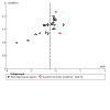
Medium‐term (6 to 12 months)
A total of five studies, with seven comparisons (572 participants), evaluated physical function in the medium term. The pooled SMD was ‐0.11 (95% CI ‐0.27 to 0.06) lower in the adjunctive therapy group plus exercise therapy compared with the placebo adjunctive therapy plus exercise therapy group (Analysis 3.2). An I2 of 0% indicates that statistical heterogeneity was not important. When converted to a MD, based on the WOMAC 0 to 68 physical function subscale (0 = best function), mean physical function was 1.29 points lower (95% CI 3.16 lower to 0.70 higher) and 4% lower (95% CI 10% lower to 2% higher) in the adjunctive therapy plus exercise group than in the placebo adjunctive therapy plus exercise group. The results therefore show no evidence of important differences in physical function between groups in the medium term. We downgraded the evidence to moderate certainty due to imprecision, associated with wide confidence intervals (Analysis 3.2).
3.2. Analysis.
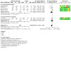
Comparison 3: Exercise and adjunctive therapy versus exercise and placebo adjunctive therapy (medium‐term), Outcome 2: Physical function
Long‐term (12 months onwards)
Two studies with 266 participants compared adjunctive therapies (acupuncture (Foster 2007) or dry needling (Sanchez‐Romero 2020)) in addition to exercise against placebo adjunctive therapy and exercise therapy in the long term. Following conversion of the SMD (0.05, 95% CI ‐0.19 to 0.29; Analysis 5.2) to a WOMAC physical function subscale (0 to 68, 0 = best function), the mean difference was 0.64 points higher (95% CI 2.43 lower to 3.71 higher) and the relative difference was 2% higher (95% CI 8% lower to 12% higher). These results provide evidence of no differences in physical function between the groups in the long term. Statistical heterogeneity was unimportant (I2 = 0%). We rated the evidence as high certainty (Analysis 5.2).
5.2. Analysis.
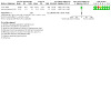
Comparison 5: Exercise and adjunctive therapy vs exercise and placebo adjunctive therapy (long‐term), Outcome 2: Physical function
Participant‐reported quality of life
Short‐term (0 to 6 months) (0 to 100 scale, higher scores represent better quality of life)
Two studies with 82 participants compared adjunctive therapies in addition to exercise therapy to placebo adjunctive therapy and exercise (Sanchez‐Romero 2020; Sardim 2020), resulting in a SMD of ‐0.08 (95% CI ‐0.51 to 0.36) (Analysis 1.3). When converted to a SF‐36 (0 to 100) scale, mean quality of life in the placebo group was 81.8 and 0.75 points lower (95% CI 4.80 lower to 3.39 higher) in the adjunctive therapy and placebo group. The absolute difference was 0.75 points worse (4.80 points worse to 3.39 points better). The relative difference was 1% lower (95% CI 7% lower to 5% higher). These results provide evidence of no important differences between the two groups. Statistical heterogeneity was unimportant (I2 = 0%). We downgraded the evidence to moderate certainty due to imprecision associated with large confidence intervals (Table 1).
1.3. Analysis.

Comparison 1: Exercise and adjunctive therapy versus exercise and placebo adjunctive therapy (short‐term), Outcome 3: Quality of life
Medium‐term (6 to 12 months) (EuroQol 5‐D 5 to 15 scale, higher scores represent better quality of life)
One study of dry needling compared adjunctive therapies plus exercise to placebo adjunctive therapies using the EuroQol 5‐D (Sanchez‐Romero 2020). The results are presented in the same units as the outcome used in their study (5 to 15 scale, higher scores represent better quality of life). The MD was ‐0.06 points in favour of the placebo adjunctive therapy group (95% CI 0.60 lower to 0.48 higher), providing evidence of no important differences between the groups (Analysis 3.3). We downgraded the evidence to low certainty as the results were based on a single study.
3.3. Analysis.
Comparison 3: Exercise and adjunctive therapy versus exercise and placebo adjunctive therapy (medium‐term), Outcome 3: Quality of life
Long‐term (12 months onwards) (EuroQol 5‐D 5 to 15 scale, higher scores represent better quality of life)
The same study of dry needling evaluated quality of life in the long term, with results reported using the EuroQol‐5D (5 to 15 scale, higher scores represent better quality of life) (Sanchez‐Romero 2020). The MD was 0.15 points higher in favour of the adjunctive therapy group (95% 0.58 lower to 0.88 higher), providing evidence of no important differences between the groups. We downgraded the evidence to low certainty for imprecision and bias as the results were based on a single study.
Adverse events
Adverse events were reported in just three of the 21 studies that evaluated adjunctive therapies against placebo adjunctive therapies, in addition to land‐based exercise. One study, which compared continuous or pulsed ultrasound to placebo (total of 90 participants), reported that no participant had an adverse event or complication and was not included in the meta‐analysis (Kapci Yildiz 2015). We pooled two studies that evaluated acupuncture (Foster 2007) and short‐wave diathermy (Rattanachaiyanont 2008) to provide a RR of 2.41 (95% CI 0.27 to 21.90), providing evidence of no important differences between the groups. An I2 of 62% indicated substantial heterogeneity. Details of the adverse events are provided in Table 7. Rattanachaiyanont 2008 specified which adverse events were due to the adjunctive therapy and the exercise intervention. We downgraded the evidence to low certainty due to inconsistency, associated with substantial heterogeneity, and imprecision due to wide confidence intervals (Table 1). No studies reported withdrawals due to adverse effects.
Adjunctive therapy in addition to exercise therapy versus exercise therapy only
We analysed the following outcomes: participant‐reported pain, participant‐reported physical function, quality of life, participant‐reported global assessment, radiographic structural changes and adverse events. Withdrawals due to adverse events were not measured in the included trials and, therefore, not analysed.
Participant‐reported pain (0 to 10 scale, higher scores correspond to greater pain)
Short‐term (0 to 6 months)
A total of 41 studies (45 comparisons) with 3322 participants were included in the meta‐analysis, which evaluated pain intensity at the end of the intervention period in studies that compared adjunctive therapy plus exercise therapy to exercise therapy only. Due to diversity of adjunctive therapies, we grouped outcomes by type of adjunctive therapy, etc. We back‐translated the pooled overall SMD (‐0.27, 95% CI ‐0.42 to ‐0.11; Analysis 2.1) to a 0 to 10 NPRS (0 = no pain). The mean pain in the control group was 3.8 and the mean difference was ‐0.41 points lower in favour of adjunctive therapy in addition to exercise therapy compared with exercise therapy only (95% CI 0.63 lower to 0.17 lower); this does not include the MCID of 1.9 points identified in osteoarthritis (Tubach 2005). The absolute difference was 0.41 points better (0.17 points better to 0.63 points better). The relative difference was 7% lower (95% CI 11% lower to 3% lower), which does not include the MCID of 15% (Tubach 2005). There was statistical heterogeneity (I2 = 75%). These results provide evidence of no differences between the groups (Analysis 2.1). We downgraded the evidence to low certainty due to unclear or high risk of selection, performance and reporting bias, and inconsistency associated with the high I2 value (Table 2). There was no evidence of publication bias (Figure 6).
2.1. Analysis.

Comparison 2: Exercise and adjunctive therapy versus exercise (short‐term), Outcome 1: Pain
6.
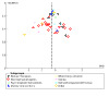
One study, with a total of 200 participants, which evaluated three different adjunctive therapies (pulsed electromagnetic (short‐wave) therapy (PEME), ultrasound therapy, interferential therapy) provided change scores only (Adhya 2015). We analysed these results in a separate meta‐analysis. The results are presented in Analysis 2.6 and showed no important differences in pain intensity measured on a visual analogue scale (VAS) (MD ‐1.32, 95% CI 0.38 lower to 3.01 higher). We downgraded the certainty of the evidence to very low certainty due to possible risk of selection, performance and reporting bias, statistical heterogeneity (I2 = 98%) and results based on one study. The results were not included in the summary of findings table and are unlikely to change the overall conclusions there in relation to short‐term participant‐reported pain (Table 2).
Medium‐term (6 to 12 months)
Nine studies, with 10 comparisons (1338 participants), assessed pain in the medium term. When all studies were pooled, we back‐translated the SMD of ‐0.23 (95% CI ‐0.49 to 0.02; Analysis 4.1) in favour of the adjunctive therapy plus exercise group to a 0 to 10 NPRS (0 = no pain). The mean pain intensity was 0.74 points lower (95% CI 1.57 lower to 0.06 higher) and the relative change was 13% lower (95% CI 28% lower to 1% higher) in the adjunctive therapies plus exercise therapy group compared with the exercise therapy only group. Heterogeneity was substantial (I2 = 80%). As the lower limit of 95% CI included the percentage change of 15% (Tubach 2005), an important difference in physical function between adjunctive electrophysical agents used in addition to exercise therapy compared with exercise therapy in the medium term can neither be confirmed nor excluded. We downgraded the certainty of the evidence to low certainty due to unclear or high risk of performance and reporting bias, and inconsistency associated with substantial heterogeneity and visual inspection of the forest plot (Analysis 4.1).
4.1. Analysis.

Comparison 4: Exercise and adjunctive therapy versus exercise (medium‐term), Outcome 1: Pain
Long‐term (12 months onwards)
Six studies (799 participants) assessed pain in the long term, and we pooled them to produce a SMD of ‐0.04 (95% CI ‐0.27 to 0.19; Analysis 6.1). When we back‐translated the SMD to a 0 to 10 NPRS (0 = no pain), the mean pain intensity was 0.08 points lower (95% CI 0.59 lower to 0.42 higher), and the relative mean difference in pain intensity was 2% lower (95% CI 11% lower to 7% higher) in the adjunctive therapies and exercise group than the exercise only group. These results provide evidence of no important differences in participant‐reported pain intensity in the long term between the groups. Statistical heterogeneity was moderate (I2 = 60%). We rated the overall certainty of this evidence as low due to inconsistency across the study findings and high/unclear risk of performance bias (Analysis 6.1).
6.1. Analysis.

Comparison 6: Exercise and adjunctive therapy versus exercise (long‐term), Outcome 1: Pain
Participant‐reported physical function (0 to 68 scale, higher scores correspond with poorer function)
Short‐term (up to 6 months)
The main outcomes used to assess physical function were the WOMAC Likert and visual analogue scales, the KOOS physical function subscale and the Lequesne Algofunctional Scale. Two studies that used physical performance tests in the form of a six‐minute walk test (6MWT) were not included in the meta‐analyses (Cheawthamai 2014; Vassao 2020). A total of 41 studies (45 comparisons) were eligible for inclusion in the meta‐analysis.
We pooled the results from 3323 participants to produce a SMD of ‐0.28 (95% CI ‐0.41 to ‐0.15; Analysis 2.2). Statistical heterogeneity was substantial (I2 = 63%). When we back‐translated the SMD to the WOMAC physical function subscale (based on a score range of 0 to 68, 0 = best function), the mean physical function in the exercise therapy only group was 18.2. The mean physical function was 2.83 points less in the adjunctive therapy plus exercise group than in the exercise only group (95% CI 4.04 lower to 1.62 lower), which does not include the MCID of six points identified in osteoarthritis (Bellamy 1992). The absolute difference was 2.83 points better (1.62 points better to 4.04 points better). The relative percentage change was 9% lower (95% CI 13% lower to 5% lower) in favour of the adjunctive therapy plus exercise therapy group. These results provide evidence of no important differences in physical function in the short term between the two groups. We downgraded the evidence to very low certainty due to unclear or high risk of selection, performance or reporting bias, inconsistency of study results, represented by statistical heterogeneity, and publication bias (Table 2).
2.2. Analysis.

Comparison 2: Exercise and adjunctive therapy versus exercise (short‐term), Outcome 2: Physical function
One study reported physical function change scores using the WOMAC and evaluated three different adjunctive therapies (PEME, ultrasound and interferential therapy (IFT)) (Adhya 2015). The authors did not report the scaling of the WOMAC, or whether the physical function subscale or total score was reported. The results are presented in Analysis 2.7 and show a MD of 6.84 (95% CI 2.96 lower to 16.63 higher). This result provides evidence of no important differences between the groups. We downgraded the evidence to very low certainty due to possible risk of selection, performance and reporting bias, inconsistency (I2 = 95%) and results based on one study. These results were not included in the overall summary of findings table for short‐term effects of adjunctive therapies on physical function, but these results are unlikely to change the overall conclusion (Table 2).
Medium‐term (6 to 12 months)
A total of nine studies (10 comparisons) with 1233 participants evaluated physical function in the medium term. When we pooled all studies, the SMD was ‐0.25 (95% CI ‐0.48 to ‐0.02) in favour of the adjunctive therapy plus exercise therapy group (Analysis 4.2). When we back‐translated the SMD to a 0 to 68 WOMAC physical function subscale, mean physical function was 2.52 points lower (95% CI 4.85 lower to 0.20 lower) in the adjunctive therapies plus exercise therapy group than the exercise therapy only group. The relative change was 8% lower (95% CI 16% lower to 1% lower) in the adjunctive therapies plus exercise therapy group compared with exercise therapy only group. Heterogeneity was substantial (I2 = 71%). These results provide no evidence of differences between the groups. We downgraded the certainty of evidence to low certainty due to unclear risk of performance bias and inconsistency of study results as represented by high I2 values and visual inspection of the forest plot (Analysis 4.2).
4.2. Analysis.

Comparison 4: Exercise and adjunctive therapy versus exercise (medium‐term), Outcome 2: Physical function
Long‐term (12 months onwards)
A total of six studies (781 participants) assessed physical function in the long term. When we back‐translated the SMD (0.03, 95% CI ‐0.17 to 0.22; Analysis 6.2) to a 0 to 68 WOMAC physical function subscale, the mean physical function was 0.30 points higher in favour of exercise therapy only (95% CI 1.72 lower to 2.22 higher) and the relative change was 1% higher (95% CI 6% lower to 7% higher). The results provide evidence of no important differences between the groups. Statistical heterogeneity was moderate (I2 = 46%). We downgraded the evidence to moderate certainty due to high or unclear risk of performance bias (Analysis 6.2).
6.2. Analysis.

Comparison 6: Exercise and adjunctive therapy versus exercise (long‐term), Outcome 2: Physical function
Participant‐reported quality of life (0 to 100 scale, higher scores correspond with higher quality of life)
Short‐term (0 to 6 months)
Ten studies (11 comparisons) with 1483 participants assessed quality of life using a variety of outcome measures including the Short‐Form (SF‐36), EuroQol 5‐D, Assessment of Quality of Life‐6D (AQOL‐6D) and the Knee Osteoarthritis Outcome Scale (KOOS) quality of life subscale as outlined in Table 8. When we back‐translated the SMD (‐0.12, 95% CI ‐0.36 to 0.12; Analysis 2.3) to the SF‐36 mental health scale (0 to 100 scale, where higher scores correspond with higher quality of life), mean quality of life in the control group was 56.1. Mean quality of life in the adjunctive therapies and exercise therapy group was 1.04 points lower (95% CI 3.12 lower to 1.04 higher). The relative change was 2% lower (95% CI 5% lower to 2% higher). The results provide evidence of no important differences between the groups. Statistical heterogeneity was substantial (I2 = 79%). The findings were based on very low‐certainty evidence, which we downgraded due to potential risk of selection and performance bias, inconsistency of results associated with a high I2 value and visual inspection of the forest plot, imprecision of results and evidence of publication bias (Table 2).
2.3. Analysis.

Comparison 2: Exercise and adjunctive therapy versus exercise (short‐term), Outcome 3: Quality of life
Medium‐term (6 to 12 months)
Four studies (784 participants) measured quality of life at six months post‐intervention. Two studies used the Assessment of Quality of Life Scale (AQOL‐6D) (Bennell 2016; Bennell 2017) and two used the Short‐Form‐36 (Brosseau 2012; Forestier 2010), with higher scores indicating better quality of life for both outcomes. When we back translated the SMD (‐0.19, 95% CI ‐0.41 to 0.04; Analysis 4.3) to the SF‐36, the mean quality of life in the adjunctive therapies and exercise therapy groups was 1.63 points lower (95% CI 3.53 lower to 0.34 higher) and the relative change was 3% lower (95% CI 7% lower to 1% higher) in the adjunctive therapy and exercise therapy groups. Study heterogeneity was moderate (I2 = 58%). The results show evidence of no important differences between the two groups in quality of life in the medium term. We downgraded the evidence to low certainty due to imprecision associated with heterogeneity and high/unclear risk of performance bias (Analysis 4.3).
4.3. Analysis.

Comparison 4: Exercise and adjunctive therapy versus exercise (medium‐term), Outcome 3: Quality of life
Long‐term (12 months onwards)
Two studies (253 participants) evaluated quality of life at 12 months (long‐term) (Bennell 2016; Bennell 2017). As both studies of psychological interventions measured quality of life on the same scale (Assessment of Quality of Life, AQOL 6‐D), we estimated the mean difference (MD), resulting in a pooled MD of 0.03 (95% CI ‐0.03 to 0.09; Analysis 4.3). The minimal clinically important difference (MCID) for the AQOL has been defined as 0.06 utility points (Hawthorne 2005). As the 95% CI did not include the MCID, the results provide evidence of no important differences between the groups in quality of life in the long term. Statistical heterogeneity was moderate (I2 = 59%). We downgraded the certainty of evidence to moderate due to inconsistency in the results associated with heterogeneity (Analysis 4.3).
Participant‐reported global assessment (dichotomised to improved/not improved)
Short‐term (0 to 6 months)
We pooled five studies (840 participants) that evaluated patient‐reported global assessment using 3‐, 6‐, 7‐ or 15‐point ordinal scales (Table 8) to provide a risk ratio (RR) of 1.37 (95% CI 1.15 to 1.62; Analysis 2.4). The absolute difference was 17% reporting more success (7% to 28% more). The relative difference was 37% more (15% more to 62% more) participants reporting success with adjunctive therapies in addition to exercise therapy compared with exercise therapy only. These findings indicate that adjunctive psychological therapies used with exercise therapy probably result in a clinically relevant difference in self‐reported global assessment. Although the I2 value was moderate (48%), visual inspection of the forest plot did not demonstrate inconsistency (Analysis 2.4). We downgraded the evidence to moderate certainty due to high risk of performance bias (Table 2).
2.4. Analysis.
Comparison 2: Exercise and adjunctive therapy versus exercise (short‐term), Outcome 4: Patient‐reported global assessment
Medium‐term (6 to 12 months)
Two studies (233 participants), led by the same author (Bennell 2016; Bennell 2017), evaluated patient‐reported global assessment in the medium term, using a seven‐point ordinal scale. The adjunctive therapies were both psychological therapies. When we pooled both studies for analysis, the RR was 1.31 (95% CI 1.06 to 1.62; Analysis 4.4), showing that 31% more participants (6% to 62%) reported a benefit in favour of adjunctive therapy and exercise therapy compared with exercise therapy only. There was no statistical heterogeneity (I2 = 0%). These findings indicate that adjunctive psychological therapies used with exercise therapy probably result in a clinically relevant difference in self‐reported global assessment. We downgraded the evidence to moderate certainty due to high risk of performance bias (Analysis 4.4).
4.4. Analysis.
Comparison 4: Exercise and adjunctive therapy versus exercise (medium‐term), Outcome 4: Patient‐reported global assessment
Long‐term (12 months onwards)
The same two studies (232 participants), which evaluated medium‐term effects using patient‐reported global assessment, also assessed long‐term effects at 12 months using the same seven‐point ordinal scale (Bennell 2016; Bennell 2017). The pooled analysis revealed a RR of 1.42 (95% CI 1.15 to 1.75; Analysis 6.4), showing that 42% more participants (15% to 75%) reported success in favour of adjunctive therapy and exercise therapy compared with exercise therapy (I2 = 0%). The findings indicate, based on moderate‐certainty evidence, that adjunctive psychological therapies used in combination with exercise therapy probably result in improved patient‐reported global assessment in the long term. We downgraded the evidence to moderate certainty due to high risk of performance bias (Analysis 6.4).
6.4. Analysis.

Comparison 6: Exercise and adjunctive therapy versus exercise (long‐term), Outcome 4: Patient‐reported global assessment
Radiographic joint structural changes (lower scores represent less joint space narrowing)
Short‐term (0 to 6 months)
One study of 156 participants, which evaluated a dietary intervention in addition to exercise therapy, measured medial and lateral joint space narrowing within the knee joint (Messier 2004). Lower scores, which represent less joint space narrowing, indicate a favourable effect on joint structures. The results showed that joint space narrowing was 0.25 mm less (MD ‐0.25, 95% CI ‐0.32 to ‐0.18) in favour of adjunctive dietary therapy in addition to exercise therapy versus exercise therapy only. The relative difference was ‐11% (95% CI ‐18% to ‐6%; Analysis 2.5). There is no known MCID for joint space narrowing, so the clinical meaningfulness of this result is unknown. We downgraded the evidence to low certainty due to unclear risk of performance bias and because the results were based on just one study (Table 2).
2.5. Analysis.

Comparison 2: Exercise and adjunctive therapy versus exercise (short‐term), Outcome 5: Radiographic joint structure changes
Adverse events
A total of eight studies (1542 participants) reported adverse events from adjunctive therapies in addition to land‐based exercise versus land‐based exercise only. Details are provided in Table 7. Three studies evaluated manual therapy interventions (Abbott 2013; Abbott 2015; Fitzgerald 2016), two evaluated psychological interventions (Bennell 2016; Bennell 2017), one evaluated spa therapy (Forestier 2010), two tested dietary interventions (Messier 2004; Messier 2013), and one assessed whole‐body vibration (Wang 2016). Two studies detailed the number of events that occurred in both the intervention period and follow‐up periods (Bennell 2016; Bennell 2017), whilst it was not clearly stated in the remaining studies. Two studies clearly differentiated whether adverse events were serious (Messier 2013 (which only reported serious adverse events); Forestier 2010). Messier 2013 reported six unrelated adverse events in the exercise only group and three unrelated serious events in the diet plus exercise group. Details are provided in Table 7. Two studies did not specify in which groups the adverse events occurred, so were excluded from the meta‐analysis (Fitzgerald 2016; Messier 2004). We pooled all adverse events in the remaining eight studies to provide a RR of 1.33 (95% CI 0.78 to 2.27; Analysis 2.8). The absolute difference was 50% more events (33% fewer to 392% more). The relative difference was 33% more events (22% fewer to 127% more). Statistical heterogeneity was not important (I2 = 31%). We downgraded the evidence to low certainty due to wide confidence intervals associated with the effect estimate, and unclear or high risk of performance bias (Table 2). No studies reported withdrawals due to adverse events.
2.8. Analysis.

Comparison 2: Exercise and adjunctive therapy versus exercise (short‐term), Outcome 8: Adverse events
Subgroup analysis
Adjunctive therapy in addition to exercise therapy compared to placebo adjunctive therapy plus exercise therapy
There were no important differences in pain, function or quality of life between adjunctive electrophysical agents, adjunctive acupuncture/dry needling agents, taping and corresponding placebo agents in the short term or medium term (Analysis 1.1; Analysis 1.2; Analysis 1.3; Analysis 3.1; Analysis 3.2).
Adjunctive therapy in addition to exercise therapy compared to exercise therapy alone
There were no important difference in pain, function, quality of life or adverse events between adjunctive manual therapies or adjunctive electrophysical agents or adjunctive psychological therapies or adjunctive dietary interventions or whole body vibration or taping or balneotherapy or acupuncture, orthotics or geotherapy with exercise therapy compared to exercise therapy in the short term, medium term or long term (Analysis 2.1, Analysis 2.2; Analysis 4.1; Analysis 6.1; Analysis 6.2). The results from two studies with 350 participants provided moderate‐certainty evidence (downgraded for bias) of a benefit in favour of psychological therapy plus exercise therapy compared with exercise therapy for patient‐reported global assessment (RR 1.32, 95% CI 1.09 to 1.58) in the short term. A single study showed low‐certainty evidence (downgraded for bias and imprecision) of benefit in patient‐reported global assessment in favour of balneotherapy with exercise compared to exercise (RR 1.83, 95% CI 1.41 to 2.39) in the short term (Analysis 2.4 Table 6).
Sensitivity analyses
We undertook sensitivity analyses for the two main outcomes of pain and physical function for both comparisons: 1) adjunctive therapy in addition to exercise therapy compared to placebo adjunctive therapy plus exercise therapy and 2) adjunctive therapy in addition to exercise therapy compared to exercise therapy alone. We undertook separate analyses for studies that we deemed to have high or unclear risk of selection bias (random allocation or allocation concealment), performance bias and detection bias, which we removed from the meta‐analysis. Overall, the results were largely unchanged from the original analysis (Table 9; Table 10; Table 11).
7. Sensitivity analyses: selection bias.
| Comparison | Outcome | All studies | Low risk for random allocation/allocation concealment | Outcome | All studies | Low risk for random allocation/allocation concealment |
| MD (95% CI), % diff (95% CI) number of studies, I2 | MD (95% CI), number of studies, I2 | MD (95% CI), % diff number of studies, I2 | MD (95% CI), number of studies, I2 | |||
| Adjunctive therapy plus exercise therapy versus placebo adjunctive therapy plus exercise therapy (short‐term) | Pain | MD ‐ 0.77 (‐1.16 to ‐0.48); % difference ‐10% (‐15 % to ‐6%) studies = 22; I2 = 70% | MD ‐0.35 (‐0.78 to 0.07); % difference ‐4% (‐10% to 1%) studies = 9; I2 = 51% | Physical function | MD ‐5.03 (‐7.61 to ‐2.57); % difference ‐12% (‐18% to ‐6%) studies = 20; I2 = 69% | MD ‐2.81 (‐4.68 to ‐0.23); % difference ‐6% (‐11% to ‐1%); studies = 8; I2 = 50% |
| Adjunctive therapy and exercise therapy versus exercise therapy (short‐term) | Pain | MD ‐0.41 (‐0.63 to ‐0.17); % difference ‐7% (‐13% to ‐5%); studies = 40; I2 = 75% | MD ‐0.55 (‐0.92 to ‐0.18); % difference ‐10% (‐17% to ‐3%); studies = 14; I2 = 68% | Physical function | MD ‐2.83 (‐4.04 to ‐1.62); % difference ‐9% (‐13% to ‐5%) studies = 39; I2 = 63% | MD ‐2.71 (‐4.64 to ‐0.65); % difference ‐9% (‐16% to ‐3%); studies = 15; I2 = 62% |
| Adjunctive therapy plus exercise therapy versus placebo adjunctive therapy plus exercise therapy (medium‐term) | Pain | MD ‐0.25 (‐0.71 to 0.23); % difference ‐5% (‐15% to 5%); studies = 6; I2 = 32% | MD ‐0.04 (‐0.37 to 0.3); % difference 0% (‐5% to 4%) studies = 3; I2 = 0% | Physical function | MD ‐1.23 (‐3.39 to 0.70); % difference ‐4% (‐11% to 2%); studies = 5; I2 = 0% | MD ‐0.7 (‐2.81 to 1.40); % difference ‐2% (‐6% to 3%); studies = 3; I2 = 0% |
| Adjunctive therapy plus exercise therapy versus exercise therapy only (medium‐term) | Pain | MD ‐0.74 (‐1.57 to 0.06); % difference ‐13% (‐28% to 16%); studies = 9; I2 = 80% | MD ‐0.48 (‐0.98 to 0.02); % difference ‐6% (‐12% to 0%); studies = 8, I2 = 82 % | Physical function | MD ‐2.52 (‐4.86 to ‐0.20); % difference ‐8% (‐16% to ‐1%); studies = 2; I2 = 0% | MD ‐3.28 (‐6.08 to ‐0.03); % difference ‐8% (‐14% to ‐1%), studies = 8, I2 = 74% |
| Adjunctive therapy plus exercise therapy versus placebo adjunctive therapy plus exercise therapy (long‐term) | Pain | MD ‐0.13 (‐1.43 to 1.17); % difference ‐1% (‐16% to 13%), studies = 2; I2 = 60% | Same as main analysis | Physical function | MD 1.36 (‐3.20 to 5.92); % difference 4% (‐10% to 19%); studies = 2; I2 = 0% | Same as main analysis |
| Adjunctive therapy plus exercise therapy versus exercise therapy only (long‐term) | Pain | MD‐0.09 (‐0.59 to 0.42); % difference ‐2% (‐11% to 7%); studies = 6; I2 = 60% | Same as main analysis | Physical function | MD ‐0.30 (‐1.72 to 2.22); % difference 1% (‐6% to 7%); studies = 6, I2 = 46% | Same as main analysis |
CI: confidence interval; MD: mean difference
8. Sensitivity analysis: performance bias.
| Comparison | Outcome | All studies | Low risk of performance bias | Outcome | All studies | Low risk of performance bias |
| MD (95% CI), % difference (95% CI) number of studies, I2 | MD (95% CI), % difference (95% CI) number of studies, I2 | MD (95% CI), % difference (95% CI) number of studies, I2 | MD (95% CI), % difference (95% CI) number of studies, I2 | |||
| Adjunctive therapy plus exercise therapy versus placebo adjunctive therapy plus exercise therapy (short‐term) | Pain | MD ‐ 0.77 (‐1.16 to ‐0.48); % difference ‐10% (‐15% to ‐6%) studies = 22; I2 = 70% | MD ‐0.61 (‐0.99 to ‐0.22); % difference ‐8% (‐12% to ‐3%); studies = 14 ; I2 = 53% | Physical function | MD ‐5.03 (‐7.61 to ‐2.57); % difference ‐12% (‐18% to ‐6%) studies = 20; I2 = 69% | MD ‐5.27 (‐8.42 to ‐2.11); % difference ‐12% (‐19% to ‐5%); studies = 13; I2 = 69% |
| Adjunctive therapy plus exercise therapy versus exercise therapy (short‐term) | Pain | MD ‐0.41 (‐0.63 to ‐0.17); % difference ‐7% (‐13% to ‐5%); studies = 40; I2 = 75% | MD ‐1.2 (‐2.17 to ‐0.23); % difference ‐26% (‐47% to ‐5%); studies = 4 ; I2 = 77% | Physical function | MD ‐2.83 (‐4.04 to ‐1.62); % difference ‐9% (‐13% to ‐5%) studies = 39; I2 = 63% | MD ‐7.18 (‐14.02 to ‐0.34); % difference ‐18% (‐36% to ‐1%); studies = 4 ; I2 = 63% |
| Adjunctive therapy plus exercise therapy versus placebo adjunctive therapy plus exercise therapy (medium‐term) | Pain | MD ‐0.25 (‐0.71 to 0.23); % difference ‐5% (‐15% to 5%); studies = 6; I2 = 32% | MD ‐0.30 (‐0.72 to 0.09); % difference ‐4% (‐9% to 1%); studies = 5; I2 = 0% |
Physical function | MD ‐1.23 (‐3.39 to 0.70); % difference ‐4% (‐11% to 2%); studies = 5; I2 = 0% | MD ‐1.52 (‐3.98 to 0.94); % difference ‐4% (‐9% to 2%); studies = 4; I2 = 0% |
| Adjunctive therapy plus exercise therapy versus exercise therapy (medium‐term) | Pain | MD ‐0.74 (‐1.57 to 0.06); % difference ‐13% (‐28% to 16%); studies = 9; I2 = 80% | MD ‐0.26 (‐0.65 to 0.11); % difference ‐6% (‐14% to 2%); studies = 1 | Physical function | MD ‐2.52 (‐4.86 to ‐0.20); % difference ‐8% (‐16% to ‐1%); studies = 2; I2 = 0% | MD ‐1.37 (‐4.96 to 2.22); % difference ‐6% (‐14% to 2%); studies = 1 |
| Adjunctive therapy plus exercise therapy versus placebo adjunctive therapy plus exercise therapy (long‐term) | Pain | MD ‐0.13 (‐1.43 to 1.17); % difference ‐1% (‐16% to 13%), studies = 2; I2 = 60% | MD ‐0.01 (‐0.43 to 0.41); % difference 0% (‐7% to 7%); studies = 1 | Physical function | MD 1.36 (‐3.20 to 5.92); % difference 4% (‐10% to 19%); studies = 2; I2 = 0% | MD ‐0.49 (‐6.59 to 5.62); % difference ‐2% (‐28% to 24%), studies = 1 |
| Adjunctive therapy plus exercise therapy versus exercise therapy (long‐term) | Pain | MD‐0.09 (‐0.59 to 0.42); % difference ‐2% (‐11% to 7%); studies = 6; I2 = 60% | No eligible studies | Physical function | MD ‐0.30 (‐1.72 to 2.22); % difference 1% (‐6% to 7%); studies = 6, I2 = 46% | No eligible studies |
CI: confidence interval; MD: mean difference
9. Sensitivity analysis: detection bias.
| Comparison | Outcome | All studies | Low risk of detection bias | Outcome | All studies | Low risk of detection bias |
| MD (95% CI), % difference (95% CI) number of studies, I2 | MD (95% CI), % difference (95% CI) number of studies, I2 | MD (95% CI), % difference (95% CI) number of studies, I2 | MD (95% CI), % difference (95% CI) number of studies, I2 | |||
| Adjunctive therapy plus exercise therapy versus placebo adjunctive therapy plus exercise therapy (short‐term) | Pain | MD ‐0.77 (‐1.16 to ‐0.48); % difference ‐10% (‐15% to ‐6%) studies = 22; I2 = 70% |
MD ‐0.48 (‐1.02 to 0.06); % difference ‐6% (‐13% to 1%); studies = 9 ; I2 = 63% | Physical function | MD ‐5.03 (‐7.61 to ‐2.57); % difference ‐12% (‐18% to ‐7%) studies = 20; I2 = 69% | MD ‐4.33 (‐8.54 to ‐0.12); % difference ‐6% (‐13% to 1%); studies = 9; I2 = 75% |
| Adjunctive therapy plus exercise therapy versus exercise therapy (short‐term) | Pain | MD ‐0.41 (‐0.63 to ‐0.17); % difference ‐7% (‐13% to ‐5%); studies = 40; I2 = 75% | MD ‐0.92 (‐1.62 to ‐0.23); % difference ‐16% (‐28% to ‐4%); studies = 4 ; I2 = 67% | Physical function | MD ‐2.83 (‐4.04 to ‐1.62); % difference ‐9% (‐13% to ‐5%) studies = 39; I2 = 63% | MD ‐1.31 (‐2.05 to ‐0.05); % difference ‐2% (‐4% to 0%); studies = 4 ; I2 = 49% |
| Adjunctive therapy plus exercise therapy versus placebo adjunctive therapy plus exercise therapy (medium‐term) | Pain | MD ‐0.25 (‐0.71 to 0.23); % difference ‐5% (‐15% to 5%); studies = 6; I2 = 32% | MD ‐0.02 (‐0.38 to 0.36); % difference 0% (‐8% to 8%); studies = 5, I2 = 0% |
Physical function | MD ‐2.52 (‐4.86 to ‐0.20); % difference ‐8% (‐16% to ‐1%); studies = 2; I2 = 0% | MD ‐0.82 (‐2.81 to 1.17); % difference ‐3% (‐9% to 4%); studies = 4, I2 = 0% |
| Adjunctive therapy plus exercise therapy versus exercise therapy (medium‐term) | Pain | MD ‐0.74 (‐1.57 to 0.06); % difference ‐13% (‐28% to 16%); studies = 9; I2 = 80% | No eligible studies | Physical function | MD ‐2.52 (‐4.86 to ‐0.20); % difference ‐8% (‐16% to ‐1%); studies = 2; I2 = 0% | No eligible studies |
| Adjunctive therapy plus exercise therapy versus placebo adjunctive therapy plus exercise therapy (long‐term) | Pain | MD ‐0.13 (‐1.43 to 1.17); % difference ‐1% (‐16% to 13%); studies = 2; I2 = 0% | Same as full analysis | Physical function | MD 1.36 (‐3.20 to 5.92); % difference 4% (‐10% to 19%); studies = 2 ; I2 = 0% |
Same as full analysis |
| Adjunctive therapy plus exercise therapy versus exercise therapy only (long‐term) | Pain | MD ‐0.09 (‐0.59 to 0.42); % difference ‐2% (‐11% to 7%); studies = 2; I2 = 0% | No eligible studies | Physical function | MD ‐0.30 (‐1.72 to 2.22); % difference 1% (‐6% to 7%); studies = 2 ; I2 = 0% | No eligible studies |
CI: confidence interval; MD: mean difference
Selection bias
There was no difference in pain or function between adjunctive therapy in addition to exercise therapy compared to placebo adjunctive therapy plus exercise therapy or between adjunctive therapy in addition to exercise therapy compared to exercise therapy alone in the short term or medium term (Analysis 7.1; Analysis 7.2; Analysis 8.1; Analysis 8.2; Analysis 9.1, Analysis 9.2; Analysis 10.2).
7.1. Analysis.

Comparison 7: Sensitivity analysis (selection bias): exercise and adjunctive therapy versus exercise and placebo adjunctive therapy (short‐term), Outcome 1: Pain
7.2. Analysis.
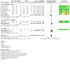
Comparison 7: Sensitivity analysis (selection bias): exercise and adjunctive therapy versus exercise and placebo adjunctive therapy (short‐term), Outcome 2: Physical function
8.1. Analysis.

Comparison 8: Sensitivity analysis (selection bias): exercise and adjunctive therapy versus exercise (short‐term), Outcome 1: Pain
8.2. Analysis.
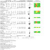
Comparison 8: Sensitivity analysis (selection bias): exercise and adjunctive therapy versus exercise (short‐term), Outcome 2: Physical function
9.1. Analysis.

Comparison 9: Sensitivity analysis (selection bias) exercise and adjunctive therapy versus exercise and placebo adjunctive therapy (medium‐term), Outcome 1: Pain
9.2. Analysis.
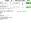
Comparison 9: Sensitivity analysis (selection bias) exercise and adjunctive therapy versus exercise and placebo adjunctive therapy (medium‐term), Outcome 2: Physical function
10.2. Analysis.
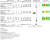
Comparison 10: Sensitivity analysis (selection bias): exercise and adjunctive therapy versus exercise (medium‐term), Outcome 2: Physical function
Performance bias
There was no difference in pain or function between adjunctive therapy in addition to exercise therapy compared to placebo adjunctive therapy plus exercise therapy or between adjunctive therapy in addition to exercise therapy compared to exercise therapy alone in the short term, medium term or long term (Analysis 11.1; Analysis 11.2; Analysis 12.1; Analysis 13.1; Analysis 13.2; Analysis 14.1; Analysis 14.2; Analysis 15.1; Analysis 15.2).
11.1. Analysis.

Comparison 11: Sensitivity analysis (performance bias) exercise and adjunctive therapy versus exercise and placebo adjunctive therapy (short‐term), Outcome 1: Pain
11.2. Analysis.

Comparison 11: Sensitivity analysis (performance bias) exercise and adjunctive therapy versus exercise and placebo adjunctive therapy (short‐term), Outcome 2: Physical function
12.1. Analysis.

Comparison 12: Sensitivity analysis (performance bias) exercise and adjunctive therapy versus exercise (short‐term), Outcome 1: Pain
13.1. Analysis.

Comparison 13: Sensitivity analysis (performance bias) exercise and adjunctive therapy versus exercise and placebo adjunctive therapy (medium‐term), Outcome 1: Pain
13.2. Analysis.

Comparison 13: Sensitivity analysis (performance bias) exercise and adjunctive therapy versus exercise and placebo adjunctive therapy (medium‐term), Outcome 2: Physical function
14.1. Analysis.

Comparison 14: Sensitivity analysis (performance bias): exercise and adjunctive therapy versus exercise (medium‐term), Outcome 1: Pain
14.2. Analysis.

Comparison 14: Sensitivity analysis (performance bias): exercise and adjunctive therapy versus exercise (medium‐term), Outcome 2: Physical function
15.1. Analysis.

Comparison 15: Sensitivity analysis (performance bias) exercise and adjunctive therapy vs exercise and placebo adjunctive therapy (long‐term), Outcome 1: Pain
15.2. Analysis.

Comparison 15: Sensitivity analysis (performance bias) exercise and adjunctive therapy vs exercise and placebo adjunctive therapy (long‐term), Outcome 2: Physical function
Detection bias
Overall, these were largely unchanged from the original analysis. There was no difference in pain or function between adjunctive therapy in addition to exercise therapy compared to placebo adjunctive therapy plus exercise therapy or between adjunctive therapy in addition to exercise therapy compared to exercise therapy alone in the short term, medium term or long term (Analysis 16.1; Analysis 16.2; Analysis 17.1; Analysis 17.2; Analysis 18.1; Analysis 18.2).
16.1. Analysis.

Comparison 16: Sensitivity analysis (detection bias): exercise and adjunctive therapy versus exercise and placebo adjunctive therapy (short‐term), Outcome 1: Pain
16.2. Analysis.
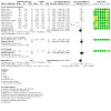
Comparison 16: Sensitivity analysis (detection bias): exercise and adjunctive therapy versus exercise and placebo adjunctive therapy (short‐term), Outcome 2: Physical function
17.1. Analysis.

Comparison 17: Sensitivity analysis (detection bias): exercise and adjunctive therapy versus exercise (short‐term), Outcome 1: Pain
17.2. Analysis.

Comparison 17: Sensitivity analysis (detection bias): exercise and adjunctive therapy versus exercise (short‐term), Outcome 2: Physical function
18.1. Analysis.

Comparison 18: Sensitivity analysis (detection bias) exercise and adjunctive therapy versus exercise and placebo adjunctive therapy (medium‐term), Outcome 1: Pain
18.2. Analysis.

Comparison 18: Sensitivity analysis (detection bias) exercise and adjunctive therapy versus exercise and placebo adjunctive therapy (medium‐term), Outcome 2: Physical function
Discussion
Summary of main results
Adjunctive therapies in addition to land‐based exercise versus placebo adjunctive therapies in addition to land‐based exercise
Compared with placebo adjunctive therapy and exercise, low‐certainty evidence indicates that adjunctive therapies used in addition to exercise therapy provide no clinically important benefits with respect to pain or physical function in the short term (up to six months post‐intervention). The results were based on 22 trials (1428 participants) evaluating pain and 20 trials (1361 participants) that measured participant‐reported physical function. We downgraded the certainty of the evidence due to inconsistency and potential publication bias. Considerably fewer trials evaluated pain (six trials with 572 participants) and physical function (five trials with 572 participants) in the medium term (6 to 12 months post‐intervention), with similar findings of no clinically important differences, based on moderate‐certainty evidence. We downgraded the evidence due to inconsistency, but we were unable to assess the possibility of publication bias due to the small number of trials. No trials evaluated long‐term effects.
Just two trials (82 participants) evaluated quality of life in the short term and also found no clinical improvements with the addition of adjunctive therapies compared with placebo therapies, used in addition to exercise therapy. We downgraded the evidence to moderate certainty due to inconsistency. We cannot rule out the possibility of publication bias, but we could not assess this fully due to the small number of trials. One trial of 62 participants also found no clinically important benefits with respect to quality of life in the medium term. We considered the certainty of the evidence associated with this finding to be low as the results are based on just one small sample size trial. No trials evaluated long‐term effects.
Of the 22 placebo trials included in this review, just three (430 participants) reported the presence of adverse events, representing 14% of the placebo‐controlled trials. As one of these reported no adverse events in any group, it was not included in the meta‐analysis. Whilst the results showed no greater risk of adverse events with adjunctive therapies, compared to their placebo counterpart, the small number of trials limits the applicability of this evidence. Adverse events included pain, sleepiness, fainting, nausea and swelling in the trial evaluating acupuncture (Foster 2007), and pain, swelling and a feeling of vasodilatation in a trial evaluating short‐wave diathermy (Rattanachaiyanont 2008). Withdrawals due to adverse events were not reported in any of the trials.
No trials that evaluated adjunctive therapies against a placebo assessed participant global assessment of treatment effects or radiographic structural changes. We rated the evidence as low certainty due to possible risk of performance bias and imprecision of the results. No studies reported withdrawals due to adverse events.
Adjunctive therapies in addition to land‐based exercise versus land‐based exercise
Low‐certainty evidence based on 38 trials indicates that adjunctive therapies used in addition to exercise therapy provide no short‐term (up to six months) clinically important benefits above exercise therapy only with respect to pain (3322 participants) and physical function (3323 participants). We downgraded the evidence to low certainty due to potential risk of selection, performance and reporting bias, and inconsistency in the results. There was no evidence of publication bias in the trials that evaluated pain, but there was possible publication bias in those that evaluated physical function.
There were also no clinically important benefits observed in the medium term (6 to 12 months). Based on low‐certainty evidence from nine studies, adjunctive therapies used in combination with exercise therapy provide no clinically important benefits over exercise only for pain (1338 participants) and physical function (1233 participants). We downgraded the certainty of the evidence due to risk of performance and reporting bias, and inconsistency associated with high statistical heterogeneity. Publication bias was not assessed due to the low number of trials. The results were similar in the long term (12 months) for both pain and physical function outcomes, with results based on six trials. We rated the evidence as low certainty for pain, due to unclear or high risk of performance bias and inconsistency, and moderate certainty for physical function due to unclear/high risk of performance bias.
Very low‐certainty evidence indicates no clinically important benefits in health‐related quality of life in the short term, based on 10 trials (1483 participants). We downgraded the evidence to very low certainty due to unclear or high risk of performance bias, inconsistency and possible risk of publication bias. Four trials, with 784 participants, evaluated quality of life in the medium term and found similar results, with the evidence rated as low‐certainty due to performance bias and imprecision. These results were also found in the long‐term evaluation, based on two trials of 253 participants. We rated the certainty of the evidence as moderate due to inconsistency. Publication bias was not assessed in the medium‐ and long‐term analyses, due to the small number of studies.
There was moderate‐certainty evidence based on five trials (840 participants) of clinically important benefits in participant‐reported global assessment in the short term. We downgraded the certainty of the evidence due to high/unclear risk of performance bias. A clinically important benefit was also found in the medium term and long term based on the same two trials (233 participants), with the evidence rated as moderate certainty, downgraded due to high risk of performance bias.
Low‐certainty evidence from one trial identified no clinically important differences in radiographic changes (measuring joint space narrowing).
Adverse events were reported in eight of the 38 studies (21%) that compared adjunctive therapies plus exercise therapy to exercise therapy. There was no evidence of clinically important differences in the number of adverse events reported (low‐certainty evidence).
Subgroup analysis
Moderate‐certainty evidence from two studies showed a benefit in favour of psychological therapy plus exercise therapy compared with exercise alone for participant‐reported global assessment in the short term. A single study showed low‐certainty evidence of benefit in favour of balneotherapy with exercise compared to exercise alone for participant‐reported global assessment in the short term. There were no important differences in pain, function or quality of life between adjunctive electrophysical agents, adjunctive acupuncture/dry needling agents, taping and corresponding placebo agents in the short term or medium term. No important differences in pain between adjuctive manual therapies or adjunctive electrophysical agents or adjunctive psychological therapies or adjunctive dietary interventions or whole body vibration or taping or balneotherapy or acupuncture, orthotics or geotherapy with exercise therapy compared to exercise therapy were found in the short term.
Overall completeness and applicability of evidence
This review includes evidence from 60 randomised controlled trials and 2 quasi‐randomised controlled trials across 22 countries assessing the effects of adjunctive therapies used in conjunction with exercise therapy compared with placebo adjunctive therapy used with exercise therapy (21 trials and 1 quasi‐RCT) or compared with exercise therapy only (39 trials and 1 quasi‐RCT). Two trials could not be included in the meta‐analysis as key outcomes could not be extracted (Jia 2005; Quirk 1985).
In this review, education was considered an adjunctive therapy, although clinical guidelines consider it as a core intervention for hip or knee osteoarthritis (Bannaru 2019; Kolasinski 2020; NICE 2014). We did not find any trials that specifically compared education as an adjunctive therapy used alongside exercise therapy with exercise therapy only. However, some trials reported an educational component of the exercise therapy or adjunctive therapy intervention (Bennell 2016; Bennell 2017; Brosseau 2012; Elboim‐Gabyzon 2013; Messier 2013). We excluded three trials that included education as an intervention in both groups (Palmer 2014; Thoumie 2018; Vader 2020). No Cochrane Reviews have been as yet conducted to evaluate the effectiveness of education and exercise as first‐line interventions for hip or knee osteoarthritis and this could be a focus of future reviews.
Numerous trials have evaluated a range of adjunctive therapies used in combination with exercise therapy (compared with exercise therapy only) for hip or knee osteoarthritis. In the main analysis, we presented the findings of all adjunctive therapies pooled together, but due to the variety of adjunctive therapies included in the review, we combined similar types of therapies together including manual therapies, electrophysical agents, psychological therapies, dietary interventions, acupuncture/dry needling, taping, whole body vibration and balneotherapy together and reported the findings in the subgroup analyses to provide a more meaningful assessment of the evidence. Where a study did not fall into these categories or only one study was eligible for meta‐analysis within these categories, it was included in an 'Other' comparison. Interventions also differed in dosage and intensity levels across these subgroups, varying from three weeks to 18 months in duration, and number of treatments varied between six and 60.
Participant‐reported pain and participant‐reported physical function were the most common outcomes assessed, with the numerical pain rating scale (NPRS) or visual analogue scale (VAS) used most commonly for measurement of pain intensity, and the Western Ontario McMaster Osteoarthritis Index (WOMAC) physical function subscale used most frequently to assess physical function. Quality of life, participant‐reported global assessment and adverse events were reported less commonly. Only 11 trials reported on adverse events, with variation in reporting detail. Messier 2013 differentiated serious adverse events into those related and unrelated to the interventions. Bennell 2016 and Bennell 2017 differentiated events into those that occurred during the intervention phase versus the follow‐up phase. Rattanachaiyanont 2008 reported on adverse events related to the adjunctive therapy versus exercise therapy. Whilst other trials reported on adverse events across different intervention groups, it was not clear if the adverse events were related to the adjunctive therapy or exercise therapy. One study reported radiographic joint structure changes (Messier 2004). Most studies assessed short‐term outcomes only, with only 14 studies assessing medium‐term outcomes (6 to 12 months post‐intervention) and six assessing long‐term outcomes (12 months or more). No studies reported withdrawals due to adverse events.
The results showed a lack of clinically important benefits of adjunctive therapies used in combination with exercise therapy compared with a placebo adjunctive therapy or exercise only, for the outcomes of pain, physical function and quality of life. Participant‐reported global assessment was the only outcome that demonstrated clinically important benefits in the short, medium and long term, when adjunctive therapy, used in addition to exercise therapy was compared to exercise therapy, but it was not used as an outcome in any of the placebo‐controlled trials. Discrepancy between the findings from the global assessment outcome and other outcomes of pain, physical function and quality of life may be due to the possibility that participant‐reported global assessment includes additional constructs compared to those captured by specific measures (Kamper 2009). Variation in baseline levels of dysfunction may also explain discordance between these outcomes. Less severe baseline dysfunction can result in smaller changes over time, which can reduce the strength of association between global scales and change scores on specific measures (Stucki 1996). However, global scales are important as, rather than focussing on specific dimensions of their health, such as pain and function, they provide participants with the opportunity to decide themselves what they consider important in relation to response to treatment (Kamper 2009).
Quality of the evidence
This review includes 62 trials that evaluated a wide range of adjunctive therapies, used in combination with exercise therapy. We found variation in the study designs of the included trials, with unclear or high risk of selection bias in 51%. We rated a total of 74% of trials as having high or unclear risk of performance bias for blinding of outcome assessment for self‐reported outcomes, whilst we rated blinding of outcome assessment for objective outcomes as a high or unclear risk of bias in 34% of trials. We rated attrition bias and reporting bias as high/unclear in 38% and 61% of trials respectively.
The overall certainty of evidence for our first comparison of adjunctive therapy plus exercise versus placebo adjunctive therapy plus exercise was high for electrophysical agents in pain and physical function outcomes, according to the GRADE approach. The use of a placebo comparator resulted in low risk of performance bias, due to blinding of participants. A grading of high certainty of evidence indicates that further research is unlikely to change the effect estimates substantially. The certainty of evidence was low for adjunctive acupuncture compared with placebo acupuncture when both were combined with exercise. We downgraded the evidence from high to low certainty due to imprecision of the results, as results were based on one trial, and performance bias. Whilst the participants were blinded, the treatment providers 'by necessity' were not (Foster 2007). A grading of low certainty indicates that future research is very likely to have an important impact on our estimates and may change the estimate. Future research should overcome performance bias.
The overall certainty of evidence for our second comparison of adjunctive therapy plus exercise versus exercise was low or moderate for pain and physical function outcomes according to the GRADE approach. We downgraded the evidence due to performance bias for all trials. Due to the nature of the interventions applied, blinding of participants or treatment providers was not possible for many of the interventions, unless a placebo intervention was compared. A grading of moderate certainty of evidence indicates that further research is likely to have an important impact on our confidence in the estimate of effect and may change the estimate, whilst low‐certainty evidence indicates that future research is very likely to have an important effect on our confidence in the estimate of effect and may change the estimate. Future research should aim to address these biases to reduce the risk associated with lack of blinding, particularly with assessment of self‐reported outcomes such as pain and physical function.
Other issues to consider when interpreting these results include 1) the diversity of adjunctive therapies included in the review and 2) the variation in treatment parameters used, resulting in clinical heterogeneity. It also should be factored into the interpretation of the results that 3) many trials assessed large numbers of outcomes and this increases the probability of finding a significant result by chance. However, the focus of the presentation of results in relation to clinical meaningfulness ensures that the results are more clinically relevant. Due to the nature of the interventions used as adjunctive therapies, there is potential for bias due to challenges in blinding the participants and/or treatment providers. However, the findings from the sensitivity analysis support the main analysis, although only 19 studies overall could be included in the sensitivity analyses related to performance bias, whilst just 13 were included in the sensitivity analyses related to detection bias (for short‐term effects). Most of these were studies that used placebo adjunctive therapies, for example electrophysical agents.
Potential biases in the review process
The trials included in this review provide the current best available evidence for the review question, and we believe we have identified all relevant trials following a thorough search of all major databases, with no language restrictions.
Many pooled results were statistically and clinically heterogenous, primarily due to the breadth of interventions included and the small sample sizes. In addition to pooling different types of adjunctive therapies, we also pooled adjunctive therapies by intervention type in the subgroup analyses. However, both between interventions and within each intervention type, there was still potential for diverse interventions; for example, numerous forms of electrophysical agents were evaluated. Because of this, we presented the subgroup results in addition to the overall results, to allow distinction between the variety of adjunctive therapies used. We did not explore the effect of treatment duration or dosage on results, which could also result in clinical heterogeneity.
For most of the comparisons, there were too few studies to be able to fully assess publication bias and asymmetry was commonly observed. However, we recognise the subjective nature of inspection of the forest plots. Funnel plot asymmetry can be due to non‐reporting bias, poor methodological quality leading to inflated effects in smaller studies, true heterogeneity, artefact (e.g. if there is a high correlation between the SMD and SE) and chance (Higgins 2019). However, it should also be noted that many of the studies reported no difference in outcomes between the adjunctive therapy plus exercise and placebo adjunctive therapy plus exercise groups or exercise only groups, indicating unlikely effect overestimation, and we used this information in assessment of the certainty of evidence according to the GRADE approach in assessment of risk of publication bias. We were able to assess publication bias in the analyses that had 10 studies or more.
We attempted to control for any biases in the following ways. Two review authors independently assessed trials for inclusion, extracted data and assessed risk of bias. Two of the review authors are authors of four included trials (Abbott 2013; Abbott 2015; Fitzgerald 2016; French 2013), and were not involved in data extraction or risk of bias assessment of their own trials. For missing data we contacted the primary authors for additional information and if that was not provided, where results were presented graphically, we extracted data using software tools (https://automeris.io/WebPlotDigitizer/). We preferentially used intention‐to‐treat data where available. Three trials with very small sample sizes (< 10 participants per group) presented results as medians and interquartile ranges (IQRs) and we converted the medians and IQRs to means and SDs as described in Dealing with missing data (Carlos 2012; Godoy 2014; Simao 2012).
Agreements and disagreements with other studies or reviews
Numerous Cochrane Reviews investigating the effects of either exercise therapy (Fransen 2014; Fransen 2015), or adjunctive therapies for hip or knee osteoarthritis (Brosseau 2003a; Brosseau 2003b; Manheimer 2010; Manheimer 2018; Rutjes 2009; Rutjes 2010) have been published, but no known reviews have comprehensively evaluated adjunctive therapies used in combination with land‐based exercise therapy to a comparator of placebo adjunctive therapies and exercise, or exercise only. Reviews varied in their eligibility criteria for adjunctive therapy, where it was compared as a stand‐alone intervention or in combination with exercise or other interventions. Likewise, there was variation in the comparator intervention, where exercise was included in some reviews. For the purposes of this discussion, only reviews conducted from 2015 onwards, in line with the most up‐to‐date evidence, where the adjunctive therapy combined with exercise was compared against placebo interventions used with exercise or exercise only, will be discussed. Adjunctive therapies as stand‐alone interventions will not be discussed.
Few reviews have evaluated manual therapy used as an adjunct to exercise therapy. Two reviews evaluated combined manual therapy and exercise therapy against no treatment or minimal treatment control, rather than against exercise therapy only (Beumer 2016; Sampath 2016), thus making the comparison with the findings from this review difficult.
Three reviews conducted on whole‐body vibration (WBV) for knee osteoarthritis included WBV with or without exercise and the comparator could have been exercise or alternative/no treatment (Li 2015; Wang 2015b; Zafar 2015). Zafar 2015 and Wang 2015b pooled all included studies in a meta‐analysis, irrespective of the comparator, which makes comparison with the findings from our review difficult. Li 2015 completed a separate meta‐analysis for studies that compared WBV combined with exercise versus exercise only. Results were pooled to provide a mean difference (MD), based on the WOMAC pain subscale of ‐51.35 (95% CI ‐141.19 to 38.50) and an MD of ‐55.05 (95% CI ‐150.48 to 40.37) based on the WOMAC physical function subscale, producing similar conclusions as in this review (albeit with different trials), that adjunctive WBV does not provide any additional benefits in pain reduction or improvement in physical function, compared with exercise only. They rated the quality of the evidence as very low for both outcomes, whilst we rated the certainty as very low for pain and low for physical function.
Two reviews evaluated dietary interventions for knee or hip osteoarthritis (Alrushud 2017; Hall 2019). Alrushud 2017 compared combined dietary and exercise interventions against comparators of usual care (including advice or physical activity alone or dietary restriction alone) or exercise (participants received an exercise programme similar to the intervention group). Data syntheses were conducted using both meta‐analysis and modified narrative synthesis. The pooled random effect of two trials (155 participants) (Messier 2000; Messier 2013) did not support the combination of diet and exercise compared with exercise only, based on a six‐minute walk test (MD 15.05 metres, 95% CI ‐11.77 to 41.87), which aligns with our findings, although based on a different physical function outcome. Hall 2019 compared diet and exercise to control, which could have included an active (non‐diet treatment) or no treatment (including placebo or waiting list) group. No distinction was made between an active control or no‐treatment control in their meta‐analysis.
Systematic reviews of electrophysical agents have commonly been completed on individual modalities. No previous reviews have compared ultrasound used in combination with exercise against exercise therapy only. Wu 2019 included trials that evaluated ultrasound against a comparator of placebo ultrasound. Concurrent hot packs or exercise therapy could also have been included with the ultrasound intervention. They completed separate meta‐analyses for pulsed ultrasound and continuous ultrasound, but did not differentiate short‐, medium‐ or long‐term effects, which varied from two weeks to one year. They also did not differentiate trials that compared ultrasound as a stand‐alone therapy and ultrasound delivered with hot packs or exercise, thus making comparison with this review's results difficult.
Ahmad 2021 examined the effects of low‐level or high‐intensity laser therapy combined with rehabilitation exercise in knee osteoarthritis with exercise alone with or without placebo laser. Whilst this is the only review to evaluate the adjunctive effect of laser therapy in addition to exercise, there are some differences to our review, which limits a head‐to‐head comparison with the results of this review. Unlike our review, it was not stipulated whether the exercise had to be identical to be included. Their meta‐analyses, which presented results for pain, function and stiffness outcomes, did not separate the control intervention comparator of placebo laser therapy and exercise, or exercise only. In our analyses, we did not perform subgroup analysis by individual electrophysical agents. They reported a SMD of ‐0.55 (95% CI ‐0.88 to ‐0.22) for pain, in favour of low‐intensity laser therapy and a SMD of ‐2.04 (95% CI ‐3.00 to ‐1.08) in favour of high‐intensity laser therapy. Similar trends in results were found for physical function: low‐intensity laser (SMD ‐0.70, 955 CI ‐1.36 to ‐0.04) and high‐intensity laser (SMD ‐3.11, 95% CI ‐5.59 to ‐0.62). These large effect sizes for high‐intensity laser were influenced by one trial not included in our review.
Zeng 2015 undertook a network meta‐analysis of RCTs, which compared six different electrical stimulation treatment regimens, including TENS, IFT and NMES with a control of sham or no intervention for pain relief of patients with knee osteoarthritis. Trials where an additional modality such as education or exercise was balanced between the experimental group and the control group were included. Their review included 27 trials, seven of which were included in this review. However, their analysis did not differentiate between comparators of no treatment or exercise therapy, so we are unable to compare the findings between their review and ours. Chen 2016, which evaluated TENS for knee osteoarthritis, also included numerous types of comparator interventions, resulting in 18 included studies that included four trials from our review. Fourteen of their 18 trials were pooled into a meta‐analysis, but were not stratified by the different comparator intervention, thus limiting the interpretation of their findings compared to this review.
Laufer 2012 included seven RCTs in their systematic review of thermal and athermal short‐wave diathermy (SWD) for knee osteoarthritis. Whilst the comparator was placebo SWD or no treatment, trials in which SWD treatment was supplemented by an additional modality (e.g. exercise) were included in the review only when the study also had a group receiving the additional intervention alone. This resulted in two of the trials in our review being included in their review. As with the other reviews, their meta‐analyses pooled all studies, irrespective of the comparator group, thus limiting comparison with this review's results.
One review has evaluated short‐wave therapy (SWT) and included variable additional modalities (including exercise therapy, routine care, other electrophysical agents or no treatment) (Wang 2017). These were pooled into one meta‐analysis limiting the ability to compare with this review.
An overview of systematic reviews evaluating acupuncture for knee osteoarthritis identified 12 systematic reviews, all conducted between 2005 and 2017 (Li 2019), and included acupuncture, electroacupuncture, auricular acupuncture, warm‐acupuncture and dry needling. A variety of comparator interventions were compared, but none of the reviews specifically compared acupuncture as an adjunct to exercise, to exercise only (or placebo acupuncture and exercise), so the results cannot be compared between our review and theirs.
Ismail 2017 evaluated the effectiveness of cognitive‐behavioural therapy (CBT) or pain coping skills training (PCST) on pain (measured with WOMAC) in adults with knee osteoarthritis in comparison with usual care. Multimodal interventions were included if they involved a component of cognitive therapy. A total of four trials were included in their review; one (which used PCST with exercise therapy) was also included in our review. Results were not pooled into a meta‐analysis, but rather presented individually for each trial. The authors concluded that there was a clinically important improvement when PCST intervention was combined with exercise based on the Bennell 2016 trial but, unlike in our analysis, in their review PCST and exercise were not compared against exercise only.
In summary, this is the first review to comprehensively compare interventions used in combination with land‐based exercise therapy to either a placebo adjunctive therapies used in addition to land‐based exercise therapy, or land‐based exercise therapy as a stand‐alone intervention. Exercise therapy is a recommended core treatment for knee or hip osteoarthritis (Bannaru 2019; Kolasinski 2020; NICE 2014). Comparison with other reviews of these adjunctive therapies, when used in combination with exercise, is difficult as previous reviews have compared exercise and adjunctive therapies to various comparisons and comparisons to exercise specifically were not conducted.
Authors' conclusions
Implications for practice.
Based on moderate‐ to very low‐certainty evidence, adjunctive therapies used in combination with land‐based exercise therapy may have little meaningful effect on pain, function or quality of life compared with either placebo adjunctive therapies used in combination with exercise therapy or exercise therapy only, in the short term, medium term or long term. Moderate‐certainty evidence indicates that psychological adjunctive therapies may have a greater impact on participant‐reported global assessment, compared with exercise therapy only, or placebo adjunctive therapies used in combination with exercise therapy. Whilst the risk of adverse events was no greater with the use of adjunctive therapies, due to the low number of trials that reported on adverse events, the risk of increased events cannot be ruled out. None of the findings from any of the subgroups analysed showed any difference between adjunctive therapies and land‐based exercise compared with either placebo adjunctive therapy and land‐based exercise, or land‐based exercise only for any outcomes.
The review highlights the risk of performance bias associated with many of the adjunctive therapies due to the inability to blind the participants, along with the risk of performance bias associated with the measurement of outcomes. Both of these may have influenced the primary outcomes of pain and function, and secondary outcomes of quality of life and participant‐reported global assessment, and should be considered in applying the findings to practice.
Implications for research.
The majority of trials included in this review included people with knee osteoarthritis, with just two trials evaluating the effects of adjunctive therapies plus exercise therapy on hip osteoarthritis. Whilst a large number of trials were included in this review, methodological flaws resulted in the downgrading of the evidence from high to moderate, low or very low certainty. There were numerous methodological flaws in the studies included in this review, related to randomisation, allocation concealment, and blinding of participants, treatment providers and outcome assessors. However, as moderate‐ to low‐certainty evidence could not demonstrate benefits of adjunctive therapies used alongside land‐based exercise in most outcomes, there is less likelihood of benefits being detected with higher‐quality trials as the effect sizes would be smaller (Kjaergard 2001). Therefore, no further trials on people with knee osteoarthritis are needed as it is unlikely that any further benefit can be demonstrated. However, due to the low number of trials conducted on individuals with hip osteoarthritis, any further research should focus on these people. The low number of studies that evaluated medium‐ and long‐term effects underscores the need for follow‐up periods beyond the post‐treatment period. We did not compare the effects of different treatment dosage, which could be a focus of future systematic reviews of the individual adjunctive therapies used in combination with land‐based exercise therapy.
Updates to this review
Due to the large volume of included studies in this review, an update should be considered in five to six years. Modifications can be made to the search terms, by excluding the terms related to swimming as an exercise (swim$), as this review focusses only on land‐based exercise, as opposed to exercise in water. In future searches, the RCT filter can also be applied in searches of the EMBASE database. Both of these modifications would reduce the burden of screening in future reviews. The utility of future updates will depend on the availability of new, well‐designed, conducted and reported studies of interventions, particularly for hip osteoarthritis, evaluating adjunctive therapies used in combination with exercise therapy for hip or knee osteoarthritis.
What's new
| Date | Event | Description |
|---|---|---|
| 18 April 2023 | Amended | Minor errors in the abstract and additional table 3 were fixed. |
History
Protocol first published: Issue 10, 2015 Review first published: Issue 10, 2022
Acknowledgements
We would like to thank Tamara Radar, Information Specialist for Cochrane Musculoskeletal, for designing the search strategy.
Appendices
Appendix 1. MEDLINE (Ovid) search strategy
1 exp osteoarthritis/
2 osteoarthr$.tw.
3 (degenerative adj2 arthritis).tw.
4 arthrosis.tw.
5 or/1‐4
6 knee/
7 exp knee Joint/
8 knee$.tw.
9 hip/
10 exp hip joint/
11 hip$.tw.
12 or/6‐11
13 5 and 12
14 exp EXERCISE/
15 exp exertions/
16 exp Physical Fitness/
17 exp Exercise Test/
18 exp Exercise Tolerance/
19 exp sports/
20 exp PLIABILITY/
21 exp Physical Endurance/
22 exertion$.tw.
23 exercis$.tw.
24 sport$.tw.
25 ((physical or motion) adj5 (fitness or therap$)).tw.
26 (physical$ adj2 endur$).tw.
27 ((strength$ or isometric$ or isotonic$ or isokinetic$ or aerobic$ or endurance or weight$) adj5 (exercis$ or train$)).tw.
28 exp physical therapy modalities/
29 physiotherap$.tw.
30 manipulat$.tw.
31 kinesiotherap$.tw.
32 exp Rehabilitation/
33 rehab$.tw.
34 (skate$ or skating).tw.
35 run$.tw.
36 jog$.tw.
37 treadmill$.tw.
38 swim$.tw.
39 bicycl$.tw.
40 (cycle$ or cycling).tw.
41 walk$.tw.
42 (row or rows or rowing).tw.
43 muscle strength$.tw.
44 or/14‐43
45 13 and 44
46 randomized controlled trial.pt.
47 controlled clinical trial.pt.
48 randomized.ab.
49 placebo.ab.
50 clinical trials as topic.sh.
51 randomly.ab.
52 trial.ti.
53 or/46‐52
54 exp animals/ not humans.sh.
55 53 not 54
56 45 and 55
Appendix 2. CINAHL and PsycINFO (EBSCO) search strategy
[S1 (MH "Osteoarthritis+")
S2 osteoarthr*
S3 degenerative arthritis
S4 arthrosis
S5 Or/ S1‐ S4
S6 knee
S7 (MH "Knee Joint+")
S8 knee*
S9 hip
S10 (MH "Hip Joint+")
S11 hip*
S12 Or /S6 ‐ S11
S13 S5 AND S12
S14 (MH "Exercise+")
S15 (MH "Physical Exertion")
S16 (MH "Physical Fitness+")
S17 (MH "Exercise Test+")
S18 (MH "Exercise Tolerance")
S19 (MH "Sports+")
S20 (MH "Pliability")
S21 (MH "Physical Endurance+")
S22 exertion*
S23 exercise*
S24 sport*
S25 physical* endur*
S26 fitness or therap*
S27 strength* OR isometric* OR isotonic* OR isokinetic* OR aerobic* OR endurance OR weight*
S28 (MH "Physical Therapy Modalities+")
S29 physiotherap*
S30 manipulat*
S31 kinesiotherap*
S32 (MH "Rehabilitation+")
S33 rehab*
S34 skate* OR skating
S35 run*
S36 treadmill*
S37 swim*
S38 bicycl*
S39 cycle* OR cycling
S40 walk*
S41 row OR rowing OR rows
S42 muscle strength*
S44 or/ S14 –S43
S45 S13 AND S44
S46 (MH "Randomized Controlled Trial+") OR (MH "Controlled Clinical Trial+")
S47 (MH "Randomized Controlled Trial+") OR (MH "Controlled Clinical Trial+")
S48 AB placebo
S49 (MH "Clinical Trials as Topic")
S50 AB randomly
S51 AB trial
S52 AB randomized OR AB randomised
S53 Or/ S46 ‐ S52
S54 animals NOT humans
S55 S53 NOT S54
S56 S45 AND S55
Appendix 3. Cochrane Library search strategy
1 MeSH descriptor: [Osteoarthritis] exp
2 osteoarthr*
3 degenerative arthritis
4 arthrosis
5 or /1‐4
6 knee*
7 knee
8 MeSH descriptor: [Knee Joint] exp
9 hip
10 hip*
11 MeSH descriptor: [Hip Joint] explode all trees
12 or/ 6‐11
13 5 and 12
14 MeSH descriptor: [Exercise] exp
15 MeSH descriptor: [Physical Exertion] exp
16 MeSH descriptor: [Physical Fitness] exp
17 MeSH descriptor: [Exercise Test] exp
18 MeSH descriptor: [Exercise Tolerance] exp
19 MeSH descriptor: [Sports] exp
20 MeSH descriptor: [Pliability] exp
21 MeSH descriptor: [Physical Endurance] exp
22 exertion*
23 exercis*
24 sport*
25 physical fitness
26 physical therap*
27 physical* endur*
28 strength* or isometric* or isotonic* or aerobic* or endurance or weight*
29 MeSH descriptor: [Physical Therapy Modalities] exp
30 physiotherap*
31 manipulat*
32 kinesiotherap*
33 MeSH descriptor: [Rehabilitation] exp
34 rehab*
35 skate* or skating
36 run*
37 jog*
38 treadmill*
39 swim*
40 bicycl*
41 cycle* or cycling
42 walk*
43 row or rows or rowing
44 muscle strength*
45 or/14‐44
46 13 and 45
Appendix 4. EMBASE (Elsevier) search strategy
1 ‘osteoarthritis’/de
2 osteoarthr*.
3 degenerative NEXT/2 arthritis
4 arthrosis.tw.
5 or/1‐4
6 ‘knee’/de
7 knee
8 knee*
9 ‘hip’/de
10 hip
11 hip*
12 or/6‐11
13 5 and 12
14 ‘exercise’/de
15 exertions
16 Physical AND Fitness
17 ‘Exercise Test’/de
18 Exercise AND Tolerance
19 'exercise'/de AND tolerance
20 ‘sports’/de
21 ‘pliability’/de
22 Physical AND Endurance/
22 exertion*
23 exercise*
24 sport$.tw.
25 (physical OR motion) NEAR/2 (fitness OR therap*)
26 physical* NEAR/2 endur*
27 physical* NEAR/2 endur*
28 strength* OR isometric* OR isotonic* OR isokinetic* OR aerobic* OR endurance OR weight*) NEAR/2 (exercise* OR train*)
29 physical AND 'therapy'/de AND modalities
30 physiotherap*
31 manipulat*.
32 kinesiotherap*
33 'rehabilitation'/de
34 rehab*
35 skat* or skating
36 run*
37 jog*
38 treadmill*
39 swim*
40 bicycl*
41 cycle* or cycling*
42 walk*
43 row OR rows OR rowing.
44 muscle AND strength*
45 or/14‐44
46 13 and 45
47 randomized AND controlled AND trial
48 controlled AND clinical AND trial
49 randomised.ab.
50 randomised
51 placebo
52 clinical AND trial*
53 trial
54 or/47‐53
55 animal* NOT human*
55 54 not 55
56 46 and 56
Appendix 5. PEDro search strategy (https://pedro.org.au/)
Abstract and Title: osteoarthritis
Therapy: electrotherapies, heat, cold OR acupuncture OR stretching, mobilisation, manipulation, massage OR orthosis, taping, splinting OR behaviour modification
Body Part: lower leg or knee/
Method: Clinical Trial
Match: AND
Abstract and Title: osteoarthritis
Therapy: electrotherapies, heat, cold OR acupuncture OR stretching, mobilisation, manipulation, massage OR orthosis, taping, splinting OR behaviour modification
Body Part: thigh or hip
Method: Clinical Trial
Match: AND
Data and analyses
Comparison 1. Exercise and adjunctive therapy versus exercise and placebo adjunctive therapy (short‐term).
| Outcome or subgroup title | No. of studies | No. of participants | Statistical method | Effect size |
|---|---|---|---|---|
| 1.1 Pain | 22 | 1428 | Std. Mean Difference (IV, Random, 95% CI) | ‐0.42 [‐0.63, ‐0.21] |
| 1.1.1 Electrophysical agents | 18 | 1086 | Std. Mean Difference (IV, Random, 95% CI) | ‐0.50 [‐0.74, ‐0.26] |
| 1.1.2 Acupuncture/dry needling | 3 | 310 | Std. Mean Difference (IV, Random, 95% CI) | 0.09 [‐0.14, 0.31] |
| 1.1.3 Taping | 1 | 32 | Std. Mean Difference (IV, Random, 95% CI) | ‐0.37 [‐1.07, 0.33] |
| 1.2 Physical function | 20 | 1361 | Std. Mean Difference (IV, Random, 95% CI) | ‐0.43 [‐0.65, ‐0.22] |
| 1.2.1 Electrophysical agents | 16 | 1019 | Std. Mean Difference (IV, Random, 95% CI) | ‐0.55 [‐0.79, ‐0.30] |
| 1.2.2 Acupuncture/dry needling | 3 | 310 | Std. Mean Difference (IV, Random, 95% CI) | 0.04 [‐0.18, 0.26] |
| 1.2.3 Taping | 1 | 32 | Std. Mean Difference (IV, Random, 95% CI) | 0.08 [‐0.61, 0.78] |
| 1.3 Quality of life | 2 | 82 | Std. Mean Difference (IV, Random, 95% CI) | ‐0.08 [‐0.51, 0.36] |
| 1.3.1 Dry needling | 1 | 62 | Std. Mean Difference (IV, Random, 95% CI) | ‐0.07 [‐0.57, 0.43] |
| 1.3.2 Electrophysical agents | 1 | 20 | Std. Mean Difference (IV, Random, 95% CI) | ‐0.11 [‐0.99, 0.77] |
| 1.4 Adverse events | 2 | 340 | Risk Ratio (M‐H, Random, 95% CI) | 2.41 [0.27, 21.90] |
1.4. Analysis.

Comparison 1: Exercise and adjunctive therapy versus exercise and placebo adjunctive therapy (short‐term), Outcome 4: Adverse events
Comparison 2. Exercise and adjunctive therapy versus exercise (short‐term).
| Outcome or subgroup title | No. of studies | No. of participants | Statistical method | Effect size |
|---|---|---|---|---|
| 2.1 Pain | 39 | 3322 | Std. Mean Difference (IV, Random, 95% CI) | ‐0.27 [‐0.42, ‐0.11] |
| 2.1.1 Manual Therapies | 6 | 608 | Std. Mean Difference (IV, Random, 95% CI) | 0.01 [‐0.30, 0.31] |
| 2.1.2 Electrophysical agents | 17 | 778 | Std. Mean Difference (IV, Random, 95% CI) | ‐0.31 [‐0.62, ‐0.00] |
| 2.1.3 Psychological interventions | 3 | 402 | Std. Mean Difference (IV, Random, 95% CI) | ‐0.11 [‐0.30, 0.09] |
| 2.1.4 Dietary interventions | 3 | 481 | Std. Mean Difference (IV, Random, 95% CI) | ‐0.21 [‐0.39, ‐0.03] |
| 2.1.5 Whole body vibration | 3 | 143 | Std. Mean Difference (IV, Random, 95% CI) | ‐0.27 [‐1.13, 0.60] |
| 2.1.6 Taping | 3 | 149 | Std. Mean Difference (IV, Random, 95% CI) | ‐0.61 [‐1.55, 0.32] |
| 2.1.7 Balneotherapy/peloid therapy | 3 | 465 | Std. Mean Difference (IV, Random, 95% CI) | ‐0.01 [‐0.32, 0.29] |
| 2.1.8 Other | 3 | 296 | Std. Mean Difference (IV, Random, 95% CI) | ‐1.06 [‐2.17, 0.06] |
| 2.2 Physical function | 39 | 3323 | Std. Mean Difference (IV, Random, 95% CI) | ‐0.28 [‐0.41, ‐0.16] |
| 2.2.1 Manual therapy | 6 | 669 | Std. Mean Difference (IV, Random, 95% CI) | ‐0.13 [‐0.45, 0.20] |
| 2.2.2 Electrophysical agents | 17 | 829 | Std. Mean Difference (IV, Random, 95% CI) | ‐0.27 [‐0.49, ‐0.04] |
| 2.2.3 Dietary interventions | 3 | 482 | Std. Mean Difference (IV, Random, 95% CI) | ‐0.28 [‐0.46, ‐0.10] |
| 2.2.4 Psychological interventions | 3 | 372 | Std. Mean Difference (IV, Random, 95% CI) | ‐0.12 [‐0.41, 0.18] |
| 2.2.5 Whole body vibration | 3 | 83 | Std. Mean Difference (IV, Random, 95% CI) | ‐0.12 [‐0.55, 0.31] |
| 2.2.6 Taping | 3 | 149 | Std. Mean Difference (IV, Random, 95% CI) | ‐0.82 [‐1.62, ‐0.02] |
| 2.2.7 Balneotherapy/peloid therapy | 2 | 443 | Std. Mean Difference (IV, Random, 95% CI) | ‐0.16 [‐0.39, 0.06] |
| 2.2.8 Other | 3 | 296 | Std. Mean Difference (IV, Random, 95% CI) | ‐0.68 [‐1.54, 0.18] |
| 2.3 Quality of life | 10 | 1483 | Std. Mean Difference (IV, Random, 95% CI) | ‐0.12 [‐0.36, 0.12] |
| 2.3.1 Psychological interventions | 3 | 433 | Std. Mean Difference (IV, Random, 95% CI) | ‐0.01 [‐0.20, 0.18] |
| 2.3.2 Balneotherapy/peloid therapy | 2 | 448 | Std. Mean Difference (IV, Random, 95% CI) | 0.15 [‐0.04, 0.34] |
| 2.3.3 Other | 5 | 602 | Std. Mean Difference (IV, Random, 95% CI) | ‐0.44 [‐1.02, 0.15] |
| 2.4 Patient‐reported global assessment | 5 | 840 | Risk Ratio (M‐H, Random, 95% CI) | 1.37 [1.15, 1.62] |
| 2.4.1 Manual therapy | 2 | 235 | Risk Ratio (M‐H, Random, 95% CI) | 1.19 [0.97, 1.46] |
| 2.4.2 Psychological interventions | 2 | 250 | Risk Ratio (M‐H, Random, 95% CI) | 1.32 [1.09, 1.58] |
| 2.4.3 Balneo (spa) therapy | 1 | 355 | Risk Ratio (M‐H, Random, 95% CI) | 1.83 [1.41, 2.39] |
| 2.5 Radiographic joint structure changes | 1 | 156 | Mean Difference (IV, Fixed, 95% CI) | ‐0.25 [‐0.32, ‐0.18] |
| 2.6 Change scores pain | 1 | 200 | Mean Difference (IV, Random, 95% CI) | 1.32 [‐0.38, 3.01] |
| 2.6.6 Electrophysical agents change scores | 1 | 200 | Mean Difference (IV, Random, 95% CI) | 1.32 [‐0.38, 3.01] |
| 2.7 Change scores physical function | 1 | 200 | Mean Difference (IV, Random, 95% CI) | 6.84 [‐2.96, 16.63] |
| 2.7.4 Electrophysical agents change scores | 1 | 200 | Mean Difference (IV, Random, 95% CI) | 6.84 [‐2.96, 16.63] |
| 2.8 Adverse events | 8 | 1542 | Risk Ratio (M‐H, Random, 95% CI) | 1.33 [0.78, 2.27] |
| 2.8.1 Manual therapies | 2 | 138 | Risk Ratio (M‐H, Random, 95% CI) | 1.02 [0.11, 9.53] |
| 2.8.2 Psychological therapies | 2 | 316 | Risk Ratio (M‐H, Random, 95% CI) | 1.08 [0.77, 1.51] |
| 2.8.3 Other | 4 | 1088 | Risk Ratio (M‐H, Random, 95% CI) | 3.42 [0.67, 17.53] |
Comparison 3. Exercise and adjunctive therapy versus exercise and placebo adjunctive therapy (medium‐term).
| Outcome or subgroup title | No. of studies | No. of participants | Statistical method | Effect size |
|---|---|---|---|---|
| 3.1 Pain | 6 | 634 | Std. Mean Difference (IV, Random, 95% CI) | ‐0.11 [‐0.31, 0.10] |
| 3.1.2 Electrophysical agents | 4 | 352 | Std. Mean Difference (IV, Random, 95% CI) | ‐0.16 [‐0.41, 0.10] |
| 3.1.3 Acupuncture/dry needling | 2 | 282 | Std. Mean Difference (IV, Random, 95% CI) | ‐0.02 [‐0.40, 0.36] |
| 3.2 Physical function | 5 | 572 | Std. Mean Difference (IV, Random, 95% CI) | ‐0.11 [‐0.27, 0.06] |
| 3.2.1 Electrophysical agents | 3 | 292 | Std. Mean Difference (IV, Random, 95% CI) | ‐0.14 [‐0.37, 0.09] |
| 3.2.2 Acupuncture/dry needling | 2 | 280 | Std. Mean Difference (IV, Random, 95% CI) | ‐0.07 [‐0.30, 0.17] |
| 3.3 Quality of life | 1 | 62 | Mean Difference (IV, Fixed, 95% CI) | ‐0.06 [‐0.60, 0.48] |
Comparison 4. Exercise and adjunctive therapy versus exercise (medium‐term).
| Outcome or subgroup title | No. of studies | No. of participants | Statistical method | Effect size |
|---|---|---|---|---|
| 4.1 Pain | 9 | 1338 | Std. Mean Difference (IV, Random, 95% CI) | ‐0.23 [‐0.49, 0.02] |
| 4.1.1 Psychological interventions | 3 | 356 | Std. Mean Difference (IV, Random, 95% CI) | ‐0.20 [‐0.41, 0.01] |
| 4.1.2 Electrophysical agents | 2 | 190 | Std. Mean Difference (IV, Random, 95% CI) | ‐0.65 [‐1.42, 0.11] |
| 4.1.3 Other | 4 | 792 | Std. Mean Difference (IV, Random, 95% CI) | ‐0.03 [‐0.38, 0.33] |
| 4.2 Physical function | 9 | 1233 | Std. Mean Difference (IV, Random, 95% CI) | ‐0.25 [‐0.48, ‐0.02] |
| 4.2.1 Psychological interventions | 3 | 357 | Std. Mean Difference (IV, Random, 95% CI) | ‐0.27 [‐0.48, ‐0.06] |
| 4.2.2 Electrophysical agents | 2 | 116 | Std. Mean Difference (IV, Random, 95% CI) | ‐0.86 [‐1.88, 0.15] |
| 4.2.3 Other | 4 | 760 | Std. Mean Difference (IV, Random, 95% CI) | ‐0.04 [‐0.25, 0.17] |
| 4.3 Quality of life | 4 | 784 | Std. Mean Difference (IV, Random, 95% CI) | ‐0.19 [‐0.41, 0.04] |
| 4.3.1 Psychological interventions | 3 | 417 | Std. Mean Difference (IV, Random, 95% CI) | ‐0.25 [‐0.57, 0.07] |
| 4.3.2 Balneotherapy | 1 | 367 | Std. Mean Difference (IV, Random, 95% CI) | ‐0.06 [‐0.26, 0.15] |
| 4.4 Patient‐reported global assessment | 2 | 233 | Risk Ratio (M‐H, Random, 95% CI) | 1.31 [1.06, 1.62] |
| 4.4.1 Psychological interventions | 2 | 233 | Risk Ratio (M‐H, Random, 95% CI) | 1.31 [1.06, 1.62] |
Comparison 5. Exercise and adjunctive therapy vs exercise and placebo adjunctive therapy (long‐term).
| Outcome or subgroup title | No. of studies | No. of participants | Statistical method | Effect size |
|---|---|---|---|---|
| 5.1 Pain | 2 | 266 | Std. Mean Difference (IV, Random, 95% CI) | ‐0.02 [‐0.26, 0.22] |
| 5.2 Physical function | 2 | 266 | Std. Mean Difference (IV, Random, 95% CI) | 0.05 [‐0.19, 0.29] |
| 5.3 Quality of life | 1 | 62 | Mean Difference (IV, Fixed, 95% CI) | 0.15 [‐0.58, 0.88] |
5.3. Analysis.

Comparison 5: Exercise and adjunctive therapy vs exercise and placebo adjunctive therapy (long‐term), Outcome 3: Quality of life
Comparison 6. Exercise and adjunctive therapy versus exercise (long‐term).
| Outcome or subgroup title | No. of studies | No. of participants | Statistical method | Effect size |
|---|---|---|---|---|
| 6.1 Pain | 6 | 799 | Std. Mean Difference (IV, Random, 95% CI) | ‐0.04 [‐0.27, 0.19] |
| 6.1.1 Manual therapy | 3 | 286 | Std. Mean Difference (IV, Random, 95% CI) | ‐0.27 [‐0.72, 0.18] |
| 6.1.2 Psychological interventions | 2 | 316 | Std. Mean Difference (IV, Random, 95% CI) | 0.13 [‐0.09, 0.35] |
| 6.1.3 Acupuncture | 1 | 197 | Std. Mean Difference (IV, Random, 95% CI) | 0.12 [‐0.16, 0.40] |
| 6.2 Physical function | 6 | 781 | Std. Mean Difference (IV, Random, 95% CI) | 0.03 [‐0.17, 0.22] |
| 6.2.1 Manual therapy | 3 | 286 | Std. Mean Difference (IV, Random, 95% CI) | ‐0.07 [‐0.49, 0.36] |
| 6.2.2 Psychological interventions | 2 | 298 | Std. Mean Difference (IV, Random, 95% CI) | 0.07 [‐0.36, 0.49] |
| 6.2.3 Acupuncture/dry needling | 1 | 197 | Std. Mean Difference (IV, Random, 95% CI) | 0.04 [‐0.24, 0.32] |
| 6.3 Quality of life | 2 | 253 | Mean Difference (IV, Random, 95% CI) | 0.03 [‐0.03, 0.09] |
| 6.4 Patient‐reported global assessment | 2 | 232 | Risk Ratio (M‐H, Random, 95% CI) | 1.42 [1.15, 1.75] |
6.3. Analysis.

Comparison 6: Exercise and adjunctive therapy versus exercise (long‐term), Outcome 3: Quality of life
Comparison 7. Sensitivity analysis (selection bias): exercise and adjunctive therapy versus exercise and placebo adjunctive therapy (short‐term).
| Outcome or subgroup title | No. of studies | No. of participants | Statistical method | Effect size |
|---|---|---|---|---|
| 7.1 Pain | 9 | 771 | Std. Mean Difference (IV, Random, 95% CI) | ‐0.19 [‐0.42, 0.04] |
| 7.1.1 Electrophysical agents | 6 | 449 | Std. Mean Difference (IV, Random, 95% CI) | ‐0.29 [‐0.60, 0.02] |
| 7.1.2 Acupuncture/dry needling | 2 | 290 | Std. Mean Difference (IV, Random, 95% CI) | 0.07 [‐0.16, 0.30] |
| 7.1.3 Taping | 1 | 32 | Std. Mean Difference (IV, Random, 95% CI) | ‐0.37 [‐1.07, 0.33] |
| 7.2 Physical function | 8 | 734 | Std. Mean Difference (IV, Random, 95% CI) | ‐0.21 [‐0.44, 0.02] |
| 7.2.1 Electrophysical agents | 5 | 412 | Std. Mean Difference (IV, Random, 95% CI) | ‐0.37 [‐0.69, ‐0.04] |
| 7.2.2 Acupuncture/dry needling | 2 | 290 | Std. Mean Difference (IV, Random, 95% CI) | 0.02 [‐0.21, 0.25] |
| 7.2.3 Taping | 1 | 32 | Std. Mean Difference (IV, Random, 95% CI) | 0.08 [‐0.61, 0.78] |
Comparison 8. Sensitivity analysis (selection bias): exercise and adjunctive therapy versus exercise (short‐term).
| Outcome or subgroup title | No. of studies | No. of participants | Statistical method | Effect size |
|---|---|---|---|---|
| 8.1 Pain | 14 | 1968 | Std. Mean Difference (IV, Random, 95% CI) | ‐0.25 [‐0.42, ‐0.08] |
| 8.1.1 Manual therapy | 4 | 480 | Std. Mean Difference (IV, Random, 95% CI) | 0.06 [‐0.30, 0.42] |
| 8.1.2 Electrophysical agents | 3 | 234 | Std. Mean Difference (IV, Random, 95% CI) | ‐0.68 [‐1.00, ‐0.36] |
| 8.1.3 Dietary interventions | 1 | 156 | Std. Mean Difference (IV, Random, 95% CI) | ‐0.28 [‐0.60, 0.04] |
| 8.1.4 Psychological interventions | 3 | 402 | Std. Mean Difference (IV, Random, 95% CI) | ‐0.11 [‐0.30, 0.09] |
| 8.1.5 Whole body vibration | 1 | 99 | Std. Mean Difference (IV, Random, 95% CI) | ‐0.96 [‐1.38, ‐0.55] |
| 8.1.6 Other | 2 | 597 | Std. Mean Difference (IV, Random, 95% CI) | ‐0.13 [‐0.29, 0.03] |
| 8.2 Physical function | 15 | 1984 | Std. Mean Difference (IV, Random, 95% CI) | ‐0.21 [‐0.36, ‐0.05] |
| 8.2.1 Manual therapy | 5 | 584 | Std. Mean Difference (IV, Random, 95% CI) | ‐0.06 [‐0.43, 0.31] |
| 8.2.2 Electrophysical agents | 3 | 264 | Std. Mean Difference (IV, Random, 95% CI) | ‐0.62 [‐0.87, ‐0.37] |
| 8.2.3 Dietary interventions | 1 | 156 | Std. Mean Difference (IV, Random, 95% CI) | ‐0.18 [‐0.50, 0.13] |
| 8.2.4 Psychological interventions | 3 | 372 | Std. Mean Difference (IV, Random, 95% CI) | ‐0.12 [‐0.41, 0.18] |
| 8.2.5 Whole body vibration | 1 | 39 | Std. Mean Difference (IV, Random, 95% CI) | ‐0.27 [‐0.90, 0.36] |
| 8.2.6 Other | 2 | 569 | Std. Mean Difference (IV, Random, 95% CI) | ‐0.05 [‐0.21, 0.12] |
Comparison 9. Sensitivity analysis (selection bias) exercise and adjunctive therapy versus exercise and placebo adjunctive therapy (medium‐term).
| Outcome or subgroup title | No. of studies | No. of participants | Statistical method | Effect size |
|---|---|---|---|---|
| 9.1 Pain | 3 | 485 | Std. Mean Difference (IV, Random, 95% CI) | ‐0.02 [‐0.20, 0.16] |
| 9.1.2 Electrophysical agents | 1 | 203 | Std. Mean Difference (IV, Random, 95% CI) | ‐0.10 [‐0.38, 0.17] |
| 9.1.3 Acupuncture/dry needling | 2 | 282 | Std. Mean Difference (IV, Random, 95% CI) | ‐0.02 [‐0.40, 0.36] |
| 9.2 Physical function | 3 | 483 | Std. Mean Difference (IV, Random, 95% CI) | ‐0.06 [‐0.24, 0.12] |
| 9.2.1 Electrophysical agents | 1 | 203 | Std. Mean Difference (IV, Random, 95% CI) | ‐0.05 [‐0.33, 0.22] |
| 9.2.2 Acupuncture/dry needling | 2 | 280 | Std. Mean Difference (IV, Random, 95% CI) | ‐0.07 [‐0.30, 0.17] |
Comparison 10. Sensitivity analysis (selection bias): exercise and adjunctive therapy versus exercise (medium‐term).
| Outcome or subgroup title | No. of studies | No. of participants | Statistical method | Effect size |
|---|---|---|---|---|
| 10.1 Pain | 8 | 1298 | Std. Mean Difference (IV, Random, 95% CI) | ‐0.26 [‐0.53, 0.01] |
| 10.1.1 Psychological interventions | 3 | 356 | Std. Mean Difference (IV, Random, 95% CI) | ‐0.20 [‐0.41, 0.01] |
| 10.1.2 Electrophysical agents | 1 | 150 | Std. Mean Difference (IV, Random, 95% CI) | ‐1.00 [‐1.65, ‐0.35] |
| 10.1.3 Other | 4 | 792 | Std. Mean Difference (IV, Random, 95% CI) | ‐0.03 [‐0.38, 0.33] |
| 10.2 Physical function | 8 | 1193 | Std. Mean Difference (IV, Random, 95% CI) | ‐0.28 [‐0.52, ‐0.03] |
| 10.2.1 Psychological interventions | 3 | 357 | Std. Mean Difference (IV, Random, 95% CI) | ‐0.27 [‐0.48, ‐0.06] |
| 10.2.2 Electrophysical agents | 1 | 76 | Std. Mean Difference (IV, Random, 95% CI) | ‐1.31 [‐2.26, ‐0.36] |
| 10.2.3 Other | 4 | 760 | Std. Mean Difference (IV, Random, 95% CI) | ‐0.04 [‐0.25, 0.17] |
10.1. Analysis.

Comparison 10: Sensitivity analysis (selection bias): exercise and adjunctive therapy versus exercise (medium‐term), Outcome 1: Pain
Comparison 11. Sensitivity analysis (performance bias) exercise and adjunctive therapy versus exercise and placebo adjunctive therapy (short‐term).
| Outcome or subgroup title | No. of studies | No. of participants | Statistical method | Effect size |
|---|---|---|---|---|
| 11.1 Pain | 14 | 837 | Std. Mean Difference (IV, Random, 95% CI) | ‐0.33 [‐0.54, ‐0.12] |
| 11.1.1 Electrophysical agents | 12 | 743 | Std. Mean Difference (IV, Random, 95% CI) | ‐0.36 [‐0.59, ‐0.13] |
| 11.1.2 Acupuncture/dry needling | 1 | 62 | Std. Mean Difference (IV, Random, 95% CI) | 0.00 [‐0.50, 0.50] |
| 11.1.3 Taping | 1 | 32 | Std. Mean Difference (IV, Random, 95% CI) | ‐0.37 [‐1.07, 0.33] |
| 11.2 Physical function | 13 | 800 | Std. Mean Difference (IV, Random, 95% CI) | ‐0.45 [‐0.72, ‐0.18] |
| 11.2.1 Electrophysical agents | 11 | 706 | Std. Mean Difference (IV, Random, 95% CI) | ‐0.52 [‐0.81, ‐0.22] |
| 11.2.2 Acupuncture/dry needling | 1 | 62 | Std. Mean Difference (IV, Random, 95% CI) | 0.03 [‐0.46, 0.53] |
| 11.2.3 Taping | 1 | 32 | Std. Mean Difference (IV, Random, 95% CI) | 0.08 [‐0.61, 0.78] |
Comparison 12. Sensitivity analysis (performance bias) exercise and adjunctive therapy versus exercise (short‐term).
| Outcome or subgroup title | No. of studies | No. of participants | Statistical method | Effect size |
|---|---|---|---|---|
| 12.1 Pain | 5 | 544 | Std. Mean Difference (IV, Random, 95% CI) | ‐0.63 [‐1.14, ‐0.12] |
| 12.1.1 Electrophysical agents | 3 | 108 | Std. Mean Difference (IV, Random, 95% CI) | ‐0.91 [‐1.63, ‐0.19] |
| 12.1.2 Taping | 1 | 57 | Std. Mean Difference (IV, Random, 95% CI) | ‐0.57 [‐1.84, 0.71] |
| 12.1.3 Balneotherapy/peloid therapy | 1 | 379 | Std. Mean Difference (IV, Random, 95% CI) | ‐0.14 [‐0.34, 0.06] |
| 12.2 Physical function | 5 | 516 | Std. Mean Difference (IV, Random, 95% CI) | ‐0.42 [‐0.82, ‐0.02] |
| 12.2.1 Electrophysical agents | 3 | 108 | Std. Mean Difference (IV, Random, 95% CI) | ‐0.41 [‐0.80, ‐0.02] |
| 12.2.2 Taping | 1 | 57 | Std. Mean Difference (IV, Random, 95% CI) | ‐0.88 [‐2.61, 0.85] |
| 12.2.3 Balneotherapy/peloid therapy | 1 | 351 | Std. Mean Difference (IV, Random, 95% CI) | ‐0.08 [‐0.29, 0.13] |
12.2. Analysis.
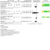
Comparison 12: Sensitivity analysis (performance bias) exercise and adjunctive therapy versus exercise (short‐term), Outcome 2: Physical function
Comparison 13. Sensitivity analysis (performance bias) exercise and adjunctive therapy versus exercise and placebo adjunctive therapy (medium‐term).
| Outcome or subgroup title | No. of studies | No. of participants | Statistical method | Effect size |
|---|---|---|---|---|
| 13.1 Pain | 5 | 414 | Std. Mean Difference (IV, Random, 95% CI) | ‐0.17 [‐0.39, 0.05] |
| 13.1.2 Electrophysical agents | 4 | 352 | Std. Mean Difference (IV, Random, 95% CI) | ‐0.16 [‐0.41, 0.10] |
| 13.1.3 Acupuncture/dry needling | 1 | 62 | Std. Mean Difference (IV, Random, 95% CI) | ‐0.28 [‐0.78, 0.22] |
| 13.2 Physical function | 4 | 354 | Std. Mean Difference (IV, Random, 95% CI) | ‐0.13 [‐0.34, 0.08] |
| 13.2.1 Electrophysical agents | 3 | 292 | Std. Mean Difference (IV, Random, 95% CI) | ‐0.14 [‐0.37, 0.09] |
| 13.2.2 Acupuncture/dry needling | 1 | 62 | Std. Mean Difference (IV, Random, 95% CI) | ‐0.07 [‐0.57, 0.43] |
Comparison 14. Sensitivity analysis (performance bias): exercise and adjunctive therapy versus exercise (medium‐term).
| Outcome or subgroup title | No. of studies | No. of participants | Statistical method | Effect size |
|---|---|---|---|---|
| 14.1 Pain | 1 | 379 | Std. Mean Difference (IV, Random, 95% CI) | ‐0.14 [‐0.34, 0.06] |
| 14.1.1 Other | 1 | 379 | Std. Mean Difference (IV, Random, 95% CI) | ‐0.14 [‐0.34, 0.06] |
| 14.2 Physical function | 1 | 351 | Std. Mean Difference (IV, Random, 95% CI) | ‐0.08 [‐0.29, 0.13] |
| 14.2.1 Other | 1 | 351 | Std. Mean Difference (IV, Random, 95% CI) | ‐0.08 [‐0.29, 0.13] |
Comparison 15. Sensitivity analysis (performance bias) exercise and adjunctive therapy vs exercise and placebo adjunctive therapy (long‐term).
| Outcome or subgroup title | No. of studies | No. of participants | Statistical method | Effect size |
|---|---|---|---|---|
| 15.1 Pain | 1 | 62 | Std. Mean Difference (IV, Random, 95% CI) | ‐0.01 [‐0.51, 0.48] |
| 15.2 Physical function | 1 | 62 | Std. Mean Difference (IV, Random, 95% CI) | ‐0.04 [‐0.54, 0.46] |
Comparison 16. Sensitivity analysis (detection bias): exercise and adjunctive therapy versus exercise and placebo adjunctive therapy (short‐term).
| Outcome or subgroup title | No. of studies | No. of participants | Statistical method | Effect size |
|---|---|---|---|---|
| 16.1 Pain | 9 | 555 | Std. Mean Difference (IV, Random, 95% CI) | ‐0.26 [‐0.55, 0.03] |
| 16.1.1 Electrophysical agents | 7 | 461 | Std. Mean Difference (IV, Random, 95% CI) | ‐0.29 [‐0.63, 0.06] |
| 16.1.2 Acupuncture/dry needling | 1 | 62 | Std. Mean Difference (IV, Random, 95% CI) | 0.00 [‐0.50, 0.50] |
| 16.1.3 Taping | 1 | 32 | Std. Mean Difference (IV, Random, 95% CI) | ‐0.37 [‐1.07, 0.33] |
| 16.2 Physical function | 9 | 551 | Std. Mean Difference (IV, Random, 95% CI) | ‐0.37 [‐0.73, ‐0.01] |
| 16.2.1 Electrophysical agents | 7 | 457 | Std. Mean Difference (IV, Random, 95% CI) | ‐0.48 [‐0.91, ‐0.05] |
| 16.2.2 Acupuncture/dry needling | 1 | 62 | Std. Mean Difference (IV, Random, 95% CI) | 0.03 [‐0.46, 0.53] |
| 16.2.3 Taping | 1 | 32 | Std. Mean Difference (IV, Random, 95% CI) | 0.08 [‐0.61, 0.78] |
Comparison 17. Sensitivity analysis (detection bias): exercise and adjunctive therapy versus exercise (short‐term).
| Outcome or subgroup title | No. of studies | No. of participants | Statistical method | Effect size |
|---|---|---|---|---|
| 17.1 Pain | 4 | 165 | Std. Mean Difference (IV, Random, 95% CI) | ‐0.77 [‐1.36, ‐0.19] |
| 17.1.1 Electrophysical agents | 3 | 108 | Std. Mean Difference (IV, Random, 95% CI) | ‐0.91 [‐1.63, ‐0.19] |
| 17.1.2 Taping | 1 | 57 | Std. Mean Difference (IV, Random, 95% CI) | ‐0.57 [‐1.84, 0.71] |
| 17.2 Physical function | 4 | 165 | Std. Mean Difference (IV, Random, 95% CI) | ‐0.51 [‐0.97, ‐0.05] |
| 17.2.1 Electrophysical agents | 3 | 108 | Std. Mean Difference (IV, Random, 95% CI) | ‐0.41 [‐0.80, ‐0.02] |
| 17.2.2 Taping | 1 | 57 | Std. Mean Difference (IV, Random, 95% CI) | ‐0.75 [‐2.37, 0.88] |
| 17.3 Change scores pain | 1 | 200 | Mean Difference (IV, Random, 95% CI) | 1.32 [‐0.38, 3.01] |
| 17.3.6 Electrophysical agents change scores | 1 | 200 | Mean Difference (IV, Random, 95% CI) | 1.32 [‐0.38, 3.01] |
| 17.4 Change scores physical function | 1 | 200 | Std. Mean Difference (IV, Random, 95% CI) | 0.52 [‐0.26, 1.30] |
| 17.4.4 Electrophysical agents change scores | 1 | 200 | Std. Mean Difference (IV, Random, 95% CI) | 0.52 [‐0.26, 1.30] |
17.3. Analysis.

Comparison 17: Sensitivity analysis (detection bias): exercise and adjunctive therapy versus exercise (short‐term), Outcome 3: Change scores pain
17.4. Analysis.

Comparison 17: Sensitivity analysis (detection bias): exercise and adjunctive therapy versus exercise (short‐term), Outcome 4: Change scores physical function
Comparison 18. Sensitivity analysis (detection bias) exercise and adjunctive therapy versus exercise and placebo adjunctive therapy (medium‐term).
| Outcome or subgroup title | No. of studies | No. of participants | Statistical method | Effect size |
|---|---|---|---|---|
| 18.1 Pain | 5 | 585 | Std. Mean Difference (IV, Random, 95% CI) | ‐0.01 [‐0.17, 0.16] |
| 18.1.2 Electrophysical agents | 3 | 303 | Std. Mean Difference (IV, Random, 95% CI) | ‐0.05 [‐0.27, 0.18] |
| 18.1.3 Acupuncture/dry needling | 2 | 282 | Std. Mean Difference (IV, Random, 95% CI) | ‐0.02 [‐0.40, 0.36] |
| 18.2 Physical function | 4 | 523 | Std. Mean Difference (IV, Random, 95% CI) | ‐0.07 [‐0.24, 0.10] |
| 18.2.1 Electrophysical agents | 2 | 243 | Std. Mean Difference (IV, Random, 95% CI) | ‐0.07 [‐0.33, 0.18] |
| 18.2.2 Acupuncture/dry needling | 2 | 280 | Std. Mean Difference (IV, Random, 95% CI) | ‐0.07 [‐0.30, 0.17] |
Characteristics of studies
Characteristics of included studies [ordered by study ID]
Abbott 2013.
| Study characteristics | ||
| Methods | 2 x 2 factorial RCT, 4 groups | |
| Participants |
Location: New Zealand Number randomised: 206 (knee OA = 113, hip OA = 93) to the following groups: exercise therapy = 50, manual therapy = 54, combined manual and exercise therapy = 51, no trial physiotherapy = 51 Mean age: 66 years (range 37 to 92) Setting: university research clinic Inclusion criteria: men and women with diagnosis of hip or knee OA based on ACR clinical criteria Exclusion criteria: rheumatoid arthritis; previous knee or hip joint replacement surgery of the affected joint; any other surgical procedure on the lower limbs in the previous 6 months; surgical procedure on the lower limbs planned in the next 6 months; initiation of opioid analgesia or corticosteroid or analgesic injection intervention for hip or knee pain within the previous 30 days; physical impairments unrelated to the hip or knee, which would prevent safe participation in exercise, manual therapy, walking or stationary cycling; inability to comprehend and complete study assessments or comply with study instructions; or stated inability to attend or complete the proposed course of intervention and follow‐up schedule |
|
| Interventions |
Exercise therapy: 9 treatment sessions of approximately 50 minutes: 7 in the initial 9 weeks of the trial and 2 ‘booster’ sessions at week 16. The exercise therapy protocol consisted of a multi‐modal, supervised programme of warm‐up/aerobic, muscle strengthening, muscle stretching, and neuromuscular control exercises. Additional exercise therapy interventions were prescribed individually for each participant on the basis of the physical examination findings, from a limited list of interventions. In addition, a home exercise programme was completed 3 times per week. The exercise therapy protocol did not allow therapist‐applied manual forces. Manual therapy: 9 treatment sessions of approximately 50 minutes: 7 in the initial 9 weeks of the trial and 2 ‘booster’ sessions at week 16. The manual therapy protocol consisted of procedures intended to modify the quality and range of motion of the target joint and associated soft tissue structures. Additional manual therapy interventions for the hip, ankle or lumbopelvic region were prescribed individually for each participant randomised to this intervention on the basis of the physical examination findings, from a limited list of interventions defined in the protocol. A home programme of joint range of motion activities was completed 3 times per week. |
|
| Outcomes | At 9 weeks, 6 months and 1 year: Primary outcome: composite WOMAC index Secondary outcomes: numerical pain rating scale (NPRS), WOMAC Pain, WOMAC Function, Patient Global Assessment, Timed Up and Go Test (TUGT), 40 m self‐paced walk test, 30‐second sit to stand test, OMERACT‐OARSI responder criteria Outcomes included in this review
|
|
| Notes | All participants continued to receive routine care offered by their own GP and other healthcare providers. GPs’, surgeons’, other practitioners’ or participants’ use of other interventions were not influenced or restricted, but was monitored. Funding source: funded by research contracts from the Health Research Council of New Zealand (HRC 07/199 and 07/200) and the NewZealand Lottery Grants Board (MR212664) |
|
| Risk of bias | ||
| Bias | Authors' judgement | Support for judgement |
| Random sequence generation (selection bias) | Low risk | Random allocation using TENELEA online randomisation service |
| Allocation concealment (selection bias) | Low risk | Online randomisation service generated and held the randomisation schedule |
| Blinding of participants and personnel (performance bias) All outcomes | Unclear risk | Not reported |
| Blinding of outcome assessment for self‐reported outcomes | Unclear risk | Not reported |
| Blinding of outcome assessment for objective outcomes | Low risk | Outcome assessors were blinded to group allocation and not involved in providing interventions |
| Incomplete outcome data (attrition bias) All outcomes | Low risk | Intention‐to‐treat analysis used. Missing data imputed by multiple imputation. Dropout rate of 6.8% (n = 13). |
| Selective reporting (reporting bias) | Low risk | Published trial protocol published and trial registration ‐ ACTRN12608000130369 |
| Other bias | Low risk | None apparent |
Abbott 2015.
| Study characteristics | ||
| Methods | Factorial RCT, 4 groups | |
| Participants |
Location: New Zealand Number randomised: 75 (exercise only = 19, exercise and booster (n = 19), exercise and manual therapy (n = 18), exercise with booster session and manual therapy (n = 19) Mean age: exercise = 64, exercise and booster = 65, exercise and manual therapy = 61, exercise with booster session and manual therapy = 64. Setting: hospital outpatient physiotherapy department Inclusion criteria: age 40 years or older and meeting the ACR clinical criteria for a diagnosis of knee OA Exclusion criteria: rheumatoid arthritis; previous knee or hip joint replacement surgery of the affected joint; any other surgical procedure on the lower limbs in the previous 6 months; surgical procedure on the lower limbs planned in the next 6 months; initiation of opioid analgesia or corticosteroid or analgesic injection intervention for hip or knee pain within the previous 30 days; physical impairments unrelated to the hip or knee that would prevent safe participation in exercise, manual therapy, walking or stationary cycling; inability to comprehend and complete study assessments or comply study instructions; or stated inability to attend or complete the proposed course of intervention and follow‐up schedule |
|
| Interventions |
Exercise therapy ‐ no booster: 12 x 45‐minute sessions over 9 weeks comprising: 1) aerobic exercise: up to 10 minutes (cycle or walk); 2) strengthening: 3 sets of 10 repetitions of knee extension, hip extension, knee flexion, resistance adjusted as appropriate; 3) stretching: 60‐second passive stretch of knee flexors, knee extensors, ankle plantar flexors; 4) neuromuscular co‐ordination control exercises: 3 sets of 2 minutes from choice (standing, weight shifting, standing balance on uneven surfaces, sidestepping, forward/backward and shuttle walking drills, stair walking) Manual therapy ‐ no booster: 12 x 30‐ to 45‐minute sessions in the first 9 weeks. Techniques used were based on a list of mandatory techniques including joint and soft tissue techniques of the knee region. List of non‐mandatory techniques were based on assessment findings (hip, ankle, lumbo‐pelvic region). Exercise therapy ‐ booster: similar exercise therapy used with 8 consecutive sessions in the first 9 weeks, 2 booster sessions at 5 months and 1 booster at 11 months for a total of 12 sessions Manual therapy ‐ booster: similar manual therapy techniques used, with 8 consecutive sessions in the first 9 weeks, 2 booster sessions at 5 months, 1 booster session at 8 months and 1 booster session at 11 months, for a total of 12 sessions |
|
| Outcomes | At 1 year: Primary outcome: WOMAC total score (version NRS 3.1) Secondary outcomes: number needed to treat (NNT) to gain: 1) additional OMERACT‐OARSI responder criteria, pain measured using numerical pain rating scale (NPRS), Timed Up and Go (TUG), 40‐metre self‐paced walk, 30‐second sit‐to‐stand, adverse events Outcomes included in this review
|
|
| Notes | Funding source: supported in part by the New Zealand Lottery Grants Board, the New Zealand Society of Physiotherapists Scholarship Trust, the Health Research Council of New Zealand, and a University of Otago Research Grant. The first author was supported in part by a Sir Charles Hercus Health Research Fellowship from the Health Research Council of New Zealand | |
| Risk of bias | ||
| Bias | Authors' judgement | Support for judgement |
| Random sequence generation (selection bias) | Low risk | Random allocation sequence generated using on online service; blocked randomisation (random blocks of 8 or 12) used |
| Allocation concealment (selection bias) | Low risk | Random allocation by person not involved in patient assessment or treatment; random sequence concealed from recruitment staff |
| Blinding of participants and personnel (performance bias) All outcomes | High risk | Due to the nature of the interventions, it was not possible to blind treatment providers to group allocation |
| Blinding of outcome assessment for self‐reported outcomes | Unclear risk | Not reported |
| Blinding of outcome assessment for objective outcomes | Low risk | Assessors were trained in assessment methods and blinded to group allocation |
| Incomplete outcome data (attrition bias) All outcomes | High risk | 9/75 (12%) dropped out. Missing data imputed for primary outcome. Only the results of those who completed analyses were reported for secondary outcomes. |
| Selective reporting (reporting bias) | Low risk | Registered trial protocol: ACTRN12612000460808 |
| Other bias | Low risk | None apparent |
Adedoyin 2002.
| Study characteristics | ||
| Methods | RCT, 2 groups | |
| Participants |
Location: Nigeria Number randomised: 30 (15 to each group: exercise + inferential therapy and exercise + placebo interferential therapy) Setting: physiotherapy department Inclusion criteria: X‐ray confirmed OA, referred to physiotherapy by physician, pain for 3 months or more Exclusion criteria: very acute inflammation, fever or tumours, pregnant women, and people wearing cardiac pacemakers or metallic implants |
|
| Interventions |
Exercise therapy: mobilisation exercise twice weekly with at least 24 hours in between Interferential therapy: treatment duration was 20 minutes. Patients were positioned on a plinth. Two pairs of electrodes were placed lateromedially and anteroposteriorly around the knee and secured with velcro. The intensity of the stimulus was gradually increased until the patients felt an appreciable sensation. Frequency was set at 100 Hz and pulse length was one‐thirtieth of a second for the first 15 minutes of treatment. The stimulus was then reduced to 80 Hz for the next 5 minutes while other parameters remained unchanged. Placebo interferential therapy: all parameters were the same as active except that intensity was not increased. Patients were positioned so that they could see the lights on the dial without being able to read the dial. |
|
| Outcomes | At 4 weeks: pain severity measured on a 10‐point VAS (0 to 10) Outcomes included in this review
|
|
| Notes | Funding support: not reported | |
| Risk of bias | ||
| Bias | Authors' judgement | Support for judgement |
| Random sequence generation (selection bias) | High risk | Patients were assigned alternately to the treatment groups |
| Allocation concealment (selection bias) | Unclear risk | Not reported |
| Blinding of participants and personnel (performance bias) All outcomes | Unclear risk | Not reported |
| Blinding of outcome assessment for self‐reported outcomes | Unclear risk | Not reported |
| Blinding of outcome assessment for objective outcomes | Low risk | Not relevant: no objective outcomes |
| Incomplete outcome data (attrition bias) All outcomes | Unclear risk | Not reported |
| Selective reporting (reporting bias) | Unclear risk | No published trial protocol |
| Other bias | Low risk | None apparent |
Adedoyin 2005.
| Study characteristics | ||
| Methods | RCT, 3 groups | |
| Participants |
Location: Nigeria Number randomised: 51 (exercise = 17, exercise + TENS = 15, exercise + IFT = 19) Mean age: 55 years Setting: physiotherapy department Inclusion criteria: X‐ray confirmed unilateral OA of the knee, had not previously received electrical stimulation, referred to physiotherapy by physician, pain for 3 months or more Exclusion criteria: cardiopulmonary dysfunction, acute inflammation, fever or tumours, pregnant women and people wearing cardiac pacemakers |
|
| Interventions |
Exercise therapy: 2 sessions a week for 4 weeks for a total of 8 sessions. Quadriceps strengthening using metallic interchangeable weights fastened to ankle of affected leg. The DeLorme and Watkins principle of 10‐repetitive maximum was used to determine and standardise the weight used by each participant. Participants sat on the edge of plinth with back fully supported and 2 hands holding the edge of the plinth, isometric exercises were performed from a start position of 0° knee flexion. Participant was helped by an investigator to lift the weight from 90° to 180° extension. Extension was sustained for 10 seconds before the leg was lowered and rested for 5 seconds. This was done for 10 bouts (1 bout comprised 10 contractions) with a rest of 2 minutes between each bout. Strengthening was followed by at least 5 minutes cycling on cycle ergometer with an initial resistance of 25 W for 6 minutes. Resistance was increased to 35 W, 45 W and 55 W in weeks 2 to 4, based on recommendations by Pollok and Wilmore. No home exercise programme was given as different rates of compliance may have affected study results. Interferential therapy (IFT): treatment duration was 20 minutes. Two treatment sessions were given per week for 4 weeks for a total of 8 sessions, with at least 24 hours between each session. Two electrodes, with moist pads, were applied to either side of the knee and aligned longitudinally with the limb and secured with Velcro straps. Beat frequency was set at 80 Hz. The intensity of the stimulus was gradually increased until the patients felt a strong but comfortable sensation. TENS: procedure and application of TENS was precisely the same as for IFT. Stimulation frequency was 80 Hz continuous, phase duration was 200 ms. Current intensity was strong but comfortable. A different therapist used IFT and TENS for all patients. |
|
| Outcomes | At weeks 1, 2, 3, 4: WOMAC total score (Likert version), 10‐point pain intensity scale Outcomes included in this review
|
|
| Notes | Patients were asked to refrain from use of analgesic drugs during the study period and patients verbally confirmed that they had complied with this request Funding support: not reported |
|
| Risk of bias | ||
| Bias | Authors' judgement | Support for judgement |
| Random sequence generation (selection bias) | Unclear risk | Not reported |
| Allocation concealment (selection bias) | Unclear risk | Not reported |
| Blinding of participants and personnel (performance bias) All outcomes | Unclear risk | Not reported |
| Blinding of outcome assessment for self‐reported outcomes | Unclear risk | Not reported |
| Blinding of outcome assessment for objective outcomes | Low risk | No objective outcomes |
| Incomplete outcome data (attrition bias) All outcomes | High risk | 46/51 completed and data analysed, completed data for 90.2%. Five dropouts (2 from exercise group, 3 from IFT group) |
| Selective reporting (reporting bias) | Unclear risk | No published trial protocol |
| Other bias | Low risk | None apparent |
Adhya 2015.
| Study characteristics | ||
| Methods | RCT, 4 groups | |
| Participants |
Location: India Number randomised: 200 (50 to the following 4 groups: exercise only, exercise + PEME, exercise + IFT, exercise + US) Mean age: not reported Setting: physiotherapy department Inclusion criteria: confirmed OA knee by radiologic investigation, no history of any knee surgery, pain duration of 3 months or more Exclusion criteria: reported bleeding disorders, local malignancy, fever, tumours, pregnant women, excessive obesity (BMI > 40) or any co‐morbidity not allowing proper exercise protocol, cardiac pacemakers, metallic implants, abnormal skin sensation, obvious deformity, history of knee surgery or knee trauma, hip or ankle instability, excessive weakness, hip or knee replacement, intra‐articular joint injection within 4 weeks of the study, inadequate communication skills in Hindi /Punjabi, Illiterate (nor have any literate caregivers), unable to comply with exercise protocol |
|
| Interventions |
Exercise therapy: all patients were given the exercise programme to follow at home daily. Isometric exercises for hamstrings, quadriceps (10 seconds hold, 20 repetitions, 3 sets each), hip abductors dynamic strengthening exercises (20 repetitions, 3 sets for each leg, with 1 kg.wt), free ROM exercises 10 repetitions. The patients wore knee caps while in weight‐bearing position for a longer time as advised (standing and walking). PEME: 3 times a week for 8 weeks; treatment dosages as suggested by previous research, 11 m waves at 27.12 MHz and 150 watts for 15 minutes with condenser electrodes US: 3 times a week for 8 weeks. The regions for application of therapeutic ultrasonic therapy were selected according to locations of tender points, the knee divided in 4 quadrants noted on orthopaedic clinical examination. US was delivered at a frequency of 1.1 MHz, duty cycle 100%, effective radiating area (ERA) 4.0 cm2, intensity 1.00 W/cm2, treatment time 7.30 minutes IFT: 3 time a week for 8 weeks. IFT was delivered at a frequency of 100 Hz and pulse length of 1/30th of a second for the first 15 minutes of their treatment session. The stimulus was then reduced to 80 Hz for the next 5 minutes while other parameters remained unchanged. The intensity of the stimulus was gradually increased until the patients felt an appreciable sensation (moderate pins and needles). |
|
| Outcomes | Post‐intervention (8 weeks): knee pain severity (VAS) after standing or walking at parallel bars for 5 minutes, OMAC total score Outcomes included in this review
|
|
| Notes | All patients were sent to clinical psychologists for routine counselling. Funding support: none |
|
| Risk of bias | ||
| Bias | Authors' judgement | Support for judgement |
| Random sequence generation (selection bias) | Unclear risk | Not reported |
| Allocation concealment (selection bias) | Unclear risk | Not reported |
| Blinding of participants and personnel (performance bias) All outcomes | Unclear risk | Not reported |
| Blinding of outcome assessment for self‐reported outcomes | Unclear risk | Not reported |
| Blinding of outcome assessment for objective outcomes | Low risk | No objective outcomes |
| Incomplete outcome data (attrition bias) All outcomes | Unclear risk | Not reported |
| Selective reporting (reporting bias) | Unclear risk | Not reported |
| Other bias | Low risk | None apparent |
Akaltun 2021.
| Study characteristics | ||
| Methods | Double‐blind, placebo RCT, 2 groups | |
| Participants |
Location: Turkey Number randomised: 40 (20 to exercise and high intensity laser therapy (HILT), and exercise and placebo HILT) Mean age: exercise + HILT = 57.8 ± 8.07; exercise + placebo HILT = 58.2 ± 11.28 Setting: physical medicine and rehabilitation clinic Inclusion criteria: grade 2 to 3 knee OA according to Kellgren‐Lawrence classification; aged 45 to 75 years, knee pain for at least 6 months, VAS score of 3 or more Exclusion criteria: history of surgery or traumatic injury, inflammatory arthritis (i.e. rheumatoid arthritis, psoriatic arthritis), intra‐articular injection in the knee in the last 6 months, history of cancer, bleeding diathesis, psychiatric disease, disease that would limit their participation in exercises (i.e. severe chronic obstructive pulmonary disease, severe heart failure, cerebrovascular event history), problems in the hip and ankle, participation in another physical therapy programme in the last 3 months |
|
| Interventions |
Exercise therapy: patients received 5 treatment sessions per week for 2 weeks (total of 10 treatments). A standard exercise programme was given to the patients by the same researcher. The exercise programme included ROM exercises, stretching, strengthening and flexibility exercises. The exercise programme started with active ROM exercises for the lower limb joints in supine and prone positions in a pain‐free range. The exercises were continued with straight leg raising, quadriceps setting, pillow squeeze, heel raising, one leg balance, step up and quadriceps strengthening exercises. Each exercise was performed 10 times/set for 3 sets in total. A 2‐minute rest period was given after each set. Patients were instructed to continue the same programme at home. HILT: patients received 5 treatment sessions per week for 2 weeks (total of 10 treatments). The 1064 nm wavelength Nd:YAG laser (BTL‐6000 high intensity laser, with a maximum power of 12 W) was used. The biostimulation and analgesic modes were used in the study. The application was made continuously with circular motions in both modes. The analgesic mode was applied for treatments 1 to 3. A total of 300 J was applied as 12 J/m2 25 cm2 at a frequency of 25 Hz in these sessions. The biostimulation mode was used for treatments 4 to 10. A total of 3000 J was applied as 120 J/cm2 in this mode. Placebo HILT: patients received 5 treatment sessions per week for 2 weeks (total of 10 treatments). The same device was applied in the form of a sham laser that did not radiate a laser beam in the placebo application. |
|
| Outcomes | Post‐intervention (2 weeks) and 6 weeks: pain severity (VAS), WOMAC physical function, stiffness and pain subscales (Likert version), flexion ROM, femoral cartilage thickness using ultrasound Outcomes included in this review
|
|
| Notes | Funding support: not reported | |
| Risk of bias | ||
| Bias | Authors' judgement | Support for judgement |
| Random sequence generation (selection bias) | Unclear risk | Insufficient information provided ("randomisation was made with envelope pulling procedure") |
| Allocation concealment (selection bias) | Unclear risk | Not reported |
| Blinding of participants and personnel (performance bias) All outcomes | Unclear risk | Not reported |
| Blinding of outcome assessment for self‐reported outcomes | Unclear risk | Not reported |
| Blinding of outcome assessment for objective outcomes | Unclear risk | Not reported |
| Incomplete outcome data (attrition bias) All outcomes | Low risk | No dropouts |
| Selective reporting (reporting bias) | Unclear risk | No trial protocol available |
| Other bias | Low risk | None apparent |
Akyol 2010.
| Study characteristics | ||
| Methods | RCT, 2 groups | |
| Participants |
Location: Turkey Number randomised: 40 (20 to each group ‐ exercise + short‐wave diathermy (SWD) and exercise) Mean age: SWD and exercise = 58 years, exercise only = 57 years Setting: inpatient physiotherapy department Inclusion criteria: bilateral knee OA based on ACR criteria. Radiographic criteria Kellgren‐Lawrence Gr 1 to 3 (Gr 4 excluded) Exclusion criteria: serious systemic medical conditions for whom exercises would be contra‐indicated; neuromuscular or dermatologic disease that involved the lower extremities, exercise programme that caused increase in muscle strength in previous months, inflammatory arthropathy, contracture, history of trauma and physiotherapy within previous 6 months, metallic implant around the knee joint, implanted cardiac pacemaker, grade 4 OA, intra‐articular injection in the previous 3 months, inability to understand how to score symptoms |
|
| Interventions |
Exercise: isokinetic muscle strengthening programme 3 times a week for 4 weeks: 10 reps of conc/conc in angular velocity 60°/s, 90°/s, 120°/s, 150°/s, 180°/s for flexors and extensors. 10‐second rest between different modes of training and 10 minutes rest between right and left knees. Short‐wave diathermy (SWD): patients were positioned supine on the treatment plinth with knees extended, with a towel wrapped around the knee. Induction coil applied circularly along the affected leg. Frequency of 27.12 mHz. Intensity based on each participant's sensation of warmth (mild but pleasant sensation of heat). SWD was applied to each knee separately for 20 minutes, so total treatment time equalled 40 minutes for both knees, 3 times a week for 4 weeks. |
|
| Outcomes | At 4 weeks (post‐treatment) and 3 months: VAS pain (0 to 10 cm), WOMAC pain, stiffness and physical function subscales (Likert version), SF‐36 quality of life, 6‐minute walk test (6MWT), isokinetic knee flexor and extensor strength, Beck Depression score Outcomes included in this review
|
|
| Notes | Use of NSAIDs, other analgesics and antidepressants was not permitted during the study period. Any pre‐treatment with these drugs had to be discontinued 7 days before the start of the study. Use of other drugs for co‐morbid diseases was permitted during the study period. Funding support: not reported |
|
| Risk of bias | ||
| Bias | Authors' judgement | Support for judgement |
| Random sequence generation (selection bias) | Unclear risk | Randomised using concealed envelopes: unclear how random sequence was generated |
| Allocation concealment (selection bias) | Unclear risk | Not reported |
| Blinding of participants and personnel (performance bias) All outcomes | Unclear risk | Not reported |
| Blinding of outcome assessment for self‐reported outcomes | Unclear risk | Not reported |
| Blinding of outcome assessment for objective outcomes | Low risk | Patients were assessed 3 times by the same physician who was blinded to treatment |
| Incomplete outcome data (attrition bias) All outcomes | Low risk | No dropouts |
| Selective reporting (reporting bias) | Unclear risk | No published protocol or trial registration |
| Other bias | Low risk | None apparent |
Al‐Rashoud 2014.
| Study characteristics | ||
| Methods | Double‐blind RCT, 2 groups | |
| Participants |
Location: Saudi Arabia Number randomised: 58 (30 to exercise + active laser, 28 to exercise + placebo laser) Setting: security forces hospital Mean age: 54 years Inclusion criteria: men and women, with knee OA according to the ACR criteria, K‐L grade 2 to 4, and knee pain and functional disability for at least 3 months. Average pain intensity of 3 or more on a 10c m VAS, ability to perform all movements in the evaluation form, ability to read and understand the patient information sheet and sign a consent form. Exclusion criteria: previous knee surgery, serious valgus or varus deformity, disease where laser treatments contraindicated (cancer, uncontrolled diabetes mellitus, hypertension, etc.) and current use of medications that might interfere with laser therapy treatment (e.g. corticosteroid injections) |
|
| Interventions |
Exercise therapy: home‐based programme of straight leg raise, 5 times daily for 11 weeks Active laser therapy: patient was in a supine position. Energy was irradiated over the joint line at 5 acupuncture points of the synovial region of the medial side of the knee and 4 points on the lateral side, at 1.2J per point for 40 seconds. Total dose per knee was 6 J per treatment. Placebo laser: same procedures were applied, but the device was inactive and only produced visible red light |
|
| Outcomes | End of laser intervention (3 weeks) and end of exercise intervention (11 weeks): Primary outcome: pain severity on a 10 cm VAS Secondary outcomes: Saudi Knee Function Scale (SKFS), active knee flexion using goniometry, patient satisfaction on a verbal numeric scale (0% to 100%) Outcomes included in this review
|
|
| Notes | Patients were allowed to take analgesics required for severe pain and to discontinue any other medication related to their knee pain (physicians were consulted at this point) Funding support: scholarship granted by the General Department of Medical Services of Ministry of Interior, Security Forces Hospital, Riyadh, Saudi Arabia |
|
| Risk of bias | ||
| Bias | Authors' judgement | Support for judgement |
| Random sequence generation (selection bias) | Low risk | Randomisation list was produced using software‐generated randomised numbers. Randomisation depended on random blocks of 10. |
| Allocation concealment (selection bias) | Unclear risk | Patients were enrolled by research assistant. Unclear who randomised patients. |
| Blinding of participants and personnel (performance bias) All outcomes | Low risk | Neither the investigator nor patient knew whether a placebo or active treatment was being administered; only the research assistant had the identifying code to determine which treatment was given. The same laser device was used for both groups and placebo and active emitters looked identical. No heat, sound or vibration was detectable. |
| Blinding of outcome assessment for self‐reported outcomes | Low risk | Neither the investigator nor patient knew whether a placebo or active treatment was being administered; only the research assistant had the identifying code to determine which treatment was given. The same laser device was used for both groups and placebo and active emitters looked identical. No heat, sound or vibration was detectable. |
| Blinding of outcome assessment for objective outcomes | Unclear risk | Unclear who administered outcomes |
| Incomplete outcome data (attrition bias) All outcomes | High risk | 58 were randomised and 49 (84.5%) were analysed, 26 in active group and 23 in the placebo group |
| Selective reporting (reporting bias) | Low risk | Clinical trial registration number ISRCTN24010862. All outcomes relevant to this review were reported. |
| Other bias | Low risk | None apparent |
Alfieri 2020.
| Study characteristics | ||
| Methods | Single‐blinded RCT, 2 groups | |
| Participants |
Location: Brazil Number randomised: 48 (24 to each group ‐ exercise + geotherapy, and exercise) Mean age: geotherapy and exercise = 65 years, exercise only = 62 years Setting: university clinic Inclusion criteria: formal diagnosis of OA Exclusion criteria: any other chronic disease, total or partial prosthesis in one or both knees or hips, heart disease, uncontrolled hypertension, rheumatoid arthritis, fibromyalgia, neurological diseases affecting mobility |
|
| Interventions |
Exercise therapy: 15 twice‐weekly sessions, comprising supervised stretching exercises for flexor and extensor muscles of hips and knees, for plantar flexors and ankle extensor muscles. Participants also performed isotonic and isometric strengthening exercises for these same muscle groups using their own body weight as resistance. This part of the intervention lasted 20 minutes, followed by a 10‐minute walking circuit, diverting from mattresses, hula‐hoops, ladders and cones, to develop co‐ordination and proprioception. Geotherapy: a poultice of Brazilian dolomite powder (300 g per person), mixed with hot water (50 ml, 37 degrees C) was applied to the knees for 25 minutes |
|
| Outcomes | Post‐intervention: pain severity (VAS), WOMAC pain, stiffness and physical function subscales (Likert version), pressure pain thresholds (PPT), Lequesne Algofunctional Index Outcomes included in this review
|
|
| Notes | Funding support: this research did not receive any specific grant from funding agencies in the public, commercial, or not‐for‐profit sectors | |
| Risk of bias | ||
| Bias | Authors' judgement | Support for judgement |
| Random sequence generation (selection bias) | Unclear risk | Not reported |
| Allocation concealment (selection bias) | Unclear risk | Not reported |
| Blinding of participants and personnel (performance bias) All outcomes | Unclear risk | Not reported |
| Blinding of outcome assessment for self‐reported outcomes | Unclear risk | Not reported |
| Blinding of outcome assessment for objective outcomes | Low risk | An evaluator was blinded to the intervention group each participant had been assigned to |
| Incomplete outcome data (attrition bias) All outcomes | Low risk | Intention‐to‐treat analysis was performed |
| Selective reporting (reporting bias) | Unclear risk | No trial protocol available |
| Other bias | Low risk | None apparent |
Alfredo 2018.
| Study characteristics | ||
| Methods | Double‐blind RCT, 2 groups | |
| Participants |
Location: Brazil Number randomised: 46 (24 in exercise + active laser, 22 in exercise + placebo laser) Mean age: exercise and active laser = 61 years; exercise and placebo laser = 62 years Setting: special rehabilitation services Inclusion criteria: men and women, aged 50 to 75 years, with knee OA according to the ACR criteria and Kellgren Lawrence Grade 2 to 4 and knee pain and functional disability for at least 3 months Exclusion criteria: cancer, diabetes, symptomatic hip osteoarthritis, or used antidepressants, anti‐inflammatory medications or anxiolytics during 6 months prior to enrolment |
|
| Interventions |
Exercise: exercise was delivered after the 3 weeks of laser (or placebo) in both groups. It was divided into 3 phases: P‐1, P‐2 and P‐3 during 8 weeks with 3 sessions a week. Each session lasted 45 minutes: 10 minutes warming‐up (treadmill, ergometer bike or rowing machine); 30 minutes 2 to 3 sets with P‐1, P‐2 or P‐3; 5 minutes stretching (hamstrings, quadriceps, adductors and gastrocnemius). Laser therapy: energy was irradiated over the joint line at 5 points of the synovial region of the medial side of the knee and 4 points on the lateral side, at 3 J per point. Total dose per knee was 27 J per treatment and used previously calibrated equipment. Placebo laser: procedures were identical but without emission of energy |
|
| Outcomes | 6 months: Primary outcome: pain severity on a 10 cm VAS Secondary outcomes: Lequesne Algofunctional Index, WOMAC pain, function and stiffness subscales, knee ROM, Isometric quadriceps muscle strength using a portable dynamometer Outcomes included in this review
|
|
| Notes | Funding support: financially supported by Fundacao de Amparo a Pesquisa do Estado de São Paulo (FAPESP) – Foundation of Research Support of São Paulo State and Coordenacao de Aperfeicoamento de Pessoal de Nıvel Superior (CAPES) – Coordination for the Improvement of Higher Level, or Education, Personnel. | |
| Risk of bias | ||
| Bias | Authors' judgement | Support for judgement |
| Random sequence generation (selection bias) | Unclear risk | Insufficient information to permit judgement ("Randomisation was performed using sealed randomly filled envelopes describing the treatment group") |
| Allocation concealment (selection bias) | Low risk | Randomisation done by investigator not involved in assessment, treatment or diagnosis |
| Blinding of participants and personnel (performance bias) All outcomes | Low risk | "Patients and the physiotherapist responsible for the evaluation were unaware of randomisation results" |
| Blinding of outcome assessment for self‐reported outcomes | Low risk | "Patients and the physiotherapist responsible for the evaluation were unaware of randomisation results" |
| Blinding of outcome assessment for objective outcomes | Low risk | A physiotherapist blinded to group allocation performed all examinations |
| Incomplete outcome data (attrition bias) All outcomes | High risk | Laser dropout = 4/24 (16.67%), placebo = 2/22 (9.1%); no ITT and no detail provided on flow diagram |
| Selective reporting (reporting bias) | Low risk | Trial registered CT01306435 |
| Other bias | Low risk | None apparent |
Alghadir 2014.
| Study characteristics | ||
| Methods | Single blind RCT, 2 groups | |
| Participants |
Location: Saudi Arabia Number randomised: 40 (20 to exercise and active laser, 20 to exercise and placebo laser) Mean age: active laser = 55 years, placebo laser = 57 years Setting: single centre, physical therapy department Inclusion criteria: knee OA according to ACR criteria; knee OA of grade II or III according to the Kellgren‐Lawrence scale, minimum score of 25 on the WOMAC total score, knee pain ≥ 4 on VAS in the previous 3 months; and willingness to participate and follow the treatment schedule Exclusion criteria: concomitant disease affecting the knee (such as rheumatoid arthritis, injury, and/or surgery to the knee) and had received physical therapy and/or intra‐articular corticosteroid or hyaluronic acid injections during the last 6 months, history of cancer, dementia, neurological deficits, heart pacemaker, diabetes mellitus, uncontrolled hypertension or morbid obesity (BMI ≥ 40) |
|
| Interventions |
Exercise therapy: home‐based exercise programme of isometric knee extension and straight leg raising 10 times/et for 3 sets, with 2‐minute rest interval. Contraction maintained for 10 seconds, rest for 5 seconds. Photographic details of the exercises were provided along with a diary log‐book. Patients were encouraged to record the number of days the exercises were performed per week. Active laser therapy: 2 treatments per week for 4 weeks for a total of 8 treatments. Hot packs wrapped in towelling were placed on the target knee(s) for 20 minutes followed by laser therapy. Patients in the active laser group received irradiation with a Ga–As laser device, wavelength of 850 nm, power of 100 mW, and spot size of 1.0 mm. Eight points were irradiated; 3 on the medial side of the knee, 3 on the lateral side of the knee, and 2 on the medial edge of the tendon of the biceps femoris muscle and semitendinosus muscle in the popliteal fossa. Each point received energy of 6 J/point for 60 s, with a total dose of 48 J/cm2 in each session. Placebo laser therapy: the same treatment was applied in the placebo group except that the machine was not switched on |
|
| Outcomes | At 4 weeks: Primary outcome: pain severity on a 10 cm VAS Secondary outcomes: WOMAC pain, stiffness and physical function subscales (Likert version), 50 foot walk test Outcomes included in this review
|
|
| Notes | All patients were advised to keep their activity level and medication (2 g paracetamol) unchanged during the study period Funding support: Deanship of Scientific Research, King Saud University |
|
| Risk of bias | ||
| Bias | Authors' judgement | Support for judgement |
| Random sequence generation (selection bias) | Low risk | Sealed, randomly filled envelopes describing the treatment groups |
| Allocation concealment (selection bias) | Unclear risk | Not reported |
| Blinding of participants and personnel (performance bias) All outcomes | Unclear risk | Placebo laser intervention but blinding not explicitly stated |
| Blinding of outcome assessment for self‐reported outcomes | Unclear risk | Not reported |
| Blinding of outcome assessment for objective outcomes | Unclear risk | Not reported |
| Incomplete outcome data (attrition bias) All outcomes | Low risk | 40/40 completed the study |
| Selective reporting (reporting bias) | Low risk | Trial registration ACTTRN12612000615886 |
| Other bias | Low risk | None apparent |
Altinbilek 2018.
| Study characteristics | ||
| Methods | Single‐blind RCT, 2 groups | |
| Participants |
Location: Turkey Number randomised: 100 (50 to exercise and osteopathic manipulative treatment (OMT), 50 to exercise only) Mean age: exercise only = 54 years, exercise + OMT = 56 years Setting: not reported Inclusion criteria: bilateral knee OA, OA based on ACR criteria, grade 2 and 3 K‐L Exclusion criteria: inflammatory arthritis, soft tissue rheumatism, any inflammation in the knee joint, higher erythrocyte sedimentation rate (ESR) and C‐reactive protein (CRP), history of knee surgery, trauma (meniscopathy or instability), intra‐articular intervention or physical therapy within the last 6 months. Patients using anti‐inflammatory drugs other than simple analgesics, those using knee braces, patients with vascular and cardiovascular disease, paresis or neuropathy, intra‐articular neoplasm, osteonecrosis, mental mood disorder and those with knee contracture |
|
| Interventions |
Exercise therapy: 2 days a week for a total of 4 sessions comprising quadriceps isometric strengthening, straight leg lifting, iliotibial band, hamstring stretching, strengthening abductor and adductor muscle of the hip. Stretching was applied as a 10‐repetition x 3 sets. The home programme was to be completed twice a day at home. OMT: OMT was delivered as 3 minutes mobilisation and 3 minutes compression for bilateral patellofemoral and tibiofemoral joint respectively with 1‐minute intervals in addition to the exercise programme. Following these applications, 2‐minute bilateral lower extremity pumping technique was performed. All these applications were taught to the patient to apply twice a day at home. |
|
| Outcomes | Post‐intervention and 4 weeks follow‐up: VAS for pain severity, 50 m walk test, WOMAC pain, stiffness and physical function subscales Outcomes included in this review
|
|
| Notes | The patients were prevented from taking nonsteroidal anti‐inflammatory drugs (NSAIDS) 1 week before beginning of the study and during the study period. They were allowed to take paracetamol up to 3 g daily for pain control. The drugs they used due to systemic diseases were continued. Funding support: none |
|
| Risk of bias | ||
| Bias | Authors' judgement | Support for judgement |
| Random sequence generation (selection bias) | Low risk | Randomisation done using online randomisation software (www.randomisation.org) |
| Allocation concealment (selection bias) | Unclear risk | Not reported |
| Blinding of participants and personnel (performance bias) All outcomes | Unclear risk | Not reported |
| Blinding of outcome assessment for self‐reported outcomes | Unclear risk | Not reported |
| Blinding of outcome assessment for objective outcomes | Low risk | All evaluations were made before treatment and at follow‐up points by a physiatrist who was blind to the treatment method. Exercises were applied and taught by another physiatrist and orthopaedic manual therapy (OMT) provided by a third physiatrist who had completed osteopathy training. |
| Incomplete outcome data (attrition bias) All outcomes | High risk | Dropout rate: exercise group = 6/50 (12%), exercise + OMT group = 9/50 (18%). No ITT. |
| Selective reporting (reporting bias) | Unclear risk | No trial protocol |
| Other bias | Low risk | None apparent |
Atamaz 2012.
| Study characteristics | ||
| Methods | Double‐blind RCT, 6 groups | |
| Participants |
Location: Turkey Number randomised: 203 (exercise + TENS = 37, exercise + sham TENS = 37, exercise + IFT = 31, exercise + sham IFT = 35, exercise + SWD = 31, exercise + sham SWD = 32) Mean age: TENS = 62 years, TENS sham = 61 years, IFT = 62 years, IFT sham = 61 years, SWD = 62 years, SWD sham = 61 years Setting: multicentre (4 centres), departments of physical medicine and rehabilitation Inclusion criteria: aged 50 to 80 years with knee OA, based on ACR criteria and radiologically confirmed K‐L Gr 2 or 3 and symptomatic with at least 4 cm of pain severity on the VAS for at last 6 months Exclusion criteria: any experience with electrotherapy, history of any contraindication for electrotherapy, had received corticosteroid therapy or chondro‐protective agents during the 30 days prior to the study or visco supplementation treatment within 6 months prior to the study, had undergone previous major surgery, such as joint replacement or arthroscopy, within 6 months prior to the study, diagnosis of joint infection, a specific condition (neoplasm, diabetes mellitus, paresis, osteonecrosis, recent trauma, etc.), ascertained/suspected pregnancy or lactation, and poor general health status that would interfere with the functional assessments during the study. These exclusion criteria were verified by history, physical examination and radiograph. |
|
| Interventions |
Exercise therapy: carried out in groups of 4 or 5 patients under the guidance of a physiotherapist 3 times a week for 3 weeks. 1) 5‐ to 6‐minute jogging period, 2) stretching exercises of the lower extremity muscles (10 minutes) in a standing position, 3) isometric quadriceps exercises (10 to 15 repetitions) in the seated position, with a towel rolled under the knee, for 10 seconds with 10‐second breaks between holds in a progressive manner, 4) chair lift and mini‐squats exercises (10 to 15 repetitions) were performed as muscle strength exercises. At the end of the 3‐week group exercise period, the physiotherapist prescribed a home‐based training programme (3 times a week) as well as group exercise. All patients also received a complete set of pre‐made exercise cards showing all exercises, to ensure that the training programme would be done properly. At each visit during the study, the patients were instructed to perform their exercises regularly. Active intervention‐ 3 separate interventions (TENS, interferential (IFT), SWD): physical therapy agents were applied 5 times a week for 3 weeks by the same physiotherapist at each centre Active TENS was administered at a frequency of 80 Hz with 10 to 30 mA intensity for 20 minutes. Four surface electrodes (5 x 5cm) were placed over the painful area in the knee region with intensity in the tactile sensation threshold Active IFT was applied for 20 minutes with an amplitude‐modulated frequency of 100 Hz generated by 4 kHz sinusoidal waves. Two electrodes (8 x 6cm) were placed over the knee region with intensity in the tactile sensation threshold. Active SWD: patients sat on a treatment chair that had an opaque screen between the chair and the SWD machine. The screen was used to blind the patient from viewing the procedure being performed on the SWD machine by the physical therapist. Each patient in the treatment group sat on a chair and placed his/her legs on a table during treatment, while receiving continuous SWD with a 10 cm diameter condenser plate operating at a frequency of 27.12 MHz, an input of 300 W, and a mean output of 3.2 W. Sham physical therapy was delivered 5 times a week for 3 weeks IFT sham treatment consisted of the placement of the same pads for the same time; however, no electrical stimulation was applied to the probes TENS sham group received sham stimulation at the same sites for the same duration and period as the TENS group SWD sham group received a sham SWD treatment, which had exactly the same treatment procedure as the treatment group, except that the power switch was off |
|
| Outcomes | 1, 3 and 6 months: Primary outcome: pain severity on 100 mm scale (VAS) Secondary outcomes: WOMAC pain and physical function subscales, Nottingham Health Profile (NHP) pain and physical function subscales, 15 m walk test, active pain‐free knee flexion Outcomes included in this review
|
|
| Notes | Patients were asked to discontinue any pre‐treatment NSAIDs 7 days before the start of the study. If they required additional analgesia due to knee pain, paracetamol was allowed on the condition that they noted intake on the study form. Funding support: Ege, Docuz Eylul and Adam Menderes Universities and Sisli Etfal education and research hospital |
|
| Risk of bias | ||
| Bias | Authors' judgement | Support for judgement |
| Random sequence generation (selection bias) | Low risk | Randomisation was performed by the principal centre using adaptive assignment into 6 groups. Patients were assigned a unique 2‐part number. Screened for eligibility at their second visit and principal centre contacted again to allocate a randomisation number. |
| Allocation concealment (selection bias) | Low risk | Principal centre (in multicentre study) did randomisation |
| Blinding of participants and personnel (performance bias) All outcomes | Low risk | All patients, investigators and analysts were blinded, with the exception of the data and safety monitoring board member, but these members did not assess the patients |
| Blinding of outcome assessment for self‐reported outcomes | Low risk | All patients, investigators and analysts were blinded, with the exception of the data and safety monitoring board member, but these members did not assess the patients |
| Blinding of outcome assessment for objective outcomes | Low risk | All patients, investigators and analysts were blinded, with the exception of the data and safety monitoring board member, but these members did not assess the patients |
| Incomplete outcome data (attrition bias) All outcomes | Low risk | Dropout: group 1 (4/37, 10.8%); group II (8/37, 21.6%), group III (1/35, 2.8%), group IV (4/31, 12.9%), group V (7/32, 21.9%), group VI (4/31, 12.9%). ITT analysis done. |
| Selective reporting (reporting bias) | Unclear risk | No published protocol or trial registration |
| Other bias | Low risk | None apparent |
Avelar 2011.
| Study characteristics | ||
| Methods | RCT, 2 groups | |
| Participants |
Location: Brazil Number randomised: 23 (12 to exercise and whole body vibration training, 11 to exercise only) Setting: not reported Mean age: exercise = 75 years; exercise and vibration training = 71 years Inclusion criteria: age 60 years or more, diagnosed with OA in at least 1 knee in accordance with the ACR clinical and radiographic criteria with radiographic OA grades 1 to 4, based on K‐L grading scale, no recent knee injury; not requiring a walking aid; self‐report of not having been submitted to any rehabilitation procedure in the previous 3 months; and not having used glucocorticoids for at least 2 months prior the study Exclusion criteria: any orthopaedic, neurological, respiratory or acute cardiac diseases that would preclude study participation and if they had any cognitive deficit as determined by the Mini‐Mental Status Examination |
|
| Interventions |
Exercise therapy: 3 times a week on alternate days for 12 weeks. Warm up on stationary bike at 70% maximum heart rate, predicted for age using a Polar HR monitor for 10 minutes. The session consisted of progressively increasing time and repetitions of the squat exercise. During each exercise repetition, an examiner instructed individuals to perform 3 seconds of isometric flexion of the quadriceps to 60 degrees and 3 seconds of isometric flexion of the quadriceps to 10 degrees. Intensity increased over 12 weeks by increasing number of repetitions and reducing resting time. Vibration platform: 3 times a week on alternate days for 12 weeks. Same exercises as exercise group, except that squatting exercises were also done on a vibration platform; frequency of 35 to 40 Hz, amplitude of 4 mm, and acceleration that ranged from 2.78 G to 3.26 G. The intensity of squatting exercise training was systematically augmented in the vibration and exercise groups over the 12‐week study period by increasing the number of repetitions and reducing the resting time. In the vibration group, acceleration was also increased by varying the vibration frequency (35 to 40 Hz). |
|
| Outcomes | Baseline = 0 (3 weeks prior to initial training), pre‐training (3 weeks), post ‐training (15 weeks): Primary outcome: WOMAC pain and function subscales Secondary outcomes: 6‐Minute Walk Test (6MWT), Chair Stand test, Berg Balance Scale (BBS), Timed Up and Go (TUG) Outcomes included in this review WOMAC pain and physical function subscales. Scales not specified, but most likely that higher scores representer higher pain and disability levels. |
|
| Notes | Funding support: supported by FAPEMIG, CNPq and CAPES | |
| Risk of bias | ||
| Bias | Authors' judgement | Support for judgement |
| Random sequence generation (selection bias) | Unclear risk | Details of the randomisation procedure not reported |
| Allocation concealment (selection bias) | Unclear risk | Not reported |
| Blinding of participants and personnel (performance bias) All outcomes | Unclear risk | Not reported |
| Blinding of outcome assessment for self‐reported outcomes | Unclear risk | Not reported |
| Blinding of outcome assessment for objective outcomes | Unclear risk | Not reported |
| Incomplete outcome data (attrition bias) All outcomes | Low risk | 2/23 dropouts (8.7%), 1 from each group. No ITT analysis. |
| Selective reporting (reporting bias) | Low risk | Protocol registered ACTNR12610000475044 |
| Other bias | Low risk | None apparent |
Bennell 2016.
| Study characteristics | ||
| Methods | Assessor‐blinded, 3‐arm, parallel‐group RCT | |
| Participants |
Location: Australia Number randomised: 222 (Pain Coping Skill Training (PCST) + exercise = 73, exercise only = 75, PCST only = 74) Mean age: exercise only = 63 years, PCST = 63 years, PCST + exercise = 65 years Setting: physiotherapy departments in University of Melbourne and University of Queensland Inclusion criteria: age 50 years or older with OA of the knee, based on ACR criteria (pain on most days in the last month and radiographic changes), knee pain ≥ 3 months, average pain during previous week ≥ 40 on 100 mm visual analogue scale (VAS), and at least moderate difficulty with daily activities (Western Ontario and McMaster Universities Osteoarthritis Index (WOMAC) physical function subscale ≥ 25 of 68 units) Exclusion criteria: systemic arthritic conditions such as rheumatoid arthritis; medical condition precluding safe exercise such as uncontrolled hypertension or heart condition; self‐reported history of serious mental illness, such as schizophrenia, or self‐reported diagnosis of current clinical depression; neurological condition such as Parkinson’s disease, multiple sclerosis or stroke; knee surgery including arthroscopy within the past 6 months or total joint replacement; awaiting or planning any back or lower limb surgery within the next 12 months; current or past (within 3 months) oral or intra‐articular corticosteroid use; physiotherapy, chiropractic or acupuncture treatment or exercises specifically for the knee within the past 6 months; walking exercise for > 30 minutes continuously daily; participating in a regular (more than twice a week) structured and/or supervised exercise programme such as attending exercise classes in a gym or use of a personal trainer; participating in or previous participation in a formal PCST programme; inability to walk unaided; inadequate written and spoken English; inability to comply with the study protocol such as inability to attend physiotherapy sessions or attend assessment appointments at the University |
|
| Interventions |
Exercise therapy: 10 individual sessions of 25 minutes duration were delivered by a physiotherapist over 12 weeks. Home exercises were prescribed 4 times per week, aiming for a dosage of 3 sets of 10 repetitions, during the 12‐week treatment phase, reducing to 3 times/week during the 9‐month follow‐up. Exercise comprised 6 exercises to strengthen the quadriceps, hamstrings and hip abductor muscles, performed 4 times weekly for 12 weeks and 3 times weekly thereafter. Weights and resistance elastic bands as well as exercise handouts were provided. Physiotherapists telephoned participants at weeks 22, 38 and 46 to enhance adherence to the exercise programme. Pain Coping Skills Training (PCST) was delivered for 10 x 45‐minute sessions over 12 weeks covering pain education and training in cognitive and behavioural pain coping skills (activity‐rest cycling, pleasant activity scheduling, problem‐solving, identifying and challenging negative thoughts, developing coping thoughts, pleasant imagery, counting backwards and auditory stimulation) and their application. Participants were asked to practice skills daily during the 12 weeks and then as needed during follow‐up. |
|
| Outcomes | At 12 and 52 weeks: Primary outcomes: VAS pain, WOMAC physical function subscale (Likert version 3.1) Secondary outcomes: VAS average pain on walking, WOMAC pain subscale, AQOL‐6D (Assessment of Quality of Life), PASE (Physical Activity Scale for the Elderly (PASE), Arthritis Self‐Efficacy Scale ASES), Pain Catastrophising Scale (PCS), coping attempts score of Coping Strategies Questionnaire (CSQ), Depression, Anxiety, Stress Scale (DASS), 20‐m fast‐paced walking test, maximum quadriceps isometric strength Outcomes included in this review
|
|
| Notes | Adherence, adverse events and co‐interventions were recorded in a logbook during treatment and via questionnaire during follow‐up. Healthcare costs and direct non‐healthcare costs over the previous month were collected via customised questionnaire at weeks 0, 4, 8, 12, 32 and 52 Funding support: supported by Australian Health Management, National Health and Medical Research Council (631717) |
|
| Risk of bias | ||
| Bias | Authors' judgement | Support for judgement |
| Random sequence generation (selection bias) | Low risk | Computer‐generated random number table |
| Allocation concealment (selection bias) | Low risk | Allocations were concealed in opaque, consecutively numbered envelopes by an independent person and stored in a locked location. A different independent person opened the next sequential envelope and informed the therapists. |
| Blinding of participants and personnel (performance bias) All outcomes | High risk | Unable to blind physiotherapists. Patients were blinded only to study hypothesis. |
| Blinding of outcome assessment for self‐reported outcomes | Unclear risk | Not reported |
| Blinding of outcome assessment for objective outcomes | Low risk | Blinded assessors collected strength and physical performance data and entered questionnaire data |
| Incomplete outcome data (attrition bias) All outcomes | Low risk | Loss to follow‐up was 9% at week 12, 18% at week 32 and 16% at week 52. ITT analysis with missing data imputed using chained equations with predictive means matching, imputing for each arm separately. |
| Selective reporting (reporting bias) | Low risk | Published trial protocol; Trial Registration Number ACTRN126100000533099 |
| Other bias | Low risk | None apparent |
Bennell 2017.
| Study characteristics | ||
| Methods | Parallel pragmatic RCT, 2 groups | |
| Participants |
Location: Australia Randomised: 168 (exercise + telephone coaching = 84, exercise = 84) Mean age: exercise = 61 years and exercise + telephone coaching = 63 years Setting: physiotherapy clinics Inclusion criteria: age 50 years+, average knee pain 4+ on an 11‐point numeric rating scale, ACR clinical criteria for knee OA, and a classification as sedentary or insufficiently physically active according to the Active Australia Survey (AAS; activity < 150 minutes or < 5 sessions in the past week) Exclusion criteria: inability to safely participate in moderate‐intensity exercise (SportsMedicine Australia stage I pre‐exercise screening questions) undertaking regular lower‐extremity strengthening exercises or receiving non‐drug management for knee pain from a health professional more than once within the past 6 months; knee surgery or intra‐articular corticosteroid injection within the past 6 months; history of joint replacement on study knee or on waiting list; systemic arthritic conditions or current or past (within 4 weeks) oral corticosteroid use; other condition affecting lower‐extremity function more than knee pain; unable to use/access a telephone; and a score of 21+ on the depression subscale of the Depression, Anxiety and Stress Scale |
|
| Interventions |
Exercise therapy: 5 individual 30‐minute sessions over 6 months (at weeks 1, 3, 7, 12 and 20). Evidence‐based progressive individualised home exercise programme comprising 4 to 6 lower‐extremity exercises (at least 3 knee extensor strengthening exercises and at least 1 hip abductor strengthening exercise from a predetermined list with 1 to 2 optional exercises based on assessment) performed 3 times per week and promoted increased general physical activity, including provision of a pedometer for optional self‐monitoring/motivation, and assistance with formulating short‐term goals Telephone coaching: 6 telephone‐delivered coaching sessions over 6 months (at, approximately, weeks 2, 4, 8, 13, 21 and 25), with the option of up to 6 additional sessions. The coaching intervention used HealthChange methodology, which integrates theories/principles commonly used in behavioural interventions, and which impact health literacy, readiness, motivation, decision‐making and self‐efficacy to treatment adherence. The approach drew on techniques used in motivational interviewing, solution‐focused counselling and cognitive‐behavioural therapy. Three crucial components of facilitating behavioural change were addressed: effective information exchange, assistance in forming a behavioural goal intention, and support in converting the intention into action and maintenance. Although participants were educated about target recommendations (30 minutes of moderate‐intensity physical activity in bouts of 10 minutes on most days and 10,000 steps per day), goals were individualised. Participants were encouraged to monitor their progress and assisted with identifying individual barriers as well as strategies to overcome barriers. Following each consultation, physiotherapists and coaches completed an online communication form outlining topics discussed and any problems, as well as other relevant information, such as goals and adherence, to facilitate 2‐way interaction. |
|
| Outcomes | 6, 12 and 18 months: 11 point NRS for pain, WOMAC physical function and pain, pain on walking, Physical Activity Scale for the Elderly (PASE), global rating of change 7‐point ordinal scale Outcomes included in this review
|
|
| Notes | Funding support: supported by the National Health and Medical Research Council (grant 631717) | |
| Risk of bias | ||
| Bias | Authors' judgement | Support for judgement |
| Random sequence generation (selection bias) | Low risk | Randomised in permuted blocks of 6 and 12 using a computer‐generated random numbers table, stratified by a physiotherapist and prepared by a biostatistician |
| Allocation concealment (selection bias) | Low risk | Allocations were sealed in opaque, consecutively numbered envelopes by an independent person and stored in a locked location. A different independent person access the envelopes to first reveal group allocation and then the coach if applicable. |
| Blinding of participants and personnel (performance bias) All outcomes | High risk | Blinded only to study hypothesis |
| Blinding of outcome assessment for self‐reported outcomes | High risk | Participants were blinded only to study hypothesis |
| Blinding of outcome assessment for objective outcomes | Low risk | Accelerometry based data recorded for physical activity at baseline and 3 months. Researchers entering data were blinded to group allocation. |
| Incomplete outcome data (attrition bias) All outcomes | Low risk | ITT analyses with missing data imputed using chained equations with predictive means matching, imputing data for each arm separately. Estimates from 20 imputed data sets were combined using Rubin's rules. |
| Selective reporting (reporting bias) | Low risk | Published trial protocol and trial registration, ANZCTR 12612000308897 |
| Other bias | Low risk | None apparent |
Brosseau 2012.
| Study characteristics | ||
| Methods | Single blind RCT, 3 groups | |
| Participants |
Location: Canada Randomised: 222 (walking programme + behavioural intervention = 75, walking programme = 81, control = 84) Mean age: 63.4 years Setting: community‐based Inclusion criteria: 1) confirmed diagnosis with mild to moderate unilateral or bilateral OA according to the ACR clinical and radiographic/magnetic resonance imagery criteria; 2) reported pain for at least 3 months; 3) expected his/her medication to change during the study period; 4) demonstrated an ability to ambulate for a minimum of 20 minutes, at their own pace with minimal reports of pain (≤ 3 out of 10 on a visual analogue pain rating scale); 4) were able to be treated as an outpatient; and 5) were available 3 times a week over a period of 12 months Exclusion criteria: 1) participated in regular physical or aerobic sports ≥ 2 times per week for > 20 minutes per session during the previous 6 months; 2) severe OA of the knee or other weight‐bearing joints of the lower extremity; 3) no written consent from their physician to participate in the study; 4) pain at rest or at night; 5) received rehabilitation treatment, corticosteroids injection, or any other pain‐related treatment besides medication for arthritis within the last 12 months; 6) uncontrolled hypertension (systolic blood pressure > 160 mm Hg confirmed by the screening initial VO2 max test at the Ottawa Heart Institute); 7) other illnesses, such as rheumatoid arthritis (judged by the patient or study physician to make participation in this study inadvisable); 8) significant cognitive deficit resulting in an inability to understand or comply with instructions; 9) surgery planned in the next year; 10) intention to move away from Ottawa region in the next year; 11) inability to communicate in English or French; or 12) unwillingness to sign informed consent. |
|
| Interventions |
Exercise therapy: supervised walking programme at 2 walking clubs (Ottawa and Quebec), 3 times a week for 12 months, supervised by physical activity specialist. It comprised 10‐minute warm‐up of light aerobic exercise, followed by 45 minutes aerobic walking, 10 minute cool down consisting of light aerobic exercise and stretching. Target intensity was 50% to 70% of each participant's pre‐determined heart rate. Two stages: progressive aerobic stage where duration and heart rate increased over time and a 'maintenance phase' where duration and heart rate remained constant for the remainder of the walking programme. Participants wore heart rate monitors. All groups were given pedometers and educational pamphlet and log book to measure walking in minutes and other physical activity. Behavioural intervention: part of a multifaceted knowledge translation intervention and was implemented using the adapted Program for Arthritis Control through Education and Exercise (PACEex) programme. The behavioural intervention was integrated into the PACEex programme and consisted of the following components: 1) short‐ and long‐term goal setting during PACEex classes; 2) an educational component, delivered by a trained instructor, involving consisting of instructional sessions about the benefits of PA, specifically walking; 3) monthly face‐to‐face counselling wherein participants received moral support/encouragement to adherence with physical activity. Potential barriers to programme adherence were identified, and self‐management strategies were reviewed to overcome those barriers; 4) goal setting and telephone counselling were provided as an additional source of social support until the end of the supervised phase. As with the face‐to‐face counselling, barriers were identified and strategies were shared in an effort to promote the long‐term maintenance of physical activity. Individual long‐term goal setting was discussed at the beginning of the PACEex programme and was followed by monthly face‐to‐face meetings throughout the first 6 months of the programme. The last 6 months of the 12‐month supervised phase consisted of participants receiving counselling via telephone discussing long‐term goals and barriers/facilitators to adhere to the walking programme. Control (dissemination strategy): participants received the educational pamphlet |
|
| Outcomes | At 3 months, 6 months, 9 months, 12 months (all during intervention period), 15 and 18 months (3 + 6 months post intervention); AIMS‐2, SF‐36, WOMAC (pain, physical function, stiffness subscales and total score), 6‐minute walk, TUG, 7‐day Physical Activity Recall (7‐day PAR) Outcomes included in this review
|
|
| Notes | Funding support: Canadian Institute of Health Research, University research Chair, Ministry of Human Resources (summer student programme), Canada | |
| Risk of bias | ||
| Bias | Authors' judgement | Support for judgement |
| Random sequence generation (selection bias) | Low risk | Central randomisation and computer‐generated numbers were used |
| Allocation concealment (selection bias) | Low risk | Central randomisation and computer‐generated numbers were used |
| Blinding of participants and personnel (performance bias) All outcomes | Unclear risk | Not reported |
| Blinding of outcome assessment for self‐reported outcomes | Unclear risk | Not reported |
| Blinding of outcome assessment for objective outcomes | Low risk | Independent evaluator was blinded for outcome assessments |
| Incomplete outcome data (attrition bias) All outcomes | Low risk | Incomplete data were included and assumed missing at random. ITT analysis was performed. Very high dropout rate across 3 groups: 39.1%, 44.3% and 51.4%. |
| Selective reporting (reporting bias) | Low risk | Trial registration IRSCTN 09193542 |
| Other bias | Low risk | None apparent |
Cakir 2014.
| Study characteristics | ||
| Methods | Double‐blind, factorial RCT, 3 groups | |
| Participants |
Location: Turkey Randomised: 60 (20 in each group: exercise and continuous US, exercise and pulsed US, exercise and sham US) Setting: department of physical medicine and rehabilitation Mean age: exercise and continuous US = 57 years, exercise and pulsed US = 58 years, exercise and sham US = 57 years Inclusion criteria: knee pain for at least 6 months, diagnosed knee OA according to ACR confirmed radiologically (K‐L Gr 2 or 3), aged 40 to 80 years Exclusion criteria: an experience of any physical therapy agent, intra‐articular corticosteroid therapy or chondro‐protective agents during the 30 days prior to the study or visco supplementation treatment within 6 months prior to the study, diagnosis of joint infection, neoplasm, diabetes mellitus, paresis, osteonecrosis, recent trauma, ascertained/suspected pregnancy or lactating and poor general health status, any history of contraindication of heat therapy or previous major surgery |
|
| Interventions |
Exercise therapy: home exercise programme (HEP) including quadriceps isometric exercise, muscle strength exercises (chair lift and mini‐squats exercises) and stretching exercises of the lower extremity muscles at least for 3 times per week. To ensure that exercises were learned properly, a complete set of pre‐made exercise cards which showed all exercises were also provided. At the following visits, the patients were instructed to regularly perform their exercises. Continuous ultrasound: delivered by a physiotherapist using a 5 cm2 head, 1 MHx, 1 W/cm2, delivered 5 times a week for 2 weeks for approx 12 minutes over the knee region with full contact in a supine position Pulsed ultrasound: same parameters except a 1:4 pulse ratio was used Sham ultrasound: same treatment procedure as for continuous and pulsed, except that the machine was switched off |
|
| Outcomes | End of treatment (2 weeks) and 6 months: WOMAC pain subscale, VAS pain at rest, VAS pain on movement, WOMAC physical function subscale, 20 m walk test, VAS disease severity Outcomes included in this review
|
|
| Notes | Patients were not allowed take NSAIDs during therapy period or within 1 week before treatment. Paracetamol use up to 2000 mg/day was allowed. Other drugs for systemic diseases were not stopped. Funding support: not reported |
|
| Risk of bias | ||
| Bias | Authors' judgement | Support for judgement |
| Random sequence generation (selection bias) | Unclear risk | Randomisation was carried out using covariate adaptive randomisation with Frane method. No further detail provided. |
| Allocation concealment (selection bias) | Unclear risk | "All patients, investigators and analysts were blinded with the exception of the safety monitoring board member, but this member did not assess the patient". Insufficient information to make decision. |
| Blinding of participants and personnel (performance bias) All outcomes | Low risk | "All patients, investigators and analysts were blinded with the exception of the safety monitoring board member, but this member did not assess the patients" |
| Blinding of outcome assessment for self‐reported outcomes | Low risk | "All patients, investigators and analysts were blinded with the exception of the safety monitoring board member, but this member did not assess the patients" |
| Blinding of outcome assessment for objective outcomes | Low risk | "All patients, investigators and analysts were blinded with the exception of the safety monitoring board member, but this member did not assess the patients" |
| Incomplete outcome data (attrition bias) All outcomes | Low risk | 2/60 (3.3%) dropouts reported. Tables show that data were analysed for all 60 participants. |
| Selective reporting (reporting bias) | Unclear risk | No published protocol or trial registration |
| Other bias | Low risk | None apparent |
Carlos 2012.
| Study characteristics | ||
| Methods | RCT, 3 groups | |
| Participants |
Location: Brazil Setting: not reported Randomised: 30 men and women, 10 in each group (exercise + continuous US, exercise + pulsed US, exercise only) Mean age: exercise = 63 years, exercise + continuous US = 64 years, exercise + pulsed US = 63 years Inclusion criteria: men and women age 50 to 75 years, grades 2 to 4 of OA, pain in the knee and reduction of functional capacity in the last 3 months Exclusion criteria: diabetes mellitus, dementia, cancer, uncontrolled systemic arterial hypertension, morbid obesity, symptomatic hip OA and patients who used anti‐inflammatory or anxiolytic drugs in the 6 months prior to the initial assessment |
|
| Interventions |
Exercise therapy: 3 times a week for 8 weeks. All groups performed the same isotonic exercise programme, respecting the limitations of each patient. Each exercise was performed in 2 series, with 30 repetitions. The duration of each exercise was 45 minutes (10 minutes of warm‐up on treadmill or exercise bike, 30 minutes of exercise, 5 minutes of stretching (hamstrings, quadriceps, hip adductors, gastrocnemius)) Continuous US: 1 MHz, average power output 7 W, 1.5 W/cm2 (spatial average) in continuous mode (100%), applied for 5 minutes, 3 times a week for 4 weeks to the medial side and 5 minutes to the lateral side of the knee Pulsed US: 1 MHz, average power output 7 W, 2.5 W/cm2 (spatial and temporal average), pulsed mode (20%), frequency of pulse repetition at 100 Hz, applied for 5 minutes, 3 times a week for 4 weeks |
|
| Outcomes | Post‐treatment: WOMAC pain, stiffness, physical function and total WOMAC, Lequesne Functional Index, Knee Range of Motion (ROM) using a universal goniometer, Timed Up and Go (TUG), quadriceps strength at 10, 60 and 90 degrees (hand‐held dynamometer) Outcomes included in this review WOMAC pain and physical function subscales. Scale not specified, the higher the scores the greater the impact on quality of life. |
|
| Notes | Patients were instructed not to use any type of analgesia or anti‐inflammatory drug during the study Funding support: not reported |
|
| Risk of bias | ||
| Bias | Authors' judgement | Support for judgement |
| Random sequence generation (selection bias) | Unclear risk | Insufficient information provided to reach a judgement |
| Allocation concealment (selection bias) | Low risk | "Subjects were randomly assigned by a researcher who was not involved in treatment or evaluation. The patient had no access to the result" |
| Blinding of participants and personnel (performance bias) All outcomes | Unclear risk | "Subjects were randomly assigned by a researcher who was not involved in treatment or evaluation. The patient had no access to the result" |
| Blinding of outcome assessment for self‐reported outcomes | Unclear risk | "Subjects were randomly assigned by a researcher who was not involved in treatment or evaluation. The patient had no access to the result" |
| Blinding of outcome assessment for objective outcomes | Low risk | "Subjects were randomly assigned by a researcher who was not involved in treatment or evaluation. The evaluating therapist had no access to the result" |
| Incomplete outcome data (attrition bias) All outcomes | Low risk | All 30 patients were reported in the analyses. Attrition rates not reported. ITT analyses not described in the statistical analyses. |
| Selective reporting (reporting bias) | Unclear risk | No published trial protocol |
| Other bias | Low risk | None apparent |
Castrogiovanni 2016.
| Study characteristics | ||
| Methods | RCT, 3 groups | |
| Participants |
Location: Italy Number randomised: 66 into 3 groups with 22 in each group: exercise + Kinesiotape (KT) with tension application (stabilising effect), exercise + KT without tension application (draining effect), exercise only Setting: orthopaedics traumatology and rehabilitation unit /physical medicine and rehabilitation centre Median age: 63 years Inclusion criteria: pain and functional impairment with knee OA (patellofemoral and tibiofemoral compartments), without concomitant chronic conditions, grade II or grade III OA of the knee based on K‐L grading scale Exclusion criteria: allergic reaction to tape or any skin problem, presence of any inflammatory arthritis, history of any injection at the knee, surgical intervention of the knee within the last 6 months, suspicion regarding other pathologies in the knee, severe obesity, grade IV OA, full instability of the knee joint, trauma to the knee during the study or when patients, failure to follow treatment instructions |
|
| Interventions |
Exercise therapy: all patients during the 3 months of the KT treatment received a moderate adapted or “tailor‐made” training programme for knee OA. They received 3 training sessions per week (1 hour each, supervised by physiatrists, kinesiologists and physical therapists) over 3 months, which were carried out in order to reduce the pressure on the affected knee, refrain from kneeling, as well as training in special exercises, such as hamstring and calf stretches and quadriceps strengthening. Training was moderately adapted or “tailor‐made” based on patient's tolerance in line with EULAR and OARSI guidelines. The programme included a 10‐minute warm‐up (biking exercise), 20‐minute mild leg exercise training (leg press, leg curl, leg extension, hip abduction, calf raises, squats and stepping exercises), 15‐minute cool‐down (treadmill walking) and 15‐minute flexibility training. Each leg was exercised separately to prevent an unequal distribution of load between the least affected and most affected sides. To guarantee safety, training loads were progressively increased, and tolerability was assessed at every training session. KT (stabilising effect): 2 ‘Y‐strips’ with a length of approximately 20 cm, an anchor of 2 cm and a single longitudinal section continuing from one end for a specified distance along the centre of the tape; 1 ‘I‐strip’ that has no cut down the middle of the tape. KT was applied with tension application. After shaving the area, patients were lying in the supine position with knee in maximal flexion. The first strip was a Y‐strip representative of the quadriceps, the tail of the strip was applied to the patella, wrapping the patella medially and laterally with 25% tension. The base of the strip was applied with paper‐off tension towards the anterior superior iliac spine. For the second strip, a Y‐strip was applied with the knee flexed at 90 degrees between the tibial tuberosity and inferior pole of the patella. The tails of the second strip were applied wrapping patella medially and laterally with 25% tension. The tails were directed towards vastus medialis and vastus lateralis. For the third strip, an I‐strip was applied with the knee flexed at 30 degrees. The strip was applied to patella mediolaterally with 75% tension in the middle and paper‐off tension at the ends. KT (draining effect): 2 ‘fan‐shaped strips’ approximately 30 cm long, an anchor of 2 cm and with 4 longitudinal sections to obtain 5 tails, were applied in the anterior region of the knee; 1 ‘fan‐shaped strip’ approximately 20 cm long, an anchor of 2 cm and with 4 longitudinal sections to obtain 5 tails were applied in the posterior region of the knee. No tension was applied to provide a lymph draining effect. After shaving the area, during the anterior application, the patients were sitting with the knee flexed to 110 degrees. The anchor of the first tape was applied 1 cm laterally to the centre line of the quadriceps muscle, so that the centre of the tape corresponds to the patella. The anchor of the second tape was placed 1 cm medially to the centre line of the quadriceps muscle, so that the centre of the tape corresponded to the patella. During the posterior application, patients were standing with knee extended. The base or anchor of the third tape was applied to the posterior aspect of the thigh so that half of the tape length corresponds to the posterior region of the knee (popliteal fossa). Tapes were replaced at each session (2 per week) and were retained during the week. Where the tape separated, the patient was referred to renew it. |
|
| Outcomes | Baseline, 15 days and 3 months (post‐treatment): pain intensity (VAS), WOMAC, Timed Up and Go (TUG), analgesic usage Outcomes included in this review
|
|
| Notes | Funding support: Finanziamento della Ricerca d'Ateneo” (FIR) 2016 (code 4E8207), University of Catania, Catania, Italy | |
| Risk of bias | ||
| Bias | Authors' judgement | Support for judgement |
| Random sequence generation (selection bias) | Unclear risk | No information on random sequence generation provided |
| Allocation concealment (selection bias) | Unclear risk | Not reported |
| Blinding of participants and personnel (performance bias) All outcomes | Low risk | Patients did not know to which group they were allocated |
| Blinding of outcome assessment for self‐reported outcomes | Low risk | Patients did not know to which group they were allocated |
| Blinding of outcome assessment for objective outcomes | Low risk | "All scores were assessed blindly by one physiastrist, one kinesiologist and one physical therapist at beginning of treatment, 15 days and at 3 months. The tests were repeated in triplicate". |
| Incomplete outcome data (attrition bias) All outcomes | Low risk | All those were randomised were followed up |
| Selective reporting (reporting bias) | Unclear risk | No trial protocol available |
| Other bias | Low risk | None apparent |
Cetin 2008.
| Study characteristics | ||
| Methods | Single‐blind RCT, 5 groups (2 groups eligible for review) | |
| Participants |
Location: Turkey Number randomised: 100 women into 5 groups of 20 (SWD + hot packs + exercise, TENS + hot packs + exercise, ultrasound + hot packs + exercise, hot packs + exercise, exercise only) Setting: Department of Physical Medicine and Rehabilitation at a university Mean age: 60 years Inclusion criteria: OA based on ACR clinical and radiographic criteria, ability to walk at least 100 m on an even surface, full or near‐full passive range of motion at each knee Exclusion criteria: no previous history of knee surgery, lower extremity arthroplasty or intra‐articular injection of hyaluronic acid or steroids in the last 6 months |
|
| Interventions |
Exercise therapy (Group 5): individual exercise delivered by physiotherapist, 20 minutes, 3 times a week for 8 weeks comprising: warm up on stationary bike for 20 cycles/minute for 5 minutes followed by isokinetic strengthening for knee flexor and extensors. Intensity increased from 1 to 5 sets during the first through 5 sessions and remained at 5 sets through the remaining 19 sessions. Each set consisted of 5 repetitions of concentric contractions of the knee extensors and flexors at angular velocities of 60, 120 and 180 degrees/second. Rest for 20 seconds between sets and for 60 seconds between left and right knee was provided. Hot pack (Group 4) provided 3 times a week for 8 weeks prior to exercise. No other detail was provided. |
|
| Outcomes | At 8 weeks (post‐treatment): Lequesne Index of Severity (disability) for knee OA, VAS pain, isokinetic strength, 50 m walk test Outcomes included in this review
|
|
| Notes | Patients were instructed to continue taking any current medications and not to start any new therapies for knee OA during the 8‐week study Funding support: not reported |
|
| Risk of bias | ||
| Bias | Authors' judgement | Support for judgement |
| Random sequence generation (selection bias) | Unclear risk | Not reported |
| Allocation concealment (selection bias) | Unclear risk | Not reported |
| Blinding of participants and personnel (performance bias) All outcomes | Unclear risk | Not reported |
| Blinding of outcome assessment for self‐reported outcomes | Unclear risk | Not reported |
| Blinding of outcome assessment for objective outcomes | Low risk | Patients were evaluated at the end of the treatment sessions by the physician who was blinded with regard to the type of treatment the patients would receive. |
| Incomplete outcome data (attrition bias) All outcomes | High risk | 15/100 (15% withdrew). Number of withdrawals per group not reported. No ITT analyses. |
| Selective reporting (reporting bias) | Unclear risk | No published protocol or trial registration |
| Other bias | Low risk | None apparent |
Cheawthamai 2014.
| Study characteristics | ||
| Methods | RCT, 2 groups | |
| Participants |
Location: Thailand Number randomised: 54 (27 to each group; exercise + self manual therapy, exercise only) Mean age: exercise = 64 years, exercise + manual therapy = 67 years Setting: not reported Inclusion criteria: females with a diagnosis of knee OA based on the ACR clinical criteria Exclusion criteria: systemic joint diseases, cerebrovascular conditions, Parkinson's disease, received corticosteroid injections within the last 30 days, had lower limb surgery, or back or limb injury within the previous 6 weeks |
|
| Interventions |
Exercise therapy: home‐based exercise comprising stretching, ROM and strengthening exercises. Stretches: 10 seconds x 3 x 5 times: (hams in supine, quads in prone and calf in standing); range of motion (ROM): 10 times per set (2 sets): knee mid‐flexion to full extension and full flexion to full extension, standing terminal knee extension, seated leg press, partial squat and step‐ups. Strengthening: static quads in knee extension. Exercise intensity (holding times and repetitions) were determined according to the participant's tolerance, severity of joint limitation and soft tissue tightness. They were instructed to walk daily with gradually increased distances. Patients were contacted by phone to ask about adverse events at 2nd, 5th and 8th weeks. Self‐manual therapy: patellar and tibiofemoral joint mobilisations, soft tissue massage (joint capsule, gastrocnemius, iliopsoas, iliotibial band and tensor fascia band). Patients were taught and practised the manual therapy techniques until they could perform correctly. |
|
| Outcomes | At 2, 4 and 12 weeks: VAS pain, KOOS, 6MWT, SF‐36, Active Knee ROM. Outcomes included in this review
|
|
| Notes | Patients were instructed to keep taking any current medication for OA and physical therapy intervention whilst enrolled in the study Funding support: not reported |
|
| Risk of bias | ||
| Bias | Authors' judgement | Support for judgement |
| Random sequence generation (selection bias) | Unclear risk | Not reported |
| Allocation concealment (selection bias) | Unclear risk | Sealed envelopes were used. Insufficient information to make judgement. |
| Blinding of participants and personnel (performance bias) All outcomes | Unclear risk | Not reported |
| Blinding of outcome assessment for self‐reported outcomes | Unclear risk | Not reported |
| Blinding of outcome assessment for objective outcomes | Unclear risk | Not reported |
| Incomplete outcome data (attrition bias) All outcomes | High risk | 11/54 (20.3%) dropout; 5/27 (18.5%) from the exercise group and 6/27 (22.2%) from the exercise and manual therapy group. No ITT analysis. |
| Selective reporting (reporting bias) | High risk | Poor reporting of satisfaction and SF‐36 outcomes. Reported significant differences only. No published trial protocol or trial registration. |
| Other bias | Low risk | None apparent |
Cheing 2002.
| Study characteristics | ||
| Methods | RCT, 4 groups (2 groups eligible for review: exercise only and TENS + exercise) | |
| Participants |
Location: Hong Kong Number randomised: 66 (16 to TENS, 18 to placebo TENS, 17 to exercise and TENS, 15 to exercise) Mean age: TENS = 65 years, placebo TENS = 64 years, exercise = 61 years, exercise + TENS = 64 years Setting: hospital Inclusion criteria: knee OA for more than 6 months, grade II or more on K‐L scale, been stable on medication for 3 weeks before entering the study, received no paramedical treatment within the previous 2 weeks Exclusion criteria: prior knee surgery and/or received a steroid injection during the previous 3 weeks, prior experience of using TENS |
|
| Interventions |
Exercise therapy: 20 sessions (5 days a week over 4 weeks) for approx 30 minutes per session. 10 isokinetic warm‐up knee extension exercises, starting from 90° of knee flexion through the available pain‐free range, at a speed of 180°/s. Three submaximal isometric quadriceps contractions of increasing intensity were followed by 6 maximal 5‐second isometric quadriceps contractions. Most patients achieved their peak torque within 5 seconds. The isometric quadriceps contractions were repeated with the knee flexed at 30°, 60° and 90° respectively. The isometric peak torque for the hamstrings was then performed with the knee flexed to 90°. Active TENS: stimulation was given in continuous trains of 140 μs square pulses at 80 Hz for 60 minutes. Four surface electrodes, 4 x 4 cm each, were placed on the following acupuncture points: spleen 9, stomach 35, extra 31, 32 and gallbladder 34 (one electrode pad covering both extra 32 and stomach 35). The intensity of TENS was adjusted to produce a tingling sensation that was approximately 3 to 4 times the subject’s sensory threshold. Each participant received the treatment around the same time of the day throughout the treatment period, to avoid the fluctuation of pain intensity during the day. In order to blind the participants from the placebo effect, all participants were told that they might or might not feel the stimulation. 20 minutes of rest was provided before exercise. Placebo TENS: same sites for same duration and period as active TENS group using identical TENS units, with internal lamp lit up when being switched on. However, the internal circuit was disconnected by the manufacturer for the purpose of the study. |
|
| Outcomes | Sessions 1, 10, 20 and 4 weeks: VAS pain Outcomes included in this review VAS pain (10 cm line, anchored with 'no pain' at the left end and 'pain as bad as it could be' at the right). Higher scores represent greater pain severity. |
|
| Notes | Funding support: research grant from the Hong Kong Polytechnic University | |
| Risk of bias | ||
| Bias | Authors' judgement | Support for judgement |
| Random sequence generation (selection bias) | Unclear risk | Not reported |
| Allocation concealment (selection bias) | Unclear risk | Not reported |
| Blinding of participants and personnel (performance bias) All outcomes | Unclear risk | Not reported |
| Blinding of outcome assessment for self‐reported outcomes | Unclear risk | Not reported |
| Blinding of outcome assessment for objective outcomes | Unclear risk | Not reported |
| Incomplete outcome data (attrition bias) All outcomes | Low risk | A total of 4/66 (6%) dropped out of the study. 2/33 (6%) dropped out in both the TENS + exercise groups for same reasons. No ITT analysis. |
| Selective reporting (reporting bias) | Unclear risk | No published protocol or trial registration |
| Other bias | Low risk | None apparent |
Chen 2014.
| Study characteristics | ||
| Methods | RCT, 4 groups | |
| Participants |
Location: Taiwan Number randomised: 120 (30 to 4 groups: shock wave therapy + exercise, pulsed US + exercise, exercise, control) Mean age: 63 years Setting: not reported Inclusion criteria: bilateral moderate knee OA (Altman III: patients over 40 years of age, knee pain, osteophytes, crepitus and morning stiffness more than 30 minutes without bony enlargement) and popliteal cyamella Exclusion criteria: not reported |
|
| Interventions |
Exercise therapy: exercise was delivered 3 times a week for 8 weeks. Patients in all groups received 20 minutes of hot packs and underwent passive range motion exercises on an electric stationary bike (20 cycles per minute) for 5 minutes to both knees before undergoing muscle strengthening exercises, which began with 60% of the average peak torque. Intensity of isokinetic exercise increased from one set to 5 sets during the 1st through to the 5th sessions, and it remained at 6 sets for the remaining 6th through to the 24th sessions. Each set consisted of 5 repetitions of concentric (Con/Ecc) contraction in angular velocities of 30 /second and 120 /second for extensors, and 5 repetitions of eccentric and concentric (Ecc/Con) contractions in angular velocities of 30 /second and 120 /second for flexors. The start and stop angles for extension exercise were 40 and 70, and the start and stop angles for flexion exercise were 70 and 40. Patients were allowed 5 seconds of rest between sets, 10 seconds of rest between different modes of training, and 10 minutes of rest between right and left knee training. Shock wave therapy: ultrasound gel was applied to the skin and the applicator couple was put into place with an impulse energy flux density of 0.03 to 0.4 mJ/mm2 (scaling from 1 to 20), a frequency of 1 Hz to 8 Hz and a pressure range of 11 to 82 MPa, 2000 impulses for each popliteal cyamella weekly from the 1st week for 6 weeks. The dose was applied according to the general therapeutic dose for calcific tendinopathy and the level of density depended on the size of the popliteal cyamella: the larger the popliteal cyamella the greater the density applied, ranging from the scales 1 to 20. The average intensity was around 15 to 18 scales. Ultrasound: the patient was kept in a prone position with bilateral knee full extension. The ultrasound was set at a frequency of 1 MHz and a spatial and temporal peak intensity of 2.5 W/cm2 and pulsed at a duty cycle of 25%. The probe was applied for 10 minutes to the popliteal cyamella as indicated by the real time 5 to 12 MHz high‐resolution linear scanner and X‐ray image of bilateral knees followed by tender point findings made during orthopaedic examination: around 10 cm2 in the total treated area. The ultrasound treatment was performed three times a week for 8 weeks. The intensity of ultrasound was adjusted to the level at which the patient felt a warm sensation or mild sting. |
|
| Outcomes | At 8 weeks and 6 months: VAS pain, Lequesne Index of disability Outcomes included in this review
|
|
| Notes | Funding support: National Science Council Taiwan (NSC: 99‐2314‐B‐037‐011‐MY3) | |
| Risk of bias | ||
| Bias | Authors' judgement | Support for judgement |
| Random sequence generation (selection bias) | Low risk | "Randomly assigned to 4 groups by a secure system of sequentially numbered opaque assigned envelopes containing treatment allocations assigned randomly by computer" |
| Allocation concealment (selection bias) | Low risk | "The doctor who assigned the patients was blinded to the treatment the patients would receive" |
| Blinding of participants and personnel (performance bias) All outcomes | Unclear risk | Not reported |
| Blinding of outcome assessment for self‐reported outcomes | Unclear risk | Not reported |
| Blinding of outcome assessment for objective outcomes | Unclear risk | Not reported |
| Incomplete outcome data (attrition bias) All outcomes | High risk | A total dropout of 10/120 (8.3%) post‐treatment: exercise = 3, exercise + SWT = 2, SWT = 2, control = 3. A total dropout of 20/120 (16.6%) at follow‐up: exercise = 5, exercise + SWT = 5, SWT = 4, control = 6. No ITT analysis. |
| Selective reporting (reporting bias) | Unclear risk | No published protocol or trial registration |
| Other bias | Low risk | None apparent |
De Matos Brunelli Braghin 2018.
| Study characteristics | ||
| Methods | Factorial RCT, 4 groups | |
| Participants |
Location: Brazil Number randomised: 128 (28 to 4 groups: laser + exercise, control, exercise, laser) Mean age: control = 61 years, laser = 58 years, exercise = 59 years, laser + exercise = 65 years Setting: not reported Inclusion criteria: age 40 to 70 years, grade 1 to 3 on K‐L radiographic grading of OA Exclusion criteria: presence of cardiovascular, neurological or musculoskeletal disease that disabled the volunteers for the performance of the exercises, uncontrolled diabetes mellitus; dizziness; evidence of secondary, inflammatory or metabolic disease; osteonecrosis and previous intra‐articular injection; surgery; within the 3 months prior to the study; use continuous anti‐inflammatory; drugs or participation in exercise therapy within the last 12 months, or use of drug treatment that could potentially have an effect on the results of the study |
|
| Interventions |
Exercise therapy: delivered as 15 supervised group sessions twice a week for 2 months, supervised by 2 experienced physiotherapists. It included a 5‐minute warm‐up; 20 minutes of strengthening exercises: 3 sets of 15 repetitions: flexion, straight leg raise (SLR), abduction SLR, and extension SLR; standing knee flexion; isometric quadriceps, aerobic exercise on a stationary bike for 20 minutes, starting at 65% to 70% of maximum heart rate (MHR) to reach 85% to 90% of MHR in the 5th week; and stretching (5 minutes). In the second and third phases of the protocol, the volunteers performed functional exercises: sit and stand from a “low” chair (3 sets of 10 repetitions); circuit (complete the circuit 10 times) with direction change of gait at every metre (4 m walk); overcome 4 obstacles and walk on a mat; and balance training: single‐leg support (eyes open; 3 reps of 30 seconds on each side); balance board (both legs, 5 reps of 30 seconds). Strengthening exercises of the lower limbs started at 30% of 1RM (maximum repetition) to reach 70% of 1RM in the 5th week of the protocol. Laser therapy: low‐level laser (Photon Lase III, DMC, Sao Carlos, Brazil), delivered twice a week for 2 months for total of 15 sessions by 2 experienced physiotherapists. Wave length of 808 nm, 0.028 cm2 spot area, 100 mW power output, fluence of 200 J/cm2, energy of 5.6 J per point, in the regions of the lateral and medial epicondyle of tibia and femur, medial and lateral joint lines, and popliteal fossa (tendon of the biceps femoris, semitendinosus and between the tendons) and on the patellar tendon region, totalling 10 isolated points, for 56 seconds per point, total energy of 56 J. Exercise was followed by laser therapy. |
|
| Outcomes | Post‐intervention (at 2 months): WOMAC pain, stiffness, function and total scales, gait measured by GAITRite portable walkway system Outcomes included in this review WOMAC pain and function subscales. Scale and direction of effect not specified, but most likely that higher scores represent greater pain and disability. |
|
| Notes | Funding support: São Paulo Research Foundation (FAPESP – process number 2013/18319‐3), Coordination for the Improvement of Higher Education Personnel (CAPES) for financial support, and FAPESP (process number 2012/01770‐1) for buying GAITRite Platinum 26' Portable Walkway System equipment | |
| Risk of bias | ||
| Bias | Authors' judgement | Support for judgement |
| Random sequence generation (selection bias) | Low risk | A person who did not participate in the assessments of training put papers with groups 1, 2, 3 or 4 inside envelopes and scrambled the envelopes and delivered them randomly to patients after the first evaluation |
| Allocation concealment (selection bias) | Unclear risk | Not reported |
| Blinding of participants and personnel (performance bias) All outcomes | High risk | Described as a single‐blind trial; only the evaluator was blinded |
| Blinding of outcome assessment for self‐reported outcomes | High risk | Described as a single‐blind trial; only the evaluator was blinded |
| Blinding of outcome assessment for objective outcomes | Low risk | "Volunteers were evaluated by a blinded evaluator" |
| Incomplete outcome data (attrition bias) All outcomes | High risk | 52/112 (46.42%) of the sample were excluded from the analysis |
| Selective reporting (reporting bias) | Unclear risk | No published trial protocol or trial registration |
| Other bias | Low risk | None apparent |
de Paula Gomes 2018.
| Study characteristics | ||
| Methods | 3‐armed placebo RCT | |
| Participants |
Location: Brazil Number randomised: 60 (20 to 3 groups: exercise and active laser, exercise and placebo laser, exercise only) Mean age: active laser and exercise = 65 years; placebo laser and exercise = 67 years, exercise only = 65 years Setting: physical therapy clinic Inclusion criteria: age 40 to 80 years with knee pain in the last 6 months, a diagnosis of unilateral knee OA according to American College of Rheumatology (ACR) criteria; Kellgren‐Lawrence grades II & III knee OA, able to stand independently and willingness to participate in the study Exclusion criteria: history of knee trauma, cognitive impairment, psychological disorder, neurological (sensory or motor) disorder, cancer, diabetes, any acute adverse health condition, signs of hip OA, cardiopulmonary disease that could impede the execution of the exercises and the use of a gait‐assistance device. None of the participants were submitted to any form of physical therapy besides that stipulated during the randomisation procedure and no participants made use of intra‐articular corticosteroids, anti‐inflammatory drugs or chondro‐protective agents in the previous year or during the study |
|
| Interventions |
Exercise therapy: 10 sessions, twice a week over 5 weeks. Each session was 50 minutes duration. 10 minutes of warm up on a treadmill with at constant velocity of 1.1 to 1.2 m/s; squats: hips in neutral, knees extended and feet parallel and shoulder‐width apart. The participant was asked to perform a squat with the leg remaining perpendicular to the ground until reaching 30 degrees knee flexion; Knee extension: the participant was seated in a chair at 90 degrees hip and knee flexion and performed knee extension from 90 to 45 degrees flexion. The exercise was performed unilaterally and resistance was applied on the anterior aspect of the distal third of the leg with the aid of a leg weight; side‐lying clam exercise: the participant was in side lying with the feet together, hips and knees flexed at approximately 45 degrees and an elastic band tied around the knees. The participant was instructed to hold the feet together and lift the knee, through abduction and lateral rotation of the hip, without movement of the trunk or pelvis; weight transference in standing; in the standing position, the participant held the toes flexed. Active laser therapy: 10 sessions twice week over 5 weeks. Each session was 60 minutes in duration. Laser was administered using a portable 9‐diode cluster device (PainAway/PainCure, Multi Radiance Medical, Solon, OH), with one 905 nm super pulsed diode laser (peak power: 8.5 W; frequency: 1000 Hz; mean power of each diode: 0.9 mW), four 875 nm LED (mean power of each diode: 17.5 mW) and four 640 nm LED (mean power of each diode: 15 mW). The portable 9‐diode cluster was used overlapping 3 quadrants of the knee (medial, lateral and posterior) in random sequence in direct contact with the knee with the participant in the seated position. Radiance was performed for 1 minute in each quadrant. The energy per quadrant was 7.85 J, generating a total energy of 23.55 J delivered per session. Placebo laser therapy: participants in this group received placebo phototherapy following the same procedures described for active phototherapy, but with the equipment switched off |
|
| Outcomes | Post‐intervention: physical function ‐ WOMAC and Lower Extremity Functional Scale (LEFS); pain intensity (NPRS), pressure pain threshold, isometric strength (GMed and quadriceps), static balance (Functional Research Test (FRT)) Outcomes included in this review
|
|
| Notes | Funding support: the study received no funding or grants. Multi Radiance Medical supplied the device for use in this study. However, had no role in the planning of this study, no influence on study design, data collection and analysis, decision to publish, or preparation of the article. | |
| Risk of bias | ||
| Bias | Authors' judgement | Support for judgement |
| Random sequence generation (selection bias) | Unclear risk | Block randomisation process, but unclear how random sequence was generated |
| Allocation concealment (selection bias) | Low risk | Sealed envelopes, performed by a researcher not involved in recruitment, evaluation or treatment |
| Blinding of participants and personnel (performance bias) All outcomes | Low risk | Participants were informed that they would receive treatment involving therapeutic exercise that may or may not involve phototherapy. Those who received phototherapy were blinded to whether they were receiving active or placebo treatment. |
| Blinding of outcome assessment for self‐reported outcomes | Low risk | Participants were informed that they would receive treatment involving therapeutic exercise that may or may not involve phototherapy. Those who received phototherapy were blinded to whether they were receiving active or placebo treatment. |
| Blinding of outcome assessment for objective outcomes | Low risk | A blinded examiner evaluated the clinical outcomes before and after the 10 treatment sessions |
| Incomplete outcome data (attrition bias) All outcomes | Low risk | 1/60 dropout (1.6%) |
| Selective reporting (reporting bias) | Low risk | Published trial protocol and trial registration, NCT02102347 |
| Other bias | Low risk | None apparent |
Elboim‐Gabyzon 2013.
| Study characteristics | ||
| Methods | RCT, 2 groups | |
| Participants |
Location: Israel Number randomised: 63 (30 to exercise + NMES group, 33 to exercise group) Mean age: 69 years Setting: orthopaedic outpatient physical therapy clinic Inclusion criteria: radiographic evidence of knee OA, K‐L grade ≥ II, age above 50, compliance with the ACR classification, knee pain for at least 3 months, with pain presenting at least 3 days a week during the last month, ability to ambulate independently for at least 10 m; and ability to follow instructions Exclusion criteria: cardiovascular, neurological or orthopaedic problems that could affect functional performance; previous knee surgery other than arthroscopy; injections to the knee joint during the previous 6 months; change in pain medication in the previous month or inability to tolerate electrical stimulation at a level of current sufficient to elicit full knee extension |
|
| Interventions |
Exercise therapy was delivered for 45‐minute sessions biweekly x 6 weeks for a total of 12 sessions in a group format. ROM exercise, knee and lower extremity muscle‐strengthening exercise, functional activities and balance training which included 5 levels of difficulty, determined by the number of repetitions; resistance (e.g. weights for knee extension resistance); time and external support. Patient education on self‐management, which included activity and exercise planning, and discussion of pain‐coping strategies was incorporated into each exercise session. NMES was delivered biweekly x 6 weeks for a total of 12 sessions. Two 13 × 7.5 cm self‐adhesive NMES electrodes were placed over rectus femoris proximal muscle belly and vastus medialis muscle belly. The patient was seated on a straight‐backed chair, with hips flexed at 90° and knees flexed as close to 90° as could be tolerated. NMES parameters: biphasic wave form, PD = 200μs; frequency = 75 Hz; ramp‐up time 2 s; on time 10 s; off time 50 s; current amplitude to tolerance (max 100 mA). Number of contractions = 10. |
|
| Outcomes | At 6 weeks: VAS pain, WOMAC total score, 10MWT, TUG, stair test, maximal quadriceps contraction and voluntary activation (computerised myometry) Outcomes included in this review
|
|
| Notes | Funding support: research grant from the Ministry of Health, State of Israel (Grant no. 3000004258) | |
| Risk of bias | ||
| Bias | Authors' judgement | Support for judgement |
| Random sequence generation (selection bias) | Unclear risk | Randomly allocated to one of 2 intervention groups using sealed envelopes. Insufficient information to make a judgement. |
| Allocation concealment (selection bias) | Low risk | "Treatment allocation was concealed" |
| Blinding of participants and personnel (performance bias) All outcomes | Unclear risk | Not reported |
| Blinding of outcome assessment for self‐reported outcomes | Unclear risk | Not reported |
| Blinding of outcome assessment for objective outcomes | High risk | "Assessor was blind to treatment allocation only at initial assessment" |
| Incomplete outcome data (attrition bias) All outcomes | High risk | 13/63 (20.6%) dropout, 8/33 (24.3%) from exercise group; 5/30 (6.7%) from NMES + exercise group. No ITT analysis. |
| Selective reporting (reporting bias) | Unclear risk | No published protocol or trial registration |
| Other bias | Low risk | None apparent |
Fitzgerald 2016.
| Study characteristics | ||
| Methods | Multicentre, 2 x 2 factorial RCT, 4 groups | |
| Participants |
Location: USA Number randomised: 300 (74 to manual therapy + booster exercise, 75 to exercise, no booster; 76 to exercise + booster, 75 to manual therapy + no booster exercise Setting: departments of physical therapy in 3 different US cities Mean age: 58 years Inclusion criteria: > 40 years of age and meeting the ACR clinical criteria for knee OA Exclusion criteria: if they did not meet the ACR criteria, were scheduled for total knee arthroplasty (TKA), had undergone total joint arthroplasty of any lower extremity joint, exhibited uncontrolled hypertension, answered “Yes” to the question at the time of recruitment: “Do you currently have back or leg pain in other areas besides your knee that affects your ability to perform physical activities?”, or had history of neurological disorders that would affect lower extremity function (stroke, peripheral neuropathy, Parkinson's disease, multiple sclerosis) |
|
| Interventions |
Exercise therapy ‐ no booster: 10‐minute aerobic (treadmill walk or stationary cycling) warm‐up followed by a series of strengthening, stretching and neuromuscular control (agility and balance training techniques) activities which are considered core exercises for the programme. In addition to core exercises, therapists had the option to select additional exercise activities, based on initial examination findings. These exercises addressed strength or flexibility in the hip and ankle if impairments were identified on initial examination. Participants receiving exercise only did not receive therapist‐applied manual forces during the exercise. Participants allocated to non‐booster groups received all 12 sessions in a 9‐week period. All participants were instructed in home exercise programmes that included exercises they performed during supervised sessions. They were encouraged to perform home programmes twice/week or more and to engage in at least 30 minutes of aerobic exercise at least 3 times/week. Exercise therapy ‐ booster: identical exercise content to exercise no booster group. Participants receiving booster sessions completed 8 sessions in the first 9 weeks, 2 booster sessions at 5 months, and 1 booster session at 8 and 11 months. The duration of an exercise therapy session averaged 45 to 60 minutes. Manual therapy ‐ no booster: techniques were based on those recommended for reducing pain and improving function in people with Knee OA. Core MT techniques included techniques specifically addressing knee joint mobility/flexibility and soft tissue manipulations of the quadriceps, rectus femoris, hamstring, and gastrocnemius muscles and peripatellar tissues. Additional but optional manual techniques were provided for hip, foot and ankle joints if indicated by deficits on initial examination. The MT added another 15 to 20 minutes per session for 12 supervised sessions.Participants allocated to non‐booster groups received all 12 sessions in a 9‐week period. Manual therapy ‐ booster: identical exercise content to manual therapy ‐ booster group. Participants receiving booster sessions completed 8 sessions in the first 9 weeks, 2 booster sessions at 5 months, and 1 booster session at 8 and 11 months. |
|
| Outcomes | At 9 weeks and 1 year Primary outcome: Western Ontario and McMaster (WOMAC) osteoarthritis index total score Secondary outcomes: Knee Pain Rating Scale, TUGT, 40 m walk, Timed Chair Rise, OMERACT‐OARSI responder criteria, pain on a 0 to 10 numerical scale, adverse events Outcomes included in this review
|
|
| Notes | Funding support: Agency for Healthcare Research and Quality (AHRQ), grant# R01HS019624‐01 | |
| Risk of bias | ||
| Bias | Authors' judgement | Support for judgement |
| Random sequence generation (selection bias) | Low risk | Computer‐generated randomisation |
| Allocation concealment (selection bias) | Low risk | Automated random group assignment was triggered in the system once baseline data and clinical assessment data were entered into the system. This ensured allocation concealment. |
| Blinding of participants and personnel (performance bias) All outcomes | High risk | "It was not possible to blind participants from treatment they received or physical therapists from interventions they provided". |
| Blinding of outcome assessment for self‐reported outcomes | Unclear risk | Not reported |
| Blinding of outcome assessment for objective outcomes | Low risk | Outcome assessors were blinded to group allocation and not involved in interventions |
| Incomplete outcome data (attrition bias) All outcomes | Low risk | 18/300 (6%) lost to follow‐up at 1 year. ITT analysis. |
| Selective reporting (reporting bias) | Low risk | Trial protocol registration NCT01314183 |
| Other bias | Low risk | None apparent |
Forestier 2010.
| Study characteristics | ||
| Methods | Zelen multicentre RCT, 2 groups | |
| Participants |
Location: France Number randomised: 462 were randomised, 11 refused both groups so 451 were studied (228 to exercise + spa therapy, 223 to exercise) Setting: 3 spa therapy resorts Inclusion criteria: painful knee OA using ACR criteria, aged greater than 50 years and/or morning stiffness lasting more than 30 minutes, and/or articular crepitation, knee x‐ray in the past 3 years, including AP, schuss, lateral and skyline views to grade the severity of OA, and pain intensity of 30 mm or greater on VAS pain scale Exclusion criteria: OA limited to the patellofemoral joint, severe depression or psychosis; a contraindication (immune deficiency, evolving cardiovascular conditions, cancer, infection) or intolerance to any aspect of spa treatment, professional involvement with a spa resort; spa treatment within the previous 6 months, knee joint corticosteroid injection within the past 3 months; massages, physiotherapy or acupuncture in the past month, non‐steroidal anti‐inflammatory drugs (NSAIDs) within the past 5 days or other analgesic drug in the previous 12 hours or a change in symptomatic slow‐acting drugs in osteoarthritis (SYSADOA) in the past 3 months |
|
| Interventions |
Exercise therapy: a home exercise programme and the importance of performing all 4 exercises, each 6 times, 3 times a day was explained to all patients by the examining physician. All patients were given a booklet about knee osteoarthritis with details of the home exercise programme. Spa therapy: the spa therapy group received 18 days of therapy over 3 weeks. The standardised knee OA therapy programme was designed by experienced spa therapy physicians. Spa mineral water and treatments are approved and controlled by the French authorities. Treatment included: mineral hydrojet sessions at 37 °C for 15 minutes, manual massages of the knee and thigh under mineral water at 38 °C by a physiotherapist for 10 minutes, applications of mineral matured mud at 45 °C to the knees for 15 minutes and supervised general mobilisation in a collective mineral water pool at 32 °C in groups of 6 patients for 25 minutes |
|
| Outcomes | 1, 3, 6 months (clinic‐based assessment), 9 months (home‐based, postal return of questionnaires) Primary outcome: WOMAC Function Subscale (0 to 100 score) Secondary outcomes: patient acceptable symptom state, VAS pain severity, knee flexion ROM, SF‐36 physical and psychological scores, effusion, opinion of patient and physician (at 6 months) and adverse events Outcomes included in this review
|
|
| Notes | Patients continued their usual treatments (analgesics, NSAIDs, SYSADOA, physiotherapy) Funding support: French Society for Spa Research (Association Française pour la Recherche Thermale, AFRETH) a non‐profit organisation, the Rhone‐Alpes Regional Council and the County Council of Savoie |
|
| Risk of bias | ||
| Bias | Authors' judgement | Support for judgement |
| Random sequence generation (selection bias) | Low risk | Randomised using a centralised computer program |
| Allocation concealment (selection bias) | Low risk | "In order to conceal the existence of the other group, randomisation was performed before written consent was obtained". Patients were told only about the group to which they were assigned. Concealment was assumed by protected computer file. |
| Blinding of participants and personnel (performance bias) All outcomes | Low risk | "Patients were ignorant of other group and spa personnel were not told which patients were participating" |
| Blinding of outcome assessment for self‐reported outcomes | Unclear risk | Not reported. Patients were unaware of the existence of the other group. |
| Blinding of outcome assessment for objective outcomes | Unclear risk | Not reported |
| Incomplete outcome data (attrition bias) All outcomes | Low risk | Total dropout at 6 months 52/451 (11.5%), 27/223 (12.1% in exercise group, 25/228 (10.9%) in spa therapy group). ITT analysis. Missing data for WOMAC and SF‐36 scores were replaced by the means of all the patients who had replied to at least half of the questions in that subscale. A total of 7.25 (16/228) changed from control to spa group and 10.5% (24/228) changed from spa group to control group. |
| Selective reporting (reporting bias) | Low risk | Trial registration NCT00348777 |
| Other bias | Low risk | None apparent |
Foster 2007.
| Study characteristics | ||
| Methods | Multicentre RCT, Zelen design, 3 groups | |
| Participants |
Location: UK Number randomised: 352 (117 to advice, exercise and true acupuncture, 119 to advice, exercise and non‐penetrating acupuncture, 116 to advice and exercise) Mean age: 63 years Setting: primary care Inclusion criteria: aged 50 years or more with knee pain and a clinical diagnosis of knee osteoarthritis, referred to physiotherapy Exclusion criteria: had not had acupuncture previously |
|
| Interventions |
Exercise therapy: exercises were individualised using PhysioTools software and oriented towards lower limb strengthening, stretching, and balance. This could include concentric, eccentric and isometric exercise; non‐weight bearing exercise; and weight‐bearing exercise plus a home exercise programme. Intensity was progressed, when appropriate, at each supervised exercise session. The package consisted of up to 6 sessions of 30 minutes (including the pre‐randomisation session) over 6 weeks. Acupuncture was delivered for 6 sessions over 3 weeks. Between 6 and 10 acupuncture points from 16 commonly used local and distal points were selected. Local points were Sp9, Sp10, St34, St35, St36, Xiyan, Gb34 and trigger points. Distal points were LI4, TH5, Sp6, Liv3, St44, Ki3, BI60 and Gb41. Sterilised disposable steel needles (30 × 0.3 mm) were used, depth of insertion was between 5 mm and 25 mm, depending on the points selected. Needles were manipulated to achieve the de qi sensation (e.g. aching, warm or tingling sensation) and the therapists recorded the sensations that patients reported. The protocol permitted 25 to 35 minutes between insertion of the last needle and stopping treatment. The therapists revisited and manipulated the needles as appropriate. If the de qi sensation was no longer present the therapists were expected to use stronger manipulation, either rotation or thrust and withdraw techniques, to elicit it. Moxibustion, cupping, herbs or electroacupuncture were not allowed. Non‐penetrating acupuncture: acupuncture delivered through needles with a blunt tip. The shaft of these needles collapses into the handle, creating an illusion of insertion as used in previous trials. The same protocol was used as for true acupuncture, thus all criteria for harnessing non‐specific effects were included (same contact time and interaction between therapist and patient, manual contact during the search for acupuncture points, and attention to elicited sensations). No attempt was made to elicit the de qi sensation, but participants were told they may experience sensations and to report what they felt. |
|
| Outcomes | At 2 weeks, 6 weeks, 6 months and 12 months: Primary outcome: pain subscale of WOMAC (Likert 3.0 version) Secondary outcomes: function subscale of WOMAC, global assessment of change compared with baseline, pain severity and unpleasantness, severity of patient nominated main functional problem, arthritis self‐efficacy, satisfaction with care and adherence with exercise. Side effects of treatment, adverse events and use of co‐interventions were also recorded. Outcomes included in this review
|
|
| Notes | Patients who were receiving NSAIDs were allowed to continue their stable dose Funding support: this study was supported by a project grant from the Arthritis Research Campaign, UK (grant H0640) and Support for Science funding secured by the North Staffordshire Primary Care Research Consortium for NHS service support costs |
|
| Risk of bias | ||
| Bias | Authors' judgement | Support for judgement |
| Random sequence generation (selection bias) | Low risk | Computer‐generated randomisation |
| Allocation concealment (selection bias) | Low risk | Treating physio telephoned an administrator at the research centre to receive a computed‐generated randomisation group for the patient. This process ensured that assessment and treatment were carried out blind to subsequent treatment allocation. |
| Blinding of participants and personnel (performance bias) All outcomes | High risk | "By necessity the physiotherapists delivering the interventions were not blind to allocation" |
| Blinding of outcome assessment for self‐reported outcomes | Unclear risk | Not reported |
| Blinding of outcome assessment for objective outcomes | Low risk | "Researchers who collected, entered and analysed data were unaware of treatment allocation" |
| Incomplete outcome data (attrition bias) All outcomes | Low risk | Total of 94% follow‐up at 6 months (94% for advice and exercise, 93% for true acupuncture, 97% for non‐penetrating acupuncture). ITT analysis. |
| Selective reporting (reporting bias) | Low risk | Published trial protocol. Trial registration ISRCTN88597683. |
| Other bias | Low risk | None apparent |
French 2013.
| Study characteristics | ||
| Methods | Multicentre, waiting list control RCT, 2 groups | |
| Participants |
Location: Ireland Number randomised: 131 (65 to exercise + manual therapy, 66 to exercise) Mean age: 62 years in exercise group, 61 years in exercise and manual therapy group Setting: 4 outpatient hospital‐based physiotherapy departments Inclusion criteria: OA of the hip according to the American College of Rheumatology (ACR) criteria, aged 40 to 80 years Exclusion criteria: previous hip arthroplasty, congenital or adolescent hip disease, clinical signs of lumbar spine disease, physiotherapy in the previous 6 months for hip symptoms, pregnancy, hip fracture, contraindications to exercise therapy, inflammatory arthritis, on waiting list for hip joint replacement within the next 7 months, intra‐articular hip corticosteroid injection in the previous 30 days or insufficient English language to complete questionnaires |
|
| Interventions |
Exercise therapy: 6 to 8 individual 30‐minute physiotherapy sessions over 8 weeks, which included flexibility and strengthening exercises delivered using a semi‐structured protocol. The protocol provided guidance on exercise prescription and progression, but could be tailored to individual patient physical assessment findings. Strengthening focused on low load exercise, commencing in non weight‐bearing positions and progressing to functional positions. The key target muscles were the gluteal muscles. A daily home exercise programme (HEP) supplemented the clinic‐based treatment. Adherence to the home programme was measured using a self‐report exercise diary. Participants were also encouraged to undertake aerobic exercise such as walking, cycling or swimming for at least 30 minutes, 5 days a week, and were given written and verbal information on the principles of aerobic conditioning such as pacing, gradually progressing intensity and time of exercise and incorporating exercise into daily life. Manual therapy: 6 to 8 sessions of manual therapy over 8 weeks. Up to 15 minutes of MT with no more than 5 MT techniques allowed during an individual session. A choice of non‐manipulative MT techniques based on pain/stiffness relationships and movement restrictions of the affected hip were available. |
|
| Outcomes | At 9 and 18 weeks: Primary outcome: WOMAC physical function subscale Secondary outcomes: pain NRS, global assessment of change, 5‐times sit‐to‐stand, 50‐foot walk test, SF‐36, hip ROM, pain medication usage, Hospital Anxiety and Depression Scale Outcomes included in this review
|
|
| Notes | Patients were asked to avoid all other interventions for the duration of the RCT, apart from routine doctor care and analgesics Funding support: supported by a Fellowship for the Therapy Professions from the Health Research Board, Ireland (grant no. CTPF‐0612) |
|
| Risk of bias | ||
| Bias | Authors' judgement | Support for judgement |
| Random sequence generation (selection bias) | Low risk | Computer‐generated randomisation |
| Allocation concealment (selection bias) | Low risk | Simple randomisation was conducted, following email notification of patient recruitment by the principal investigator to an independent randomiser. Group allocation was communicated via email by the independent randomiser to the treating therapists. |
| Blinding of participants and personnel (performance bias) All outcomes | High risk | "Lack of blinding of patients and physiotherapists" |
| Blinding of outcome assessment for self‐reported outcomes | High risk | "Lack of blinding of patients and physiotherapists" |
| Blinding of outcome assessment for objective outcomes | Low risk | A single assessor, blinded to group allocation and measurement data from previous assessment points, carried out all outcome assessments |
| Incomplete outcome data (attrition bias) All outcomes | Low risk | Overall dropout rate of 14.5% at 18 weeks (15.2% in exercise group, 13.8% in exercise and manual therapy group). ITT analysis with multiple imputation. |
| Selective reporting (reporting bias) | Low risk | Published trial protocol. Trial registration NCT007096566. |
| Other bias | Low risk | None apparent |
Godoy 2014.
| Study characteristics | ||
| Methods | Blinded RCT, 2 groups | |
| Participants |
Location: Spain Number randomised: 18 women (9 in each group; exercise + massage therapy, exercise) Setting: elderly home care and day centre Inclusion criteria: radiological evidence and/or clinical signs of knee OA, knee pain most days within the last month, disabling knee pain during at least one of the following activities: going up or down stairs, walking at a pace of 0.4 km and standing up or sitting down on the toilet or bed Exclusion criteria: rheumatoid arthritis or other inflammatory joint disease, surgery of the affected knee in the last year, intra‐articular injection within the last 6 months, cognitive impairment that may bias the research |
|
| Interventions |
Exercise therapy: individual treatment, twice a week for 6 weeks. Stretching, range of motion and muscle strengthening exercise. Capsule stretching in a sitting, non‐weight bearing position with a 1 kg ankle weight for 5 minutes; knee joint extension from 30 degrees flexion in supine for 5 seconds, extension straight leg raising exercise with 5 second hold for 10 repetitions. Massage therapy: individual treatment, twice a week for 6 weeks. Pressure, muscle stripping and kneading techniques applied to the different heads of the quadriceps for 5 minutes and hamstrings for 5 minutes. |
|
| Outcomes | Post‐treatment, 1 month and 3 months: WOMAC Index (Spanish version), verbal analogue scale for pain, Get Up and Go test Outcomes included in this review
|
|
| Notes | No changes in drug administration including NSAIDs during the study Funding support: not reported |
|
| Risk of bias | ||
| Bias | Authors' judgement | Support for judgement |
| Random sequence generation (selection bias) | Low risk | Randomisation prepared using statistical random allocation software |
| Allocation concealment (selection bias) | Low risk | Patients took one of the opaque, sealed envelopes that had been placed in a ballot box previously organised according to the random allocation. Envelopes were prepared and sealed by a person unaffiliated to the research study |
| Blinding of participants and personnel (performance bias) All outcomes | Unclear risk | Not reported |
| Blinding of outcome assessment for self‐reported outcomes | Unclear risk | Not reported |
| Blinding of outcome assessment for objective outcomes | Low risk | Blinded assessor (physical therapist) |
| Incomplete outcome data (attrition bias) All outcomes | Low risk | 1/18 (5.6%). ITT analysis. |
| Selective reporting (reporting bias) | Unclear risk | No trial registration or published protocol |
| Other bias | High risk | Participants in the adjunctive therapy (massage) plus exercise group appeared to have higher baseline pain (VAS) and poorer physical function (WOMAC) than the exercise only group: VAS (median (interquartile range (IQR)): exercise + massage = 5 (4.5 to 6), exercise = 2 (1.50 to 7); WOMAC: exercise + massage = 41.81 (33.31 to 52.84), exercise = 30.31 (19.37 to 40.98) |
Gunaydin 2020.
| Study characteristics | ||
| Methods | RCT, 3 groups | |
| Participants |
Location: Turkey Number randomised: 60 (22 to Kinesiotape (KT) + exercise, 18 to extracorporeal shock wave therapy (ESWT) + exercise, 20 to exercise) Setting: university school of physiotherapy and rehabilitation Mean age: 59 years Inclusion criteria: unilateral or bilateral knee OA diagnosis (grade 1 to 3 according to Kellgren‐Lawrence (KL) criteria), presence of pain for more than 1 month; presence of bone densitometry test within the last 6 months and willingness to participate Exclusion criteria: previous knee operation; receiving medication, higher than KL stage 3; presence of osteoporosis; having perception and co‐ordination disorders or any systemic disease |
|
| Interventions |
Exercise therapy: a home exercise programme, prescribed by a physiotherapist was undertaken over 12 weeks KT: study participants selected one of the tape colours without any mechanical or structural differences. Participants lay supine with the hip flexed at 30◦ and the knee flexed at 60◦. The application started approximately 10 cm inferior to the anterior superior iliac spine, divided into 2 tails at the junction between quadriceps femoris tendon and the patella, and ended rounding the patella with no stretch. Another ‘Y’ cut tape starting from the patellar tendon and ending at the proximal edge of the patella was applied. Afterward, 2 ‘I’ bands were cut and applied with medial and lateral mechanical correction of the patella with 75% stretching. Taping was repeated twice a week for 6 weeks. ESWT: ESWT was applied once a week for 6 weeks. During the treatment, the participants lay, and the affected knee was flexed at 90°. The intervention area on the tibiofemoral and patellofemoral joints was identified with a pen prior to treatment delivery. The probe was then placed on the marked area after gel application, with an average of 2000 beats at a frequency of 6 to 8 Hz used per session. During the application, peroneal nerve and vein structures were avoided. |
|
| Outcomes |
Outcomes: baseline, 6 and 12 weeks; pain severity (VAS) at rest, with sleep and with squatting, knee injury and osteoarthritis outcome score (KOOS), 10 m walk test, Timed Up and Go Outcomes included in this review
|
|
| Notes | Funding support: not reported | |
| Risk of bias | ||
| Bias | Authors' judgement | Support for judgement |
| Random sequence generation (selection bias) | Unclear risk | Not reported |
| Allocation concealment (selection bias) | Unclear risk | Not reported |
| Blinding of participants and personnel (performance bias) All outcomes | Unclear risk | Nor reported |
| Blinding of outcome assessment for self‐reported outcomes | Unclear risk | Not reported |
| Blinding of outcome assessment for objective outcomes | Unclear risk | Not reported |
| Incomplete outcome data (attrition bias) All outcomes | High risk | No ITT analysis. Control group dropout = 0%. KT dropout = 9%, ESWT dropout = 56%. |
| Selective reporting (reporting bias) | Unclear risk | No trial protocol available |
| Other bias | High risk | Participants in the adjunctive therapy (taping or shock wave therapy) plus exercise group appeared to have higher baseline pain (VAS) and function (KOOS) than the exercise only group. VAS (mean ± SD). Taping + exercise = 3.48 ± 2.69; shock wave therapy + exercise = 5.25 ± 1.58; exercise only = 2.32 ± 2.87. KOOS: taping + exercise = 45.62 ± 15.12; shock wave therapy + exercise = 36.53 ± 13.22; exercise therapy only = 53.66 ± 13.22 |
Gur 2003.
| Study characteristics | ||
| Methods | Double‐blind RCT, 3 groups | |
| Participants |
Location: Turkey Participants: 90 participants (30 in each group) Setting: not reported Inclusion criteria: OA according to ACR criteria, and radiographic evidence of knee OA (K‐L grade II, III or IV. Outpatients with clinically and X‐ray verified uni‐ or bilateral OA of the knee suffering from exercise‐induced pain of at least 6 months duration. Exclusion criteria: cancer, any acute disease, uncontrolled diabetes mellitus, untreated hypertension, neurological deficits (motor or sensory), psychotic disorders, dementia, mental disorders. Patients who had received intra‐ or periarticular injection therapy or physiotherapy during the past 6 weeks, secondary OA due to inflammatory joint diseases and patients with routine medical examinations related to other causes for knee‐related pain (e.g. hip OA, arterial insufficiency, lumbar root compression) |
|
| Interventions |
Exercise therapy: a straight leg raising isometric quadriceps exercise to be done at home. Patients were instructed to lie on their back with the affected leg to be kept straight and the opposite knee flexed. Then, the leg was raised straight, approximately 20 cm off the ground, and maintained for 10 seconds.10 repetitions on both legs (90 exercises per day) for 14 weeks. The number of repetitions was counted and recorded daily in a form provided to the patients. Every follow‐up, the patients were seen in the outpatient clinic, and whether they had performed the exercise correctly was checked. No special instructions were given to limit their daily activities. Exercise could be discontinued within the 14‐week period. Active laser therapy: in all groups, treatment was applied to 2 points at anterolateral and anteromedial portals of the knee. The anterolateral portal was located approximately 1 cm above the lateral joint line and approximately 1 cm lateral to the margin of the patellar tendon. The anteromedial portal was located 1 cm above the medial joint line and 1 cm above the edge of the patellar tendon. All patients received a total of 10 treatments. In Group I, 5‐minute stimulation time, 200‐nanosecond maximum pulse duration, 2.5 kHz pulse frequency, 20 W maximum output per pulse, 10 mW average power, 1 cm2 surface, 3 J total energy and 30 J accumulated dose were applied. In Group II, 3‐minute stimulation time, 200 nanosecond maximum pulse duration, 2.8 kHz pulse frequency, 20 W maximum output per pulse, 11.2 mW average power, 1 cm2 surface, 2 J total energy and 20 J accumulated dose were applied. Placebo laser therapy: in Group III, the placebo laser emitter was similar to the infrared emitter in appearance but did not emit light All groups received 10 treatments over 2 weeks |
|
| Outcomes | At 4, 8 and 12 weeks after the last therapy: pain at rest, pain with movement, pain on flexion of the knee (VAS), active knee flexion (goniometry), duration of morning stiffness, painless walking distance (m) and duration (mins), WOMAC Outcomes included in this review
|
|
| Notes | Patients already on treatment for more than 6 weeks continued their medication with drugs that could interfere with the intensity of pain, such as antidepressants, minor tranquillisers or analgesics Funding support: not reported |
|
| Risk of bias | ||
| Bias | Authors' judgement | Support for judgement |
| Random sequence generation (selection bias) | Unclear risk | "Patients were randomly assigned to three treatment groups by one of the non‐treating authors by drawing 1 of 90 envelopes labelled A, B or C". Unclear how random sequence was generated. |
| Allocation concealment (selection bias) | Unclear risk | Insufficient detail on how allocation was concealed in the envelopes |
| Blinding of participants and personnel (performance bias) All outcomes | Low risk | Participants and physicians were unaware of the code for active or placebo laser, until the data analysis was complete, but the therapist was aware of the code |
| Blinding of outcome assessment for self‐reported outcomes | High risk | Participants and physicians were unaware of the code for active or placebo laser, until the data analysis was complete, but the therapist was aware of the code |
| Blinding of outcome assessment for objective outcomes | Unclear risk | Not reported |
| Incomplete outcome data (attrition bias) All outcomes | Low risk | No dropouts |
| Selective reporting (reporting bias) | Unclear risk | No published trial protocol or trial registration |
| Other bias | Low risk | None apparent |
Imoto 2013.
| Study characteristics | ||
| Methods | RCT, 2 groups | |
| Participants |
Location: Brazil Participants: 100 participants (50 in both groups: exercise + NMES group and exercise) Setting: speciality ambulatory clinic at a secondary care facility Inclusion criteria: age 50 to 75 years, knee OA according to ACR criteria using history, physical examination and radiographic findings, knee x‐rays in the last 6 months, and OA grade 2 or more based on K‐L radiographic classification Exclusion criteria: pacemaker, unstable cardiac status, attendance in a physical activity programme more than twice a week, inability to ride a stationary bicycle, inability to walk or previous knee arthroplasty |
|
| Interventions |
Exercise therapy: total time was 40 minutes which included 10 minutes on the bike and hamstring stretching (3 times x 30 seconds). Quads strengthening was based on 50% to 60% of a test using 10 maximum repetitions, instead of 1RM, to avoid possibility of injury, caused by excessive strain. NMES: patient seated with hip and knee in 90° flexion and was instructed to perform a quads contraction when NMES was received. NMES was applied via 2 self‐adhesive electrodes (7.3 x 13 cm), which were positioned over the rectus femoris and vastus medialis using the following parameters via a pulsed rectangular symmetrical biphasic current; frequency 50 Hz, pulse duration 250 microseconds, on:off time 10:30 seconds. Intensity was maximum tolerated by the patient. Treatment time 20 minutes. Interventions were delivered for both groups by the same physical therapist, twice a week, for 8 weeks, with each session lasting about 40 minutes. |
|
| Outcomes | Post‐intervention Primary outcome: Timed Up and Go (TUG) Secondary outcome: numerical rating scale for pain intensity, Lequesne Index, Activities of Daily Living (ADL) scale Outcomes included in this review
|
|
| Notes | All patients were advised to use ice packs if they had any swelling of the knee and hot packs if they had any pain without inflammation Funding support: Fundação de Apoio à Pesquisa do Estado de São Paulo (FAPESP) – Grant number 07575636 |
|
| Risk of bias | ||
| Bias | Authors' judgement | Support for judgement |
| Random sequence generation (selection bias) | Low risk | Computer‐generated randomisation |
| Allocation concealment (selection bias) | Low risk | The allocation codes were sealed in opaque envelopes by a third person not involved in the study to avoid selection bias |
| Blinding of participants and personnel (performance bias) All outcomes | Unclear risk | Not reported |
| Blinding of outcome assessment for self‐reported outcomes | Unclear risk | Not reported |
| Blinding of outcome assessment for objective outcomes | Low risk | Patients were evaluated before and after intervention by a physical therapist blinded to group assignment |
| Incomplete outcome data (attrition bias) All outcomes | Low risk | Statistical analysis was performed on an intention‐to‐treat (ITT) basis. |
| Selective reporting (reporting bias) | Low risk | Registered trial protocol ACTRN012607000357459 |
| Other bias | Low risk | None apparent |
Jia 2005.
| Study characteristics | ||
| Methods | RCT, 3 groups (2 groups eligible for review) | |
| Participants |
Location: China Participants: 120 people (40 in acupuncture group, 40 in exercise group, 40 in acupuncture + exercise group) Setting: acupuncture rehabilitation department in a medical hospital Inclusion criteria: American Rheumatism Association criteria for diagnosis of OA (knee pain and osteophyte formation). Patients met at least one of the following: a) age > 50 years, b) morning stiffness for 39 minutes or less c) patients have not received other treatments or discontinued other treatments for at least 3 weeks d) participants informed and consented for participation Exclusion criteria: patients suffering from inflammatory arthritis, gout or acute knee trauma |
|
| Interventions |
Acupuncture: treatment on acupoints, Yinlingquan, Yanglingquan, Neixiyan, Waixiyan, Zusanli, Xuehai, Liangqiu, Ashixue, using 28', 6.6 cm (2 inch) needles on 5 or 6 of above acupoints until the patient felt a tingling sensation for 30 minutes, during which, needles were moved every 10 minutes. Treatment was applied once per day, 15 days for a course, with 4 days rest between every treatment. There were a total of three courses of treatment. Exercise therapy: patients in the exercise group were treated by professional therapists for the limb passive activity, including specific knee muscle relaxation, knee flexion and extension activities, as well as patella activities. It was followed by muscle training (quadriceps contraction exercise, supine straight leg raising exercise), knee movement training (supine knee passive flexion training) in resting position, electric power assisted bicycle exercise (training load was adjusted to reflect patient’s pathogenesis, 1 set per day with 2 to 4 reps each set, 10 to 15 minutes per rep, 5 minutes interval). The exercise was performed once per day for 15 days per course, with 4 days rest between every treatment. There were a total of 3 courses of treatment. Acupuncture was administered in the morning and exercise in the afternoon |
|
| Outcomes | 3 months and 6 months after completion of treatment based on 3 categories: Class I: no recurrence of symptoms Class II: relapse to a lesser degree Class III: symptoms persisted, necessitating NSAIDs Performance indicators (as assessed by physician) were stiffness, pain with walking, pain at night and pain at rest, swelling Outcomes included in this review None could be used for meta‐analysis due to the reporting of changes by classes I‐III above |
|
| Notes | All patients received a NSAID: rofecoxib (25 mg, taken 30 minutes after breakfast, 1/day) for a total of 4 weeks Funding support: not reported |
|
| Risk of bias | ||
| Bias | Authors' judgement | Support for judgement |
| Random sequence generation (selection bias) | Unclear risk | Randomisation method not described |
| Allocation concealment (selection bias) | Unclear risk | Not reported |
| Blinding of participants and personnel (performance bias) All outcomes | Unclear risk | Not reported |
| Blinding of outcome assessment for self‐reported outcomes | Unclear risk | Not reported |
| Blinding of outcome assessment for objective outcomes | High risk | "A full‐time physician performed assessment of the condition‐ there was no blinding" |
| Incomplete outcome data (attrition bias) All outcomes | Unclear risk | Not reported |
| Selective reporting (reporting bias) | Unclear risk | No published protocol or trial registration |
| Other bias | Low risk | None apparent |
Kapci Yildiz 2015.
| Study characteristics | ||
| Methods | Placebo RCT, 3 groups | |
| Participants |
Location: Turkey Number randomised: 90 (30 per group ‐ exercise + placebo US, exercise + pulsed US and exercise + continuous US) Setting: not reported Mean age: exercise and continuous US = 56 years, exercise and pulsed US = 55 years, placebo US = 58 years Inclusion criteria: knee pain, diagnosed with bilateral stage 2 or 3 primary knee OA according to Kellgren–Lawrence criteria Exclusion criteria: secondary knee OA; active synovitis, symptomatic hip, foot and ankle disease, neurologic deficits in a lower extremity, recent knee trauma, history of intraarticular steroid and/or hyaluronate injection in the past 6 months, history of knee surgery or arthroscopy to the knee joint in the last year and application of physical treatment to the knee in the last 3 months |
|
| Interventions |
Exercise therapy: home‐based quadriceps isometric exercises and strengthening exercises, for 10 repetitions of the set, 3 times a day for 8 weeks from the beginning of the treatment. To ensure that exercises were learned properly, exercise cards including the exercises were also handed out. Continuous US (frequency: 1 MHz, intensity: 1.5 W/cm2, duration: 5 minutes) Pulsed US (frequency: 1 MHz, intensity: 1.5 W/cm2, mode: 1/5, duration: 5 minutes) Placebo US: exact same treatment but power switch was off. All treatments were applied to the anterior, medial, and lateral areas of the knees bilaterally. All treatments were applied for 5 days a week for 2 weeks by the same 5 cm2 head US device and physiotherapist. |
|
| Outcomes | At 2 months: VAS pain severity, Lequesne functional index, SF‐36 (results were not reported) Outcomes included in this review
|
|
| Notes | Patients were informed that they could take 500 mg of paracetamol up to 3 times a day in case of pain during treatment Funding support: not reported |
|
| Risk of bias | ||
| Bias | Authors' judgement | Support for judgement |
| Random sequence generation (selection bias) | Unclear risk | "Previously prepared and randomly enumerated closed envelopes that contained the treatment methods were used for the randomisation". Insufficient information provided to make judgement. |
| Allocation concealment (selection bias) | Unclear risk | Not reported |
| Blinding of participants and personnel (performance bias) All outcomes | Low risk | "Placebo‐controlled double‐blind study" |
| Blinding of outcome assessment for self‐reported outcomes | Unclear risk | Not reported |
| Blinding of outcome assessment for objective outcomes | Low risk | No objective outcomes |
| Incomplete outcome data (attrition bias) All outcomes | Low risk | No participant left the study |
| Selective reporting (reporting bias) | Unclear risk | No published protocol or trial registration |
| Other bias | Low risk | None apparent |
Karadag 2019.
| Study characteristics | ||
| Methods | RCT, 4 groups | |
| Participants |
Location: Turkey Number randomised: 60 (15 to the following four groups: exercise, heat, exercise + heat, control) Setting: hospital‐based physical therapy and rehabilitation polyclinic Mean age: 58 years Inclusion criteria: bilateral knee OA for at least 6 months, based on the ACR criteria, no communication and psychiatric problem, whose VAS pain scores were 4 and above, no lower limb acute trauma, inflammation or oedema, no malignancy, circulatory disorder and peripheral vascular disease, did not receive intra‐articular steroid treatment or physical therapies in the last 6 months Exclusion criteria: not reported |
|
| Interventions |
Exercise therapy: patients were shown 7 movements specified by the consultant physiotherapist to strengthen their muscles (in standing, sitting, lying positions). They were delivered via brochures and were asked to do these exercises at home for 10 minutes twice a day (morning‐evening), 5 days a week for 4 weeks. All patients were called 3 times a week and asked if they performed the practices, and the patients who did not perform the practices regularly were excluded from the sample group. Heat therapy: the patients were given 2 hot‐packs to be applied to both knees and were recommended to use them for 20 minutes twice a day for 5 days a week. The patients were informed to apply hot‐packs in a sitting position with legs stretched out and by putting them in their cases after keeping hot‐packs in boiling water for 5 minutes. |
|
| Outcomes | Baseline and 4 weeks: pain severity (VAS), WOMAC pain, stiffness and physical function subscales (Likert version) Outcomes included in this review
|
|
| Notes | Funding support: Scientific Research Projects Unit of Erciyes University (Grant/Award Number: TSA‐2013‐4788) | |
| Risk of bias | ||
| Bias | Authors' judgement | Support for judgement |
| Random sequence generation (selection bias) | Unclear risk | Insufficient information on how the random sequence was generated |
| Allocation concealment (selection bias) | Unclear risk | Not reported |
| Blinding of participants and personnel (performance bias) All outcomes | Unclear risk | Not reported |
| Blinding of outcome assessment for self‐reported outcomes | Unclear risk | Not reported |
| Blinding of outcome assessment for objective outcomes | Unclear risk | Not reported |
| Incomplete outcome data (attrition bias) All outcomes | High risk | No ITT analysis. Total dropout = 14%. |
| Selective reporting (reporting bias) | Unclear risk | No trial protocol available |
| Other bias | Low risk | None apparent |
Karakas 2020.
| Study characteristics | ||
| Methods | Double‐blind RCT, 2 groups | |
| Participants |
Location: Turkey Number randomised: 96 (48 to pulsed US + exercise, 48 to placebo pulsed US + exercise) Setting: university department of physical medicine and rehabilitation Mean age: exercise and pulsed US = 59 years; exercise and placebo pulsed US = 61 years Inclusion criteria: diagnosis of knee OA according to the American College of Rheumatology (ACR) diagnostic criteria; grade ⩽ 3 according to the Kellgren‐Lawrence staging, and aged 45 to 75 years Exclusion criteria: presence of systemic inflammatory arthritis, taking oral steroids in the last 3 months, intra‐articular corticosteroid injection for knee in the last 6 months, neurological deficit in the lower extremity, history of knee surgery, presence of central or peripheral nervous system disease, and patients whose therapeutic ultrasound administrations are contraindicated (large and severe skin wounds, open wounds at risk of infection, pregnancy period, coexistence of malignancy) |
|
| Interventions |
Exercise therapy: both groups were given a standard home exercise programme consisting of knee joint range of motion and isometric strengthening. The home exercise programme was given to each patient before starting the treatment and constantly checked when they came in for treatment. Pulsed ultrasound: 24 sessions, comprising 3 sessions a week for 8 weeks (1 Mhz, 1 w/cm2, 1:4 ratio, 10 minutes). The pulsed therapeutic ultrasound (Enraf Nonius Sonopuls 492® device) was administered by a researcher while patients were in supine with their knees flexed in order to cover the knee joint, medial and lateral joint spacing, and suprapatellar regions. The duration of ultrasound application was estimated for each patient (total treatment time = planned local exposure time × tissue area/effective radiating area). For this study, the average local exposure time was planned to be 1 minute, and the effective radiating area of the transducer head is 5 cm². For a patient with an area of knee pain of 50 cm², for example, the required total treatment time was 1 minute × (50 cm² /5 cm²) = 10 minutes. Placebo pulsed ultrasound: placebo US was provided for the same period |
|
| Outcomes | Baseline, 8 weeks and 12 weeks: knee pain severity at rest (VAS), WOMAC pain, stiffness and physical function subscales, Timed Up and Go (TUG), femoral cartilage thickness and synovial sac thickness (measured by ultrasound) Outcomes included in this review
|
|
| Notes | In both groups, patients were only allowed to take paracetamol when there was pain. The use of any analgesics except paracetamol was avoided during the treatment and until the end of the 4 weeks following the completion of the US therapy. Funding support: not reported |
|
| Risk of bias | ||
| Bias | Authors' judgement | Support for judgement |
| Random sequence generation (selection bias) | Low risk | Block randomisation was used |
| Allocation concealment (selection bias) | Unclear risk | Not reported |
| Blinding of participants and personnel (performance bias) All outcomes | Unclear risk | Not reported |
| Blinding of outcome assessment for self‐reported outcomes | Unclear risk | Not reported |
| Blinding of outcome assessment for objective outcomes | Low risk | Researchers who performed pain, function and ultrasound assessments were blinded |
| Incomplete outcome data (attrition bias) All outcomes | Low risk | ITT analyses was performed |
| Selective reporting (reporting bias) | Low risk | Outcomes were reported on a clinical trials register (NCT03705039) |
| Other bias | Low risk | None apparent |
Kheshie 2014.
| Study characteristics | ||
| Methods | RCT, 3 groups | |
| Participants |
Location: Saudi Arabia Number randomised: 53 (20 to exercise and high intensity laser therapy (HILT), 18 to exercise and low level laser therapy (LLLT) and 15 to exercise and placebo laser Setting: university physical therapy department Mean age: 55 years Inclusion criteria: painful knee OA for at least 6 months with degenerative changes (Grade 2 to 3 or less) based on the Kellgren and Lawrence radiographic grading scale, no limitation of range of motion except for minimum tightness in the knee joint, did not engage in any high‐joint‐loading exercises such as hiking or tennis and had not undergone any specific treatments 3 months before entering the study, had a minimum score of 25 on the WOMAC total score, and had knee pain ≥ 4 on the visual analog scale (VAS) in the previous 3 months Exclusion criteria: other musculoskeletal problems of the knee joint (such as fracture, tendon or ligament tears, meniscus injury, rheumatoid arthritis, or knee surgery), musculoskeletal problems associated with the hip or ankle/foot joints, central or peripheral neuropathy, or had received physical therapy and/or intra‐articular corticosteroid or hyaluronic acid injections during the last 6 months |
|
| Interventions |
Exercise therapy: active range of motion (ROM) exercises, muscle strengthening, and flexibility exercises. A pre‐exercise ROM for the hip, knee and ankle joints of both lower limbs from supine and prone lying positions in a pain‐free range was performed, and then all patients started the exercise session with a 10‐minute warm‐up exercise on the treadmill. Then, each patient performed the quadriceps muscle strengthening exercise 10 times/set, for three sets with a 2‐min rest interval in the form of straight leg raising exercise and followed by 5 minutes of self‐stretching for the hamstring and calf muscles. These exercises were repeated at home. Each patient received a handout attached with photographic details of the home based exercises while the patients were encouraged regarding compliance with the exercise. HILT: HILT was applied for 15 minutes per session for a total of 12 weeks (2 sessions/week for 6 weeks) using a pulsed Nd:YAG laser (HIRO 3 device, ASA, Arcugnano, Italy). A degenerative joint disease (DJD) handpiece was positioned in contact and perpendicularly with the patient in supine lying with the knee flexed at 30°. Scanning was performed transversely and longitudinally in the anterior, medial and lateral aspects of the knee joint with emphasis on the joint line between the tibial and femoral epicondyles. Total energy delivered to the patient during one session was 1,250 J through three phases of treatment. In the initial phase, the laser fluency was set to two successive subphases of 710 and 810 mJ/cm2 for a total of 500 J. In the intermediate phase, the handpiece was applied on the joint line just proximal to the medial and lateral tibial condyles with 25 J, a fluency of 610 mJ/cm2, and a time of 14 s for each point and a total of 250 J in this phase. The final phase was the same as the initial phase except that slow manual scanning was applied. The application time for all 3 phases was approximately 15 minutes with the total energy delivered to the patient during one session of 1250 J. LLLT: all participants attended the physical therapy department 2 times per week over a period of 6 weeks (total of 12 treatments). A Gallium‐arsenide diode (GaAs) laser (BTL‐5000 laser) infrared probe with a wavelength of 830 nm, output power of 800 mW, average energy density of 50 J/cm2, frequency of 1 KHz and duty cycle of 80% was used. Patients assumed a supine position lying on the treatment bed while the affected knee was slightly flexed and supported with a pillow. In all cases, the cluster laser was in direct contact and perpendicular to the affected knee with a time of application of 32 minutes and 33 s per session and a total energy of 1250 J. Calibration of laser equipment was done by the manufacturing company through a thermal power meter. Placebo Laser: The patient attended the physical therapy clinic 2 times a week for 6 weeks and received sham laser |
|
| Outcomes | Baseline and post‐treatment (6 weeks): pain intensity (PAS) and WOMAC pain, stiffness and physical function scales Outcomes included in this review
|
|
| Notes | Funding support: Institute of Scientific Research and Revival of Islamic Heritage at Umm Al‐Qura University (project # 43209019) | |
| Risk of bias | ||
| Bias | Authors' judgement | Support for judgement |
| Random sequence generation (selection bias) | Low risk | Randomisation was performed using SPSS software program |
| Allocation concealment (selection bias) | Unclear risk | Not reported |
| Blinding of participants and personnel (performance bias) All outcomes | Low risk | "Participants did not know which group they were assigned to or what treatment they would be offered" |
| Blinding of outcome assessment for self‐reported outcomes | Low risk | "Participants did not know which group they were assigned to or what treatment they would be offered" |
| Blinding of outcome assessment for objective outcomes | Low risk | Only self‐reported outcomes were assessed |
| Incomplete outcome data (attrition bias) All outcomes | Low risk | No ITT analysis. Low dropout rate of 9.4%. |
| Selective reporting (reporting bias) | Unclear risk | No published trial protocol |
| Other bias | Low risk | None apparent |
Kholvadia 2019.
| Study characteristics | ||
| Methods | Single‐blind (3 groups); 2 groups eligible for inclusion | |
| Participants |
Location: South Africa Number randomised: 111 (39 to exercise, 40 to low‐level laser therapy (LLLT), 16 to exercise and LLLT) Setting: biokinetics rehabilitation centre Mean age: 62 years Inclusion criteria: male and female patients aged 40 to 75 years with knee osteoarthritis Exclusion criteria: pregnancy, diagnosed with cancer or epilepsy, physically unable to complete one or more tests in the battery of physical tests required for the study |
|
| Interventions |
Exercise therapy: the exercise programme was conducted 3 times per week and consisted of 12 sessions based on rehabilitation guidelines. The exercise protocol included four different types of exercises, i.e. flexibility (quadriceps, calf muscles and hamstrings), stability (quad setting, single leg balance), strength and endurance (supine and prone straight leg raises, abductor squeezes, step‐ups, calf raises), designed to maintain and improve knee functionality through improved muscular strength, ROM and locomotor function of the knee joint. The programme was self‐paced, starting at a low intensity and became progressively more challenging. LLLT: participants in the LLLT group were exposed to 3 different arrays as part of the LLLT protocol over a period of 12 sessions, with each session progressing from 35 to 45 minutes. Treatment was delivered 3 times a week after exercise therapy. There are no frequency protocol guidelines on the use of LLLT for KOA, therefore sessions were scheduled for times a week to maintain uniformity across all 3 intervention groups. A circumferential application method was employed. Three placements with medial and lateral applications overlapping at the patella’s surface were used. The participant’s knee was set at 60° to 70° for optimal penetration of light from the light‐emitting diodes (LEDs). |
|
| Outcomes | Baseline, post‐intervention, 1 month post‐intervention and 3 months post‐intervention: WOMAC (Likert version), 1‐minute timed sit‐to‐stand test, knee flexion and extension range of motion using goniometry, knee circumference using a measuring tape Outcomes used in this review WOMAC |
|
| Notes | Funding support: not reported | |
| Risk of bias | ||
| Bias | Authors' judgement | Support for judgement |
| Random sequence generation (selection bias) | Unclear risk | Not reported |
| Allocation concealment (selection bias) | Low risk | Use of sealed envelopes |
| Blinding of participants and personnel (performance bias) All outcomes | Unclear risk | Study reported that "participants were unaware of the diverse interventions modes of the study", but as there was no placebo laser, it is unclear how participants were blinded to receipt of intervention |
| Blinding of outcome assessment for self‐reported outcomes | Unclear risk | Not reported |
| Blinding of outcome assessment for objective outcomes | Unclear risk | Not reported |
| Incomplete outcome data (attrition bias) All outcomes | High risk | 20% dropout in combined exercise therapy and LLLT group |
| Selective reporting (reporting bias) | Unclear risk | No publication of trial protocol |
| Other bias | Low risk | None apparent |
Leon‐Ballesteros 2020.
| Study characteristics | ||
| Methods | Placebo RCT, 2 groups | |
| Participants |
Location: Mexico Number randomised: 32 (16 to exercise + Kinesiotape (KT), 16 to exercise + placebo KT) Setting: not reported Mean age: exercise + KT = 57 years, exercise + placebo KT = 60 years Inclusion criteria: women aged 50 to 70 years, with BMI between 25 and 34.9, and diagnosed with bilateral knee OA according to European League Against Rheumatism (EULAR) criteria, classified as grade 2 or 3 by the Kellgren and Lawrence radiographic scale Exclusion criteria: knee joint replacement: pain associated with other knee injuries; strengthening therapy at the time of intervention; < 90◦ knee flexion; known sensitivity to tape materials, or contraindication for exercise |
|
| Interventions |
Exercise Therapy: exercises were performed at home. Dynamic‐type strengthening exercise (6 to 8 per Omni Perceived Exertion Scale‐Resistance Exercise Scale (OMNI‐RES)) in their home, with a volume of 3 sets of 15 unilateral repetitions of extension and flexion for both knees (2‐second duration per movement, 30‐second rest interval), twice a day, 3 days a week for 6 weeks. For calculation of 1‐repetition maximum (1RM), a red, green or blue band test (Theraband®) was performed, requiring the patient perform at least 10 repetitions of extension. The band was tied to a belt worn by the participant, to provide stabilisation. The participant leaned against a wall, sitting on the floor with both knees in extension. They were asked to flex and adjust the elastic band to generate tension against the extension of the knee. The band was adjusted to the level of effort requested according to the OMNI‐RES scale. The patient was also asked to perform stretching exercises for quadriceps and hamstring muscles, lasting 15 s per muscle group, twice a day and 6 days which lasted a week. To increase adherence to the intervention, a daily control register was issued participants in which they recorded the exercises they performed on the corresponding days, the time to complete them and any adverse events. KT: the KT was applied according to the manufacturer’s specifications (Kinesio®) during each follow‐up visit during the study. Tape was applied by a single applier, who was certified by the manufacturing company for a basic course and declared no conflict of interest. With the knee flexed at 90◦, the base of the black “I” strip was adhered on the leg midline about 15 cm above the interarticular line (IAL). No tension was applied, and the strip was spread over the same midline to about 5 cm to 7.5 cm below the IAL. The Y‐strip was then applied, also without tension, and each tail extended to the sides of the midline. After applying the KT to each participant, generation of the “convolution” effect on the skin was confirmed, by extending both knees. Placebo KT: This was applied under the same conditions, on different days to the experimental group. A single black “I” strip with high tension (> 50%) was applied with the knees flexed at 90 in weightbearing. |
|
| Outcomes | Baseline, 2, 4 and 6 weeks: pain severity (VAS) and WOMAC pain, stiffness and physical function subscales (Likert version) Outcomes included in this review
|
|
| Notes | Funding support: not reported | |
| Risk of bias | ||
| Bias | Authors' judgement | Support for judgement |
| Random sequence generation (selection bias) | Low risk | Random sequence generation at www.randomisation.com was performed by member of research team |
| Allocation concealment (selection bias) | Low risk | Opaque envelopes delivered by another member of the team (blinded) who telephone‐transmitted assignment to unblinded sports doctor to give participants instructions |
| Blinding of participants and personnel (performance bias) All outcomes | Low risk | For the placebo tape, the same material was used but without the specifications of the therapeutic tape. An unblinded assessor applied the tape. |
| Blinding of outcome assessment for self‐reported outcomes | Low risk | For the placebo tape, the same material was used but without the specifications of the therapeutic tape. An unblinded assessor applied the tape. |
| Blinding of outcome assessment for objective outcomes | Low risk | The assessor was blinded to group allocation. "To maintain the sample blind, in each evaluation the participants were asked to wear long pants or skirts to prevent them showing the applied technique". |
| Incomplete outcome data (attrition bias) All outcomes | Low risk | ITT analysis was performed |
| Selective reporting (reporting bias) | Unclear risk | No published trial protocol |
| Other bias | Low risk | None apparent |
Messier 2000.
| Study characteristics | ||
| Methods | Pilot single‐blind RCT, 2 groups | |
| Participants |
Location: USA
Number randomised: 24 (13 in diet + exercise group, 11 in exercise group) Mean age: exercise = 69 years, diet and exercise = 67 years Setting: university health and exercise science centre Inclusion criteria: age 60 years or more, calculated BMI of 28 or more, knee pain on most days of the month; self‐reported difficulty in at least one of the following activities ascribed to knee pain: walking one quarter mile (3 to 4 city blocks); climbing stairs, bending, stooping, or kneeling, shopping, housecleaning, or other self‐care activities; getting in and out of bed; standing up from a chair; lifting and carrying groceries; or getting in and out of the bathtub, radiographic evidence of tibiofemoral OA as determined by a single observer and based on weight‐bearing anteroposterior X‐rays, willingness to undergo testing and intervention procedures Exclusion criteria: serious medical condition that prevented safe participation in an exercise programme, planned to leave the area or be admitted to a nursing home within the next 6 months, unable to walk at least 420 feet in 6 minutes without a cane or other assistive device, unable to walk on a treadmill without a cane or other assistive device, participating in a regular exercise programme more than once a week for 20 minutes per session, participating in another research study, were unable to participate in most of the facility‐based intervention, would not be able to complete the protocol, in the opinion of the clinical staff, because of frailty, illness, or other reasons. |
|
| Interventions |
Exercise therapy: exercise was performed for 1 hour a day, 3 times a week for 6 months. It included a combination of aerobic walking and strength training exercises. Exercise sessions began and ended with 5‐minute warm‐up and cool‐down periods. The exercise phase included two 10‐minute walking sessions separated by 20 to 30 minutes of strength training. Participants were provided with an aerobic exercise prescription that included walking within a heart rate range of 50% to 75% of heart rate reserve. Each strength training session consisted of 10 to 12 repetitions of the following exercises: leg extension, toe raise, leg curl, military press, upright row, chest fly and pelvic tilt. Upper body exercises were performed with dumbbells and lower body exercises with cuff weights. A 60‐ to 90‐second rest interval separated each exercise. All exercise sessions were supervised by American College of Sports Medicine certified exercise leaders. Exercise and attendance logs were used to gather data and monitor progress. Diet: a nutrition class was provided for 1 hour a week for 6 months. Cognitive‐ behavioural modification strategies were used to promote behaviour change. After an introductory session, three group sessions and one individual session were held monthly. Each group session was used to review a topic, followed by food‐tasting of several well‐balanced, low‐fat, nutritious meals, prepared with widely available food products. The individual sessions were used to answer questions, solve problems and set goals. Weight for those in the exercise + diet (E+D) group was measured weekly and recorded to the nearest 0.1 Ib. Participants were provided a packet containing 4‐day food records along with the necessary instructions for proper completion, and a calendar marked with the days in which the record was to be kept. Food records were completed at baseline and 3 and 6 months by all participants. In addition, the E+D group kept food records 4 days per week for the duration of the trial. |
|
| Outcomes | At 3 and 6 months: frequency and intensity of knee pain during ambulation and transfer (6 activities). Self‐reported physical function: Fitness Arthritis and Seniors Trial (FAST) Functional Performance Inventory; 6MWT, Timed Stair Climb, kinematic and kinetic analyses of gait, knee flexor and extensor concentric strength (isokinetic dynamometer), synovial fluid biomarkers (proteoglycan, keratan sulphate, Interleukin‐Iß) Outcomes included in this review
|
|
| Notes | Funding support: grants P60 AG 10484‐01, SP60 AG10484‐07 and M01‐RR00211 from the National Institutes of Health | |
| Risk of bias | ||
| Bias | Authors' judgement | Support for judgement |
| Random sequence generation (selection bias) | Unclear risk | "Randomised into 2 groups". Insufficient information to make judgement. |
| Allocation concealment (selection bias) | Unclear risk | "Randomised into 2 groups". Insufficient information to make judgement. |
| Blinding of participants and personnel (performance bias) All outcomes | Unclear risk | Not reported |
| Blinding of outcome assessment for self‐reported outcomes | Unclear risk | Not reported |
| Blinding of outcome assessment for objective outcomes | Low risk | "Data collection staff was blinded to the group assignment of the participants" |
| Incomplete outcome data (attrition bias) All outcomes | Unclear risk | 3/24 (12.5%) dropped out of the study (1 from exercise and diet group and 1 from exercise group). ITT analyses not reported. |
| Selective reporting (reporting bias) | Unclear risk | No trial registration or published protocol |
| Other bias | Low risk | None apparent |
Messier 2004.
| Study characteristics | ||
| Methods | Single‐centre RCT, 4 groups (2 groups eligible for review) | |
| Participants |
Location: USA Number randomised: 316 (78 to healthy lifestyle, 82 to diet only, 80 to exercise only, 76 to diet + exercise) Mean age: 69 years Setting: older Americans independence centre in a university Inclusion criteria: radiographic evidence of grade I–III tibiofemoral or patellofemoral OA based on weight‐bearing anteroposterior and sunrise view radiographs, age 60 years or more; calculated BMI of 28 kg/m2 or more, knee pain on most days of the month; sedentary activity pattern with < 20 minutes of formal exercise once weekly for the past 6 months, self‐reported difficulty in at least one of the following activities ascribed to knee pain: walking one‐quarter of a mile (3 to 4 city blocks), climbing stairs, bending, stooping, kneeling (e.g. to pick up clothes), shopping, house cleaning or other self‐care activities, getting in and out of bed, standing up from a chair, lifting and carrying groceries, or getting in and out of the bathtub, willingness to undergo testing and intervention procedures. Exclusion criteria: serious medical condition that prevented safe participation in an exercise programme, including symptomatic heart or vascular disease (angina, peripheral vascular disease, congestive heart failure), severe hypertension, recent stroke, chronic obstructive pulmonary disease, severe insulin‐dependent diabetes mellitus, psychiatric disease, renal disease, liver disease, active cancer other than skin cancer, anaemia, a Mini‐Mental State Examination score of <v24, inability to finish the 18‐month study or unlikely to be compliant, inability to walk without a cane or other assistive device, participation in another research study, reported alcohol consumption of 14 drinks per week, ST segment depression of at least 2 mm at an exercise level of 4 METS or less, hypotension, or complex arrhythmias during a graded exercise test, inability to complete the protocol, in the opinion of the clinical staff, because of frailty, illness or other reasons. |
|
| Interventions |
Exercise therapy: the 3 days/week exercise programme consisted of an aerobic phase (15 minutes), a resistance‐training phase (15 minutes), a second aerobic phase (15 minutes), and a cool down phase (15 minutes). Participants were provided with an aerobic exercise prescription that included walking within a heart rate range of 50% to 75% of heart rate reserve. The resistance‐training portion of the programme consisted of 2 sets of 12 repetitions of the following exercises: leg extension, leg curl, heel raise and step‐up. Cuff weights and weighted vests were used to provide resistance. A 1– to 1.5‐minute rest interval separated each exercise. Following 2 orientation sessions, participants began the exercise programme using the lowest possible resistance. Resistance was increased after the participant performed 2 sets of 12 repetitions for 2 consecutive days. For participants in the home‐based programme, weights were exchanged at the participant’s request or after a determination was made during face‐to‐face or telephone contact to increase the weights. Telephone contacts were made every other week during the first 2 months of home‐based exercise, every third week during the following 2 months, and monthly thereafter. Exercise and attendance logs were used to gather data and monitor progress. All medical questions regarding the safety of exercise were referred to the participant’s personal physician. The first 4 months of the 18‐month intervention were facility‐based, after which patients could choose a home‐based programme or a combined home and facility programme with a 2‐month transition phase Diet: was based on principles from the group dynamics literature and social cognitive theory and divided into 3 phases: intensive (months 1 to 4), transition (months 5 to 6) and maintenance (months 7 to 18). The major emphasis of the intensive phase was to heighten awareness of the importance of and need for changing eating habits in order to lower calorie intake. Behaviour change was facilitated using self‐regulatory skills. These skills included self‐monitoring, goal setting, cognitive restructuring, problem‐solving and environmental management. One introductory individual session was followed by 16 weekly sessions (3 group sessions and 1 individual session each month). Each group session included problem‐solving, the review of a specific topic and tasting of several well‐balanced, low‐fat, nutritious foods prepared with widely available ingredients. The individual sessions were used to review individual progress, solve problems, answer questions and set goals. |
|
| Outcomes | At 6 and 18 months: Primary outcome measure: WOMAC physical function subscale (Likert version) Secondary outcomes: weight, WOMAC pain subscale (Likert version), 6MWT, stair climb, joint space narrowing on x‐ray, adverse events Outcomes included in this review
|
|
| Notes | Patients were instructed to continue use of all medications and other treatments as prescribed by their personal physicians and were referred to their for any medical evaluations or personal care during the trial Funding support: supported by National Institute of Health grants 5P60‐AG‐10484‐07 and M01‐RR‐00211 |
|
| Risk of bias | ||
| Bias | Authors' judgement | Support for judgement |
| Random sequence generation (selection bias) | Low risk | Computer‐generated randomisation |
| Allocation concealment (selection bias) | Low risk | Web‐based allocation |
| Blinding of participants and personnel (performance bias) All outcomes | Unclear risk | Not reported |
| Blinding of outcome assessment for self‐reported outcomes | Unclear risk | Not reported |
| Blinding of outcome assessment for objective outcomes | Low risk | "All staff involved in data collection were blinded to treatment assignment of the participants" |
| Incomplete outcome data (attrition bias) All outcomes | Low risk | 252/317 (80%) completed the study. Dropouts included 16/80 (20%) from the exercise group and 18/76 (24%) from the exercise + diet group. ITT analysis was performed. |
| Selective reporting (reporting bias) | Low risk | Trial registration NCT00979043 |
| Other bias | Low risk | None apparent |
Messier 2013.
| Study characteristics | ||
| Methods | Single‐blind RCT, single‐centre, 3 groups (2 groups eligible for review) | |
| Participants |
Location: USA Number randomised: 454 (150 to exercise, 152 to diet, 152 to exercise and diet) Setting: university and school of medicine Mean age: 66 years Inclusion criteria: ambulatory, community‐dwelling persons age 55 years or older with the following: Kellgren‐Lawrence grade 2 or 3 (mild or moderate) radiographic tibiofemoral OA or tibiofemoral plus patellofemoral OA of one or both knees, pain on most days due to knee OA, body mass index (BMI) of 27 to 41 kg/m2 and a sedentary lifestyle (< 30 minutes per week of formal exercise for the past 6 months) Exclusion criteria: significant co‐morbid disease that would pose a safety threat or impair ability to participate, previous acute knee injury, patellofemoral OA in the absence of tibiofemoral OA, unwillingness or inability to change eating and physical activity habits due to environment; cannot speak and read English, excess alcohol use (> 21 drinks per week), inability to finish 18‐month study or unlikely to be compliant (e.g. lives > 50 miles from site or planning to leave area ≥ 3 months during the next 18 months), conditions that prohibit knee MRI, diagnosis of dementia or a Modified MiniMental State exam (3MSE) score, significant depression (CES‐D > 17) |
|
| Interventions |
Exercise therapy: 1 hour on 3 days/week for 18 months. During the first 6 months, participation was centre‐based. After 6‐month follow‐up testing and a 2‐week transition phase, participants could remain in the facility programme, opt for a home‐based programme, or combine the two. The programme consisted of aerobic walking (1 5minutes), strength training (20 minutes), a second aerobic phase (15 minutes) and cool‐down (10 minutes). Diet: the goal of this intervention was a mean group loss of at least 10% of baseline weight, with a desired range between 10% and 5%. The diet was based on partial meal replacements, including up to 2 meal‐replacement shakes per day For the third meal, participants followed a weekly menu plan and recipes that were 500 kcal to 750 kcal, low in fat and high in vegetables. Daily caloric intake was adjusted according to the rate of weight change between intervention visits. The initial diet plan provided an energy‐intake deficit of 800 to 1000 kcal/day as predicted by energy expenditure (estimated resting metabolism × 1.2 activity factor) with at least 1100 kcal for women and 1200 kcal for men. The calorie distribution goal was 15% to 20% from protein, less than 30% from fat and 45% to 60% from carbohydrates, consistent with the Dietary Reference Intakes for Energy and Macronutrients and successful weight loss programmes. As follow‐up progressed, fewer meal replacements were consumed. Bodyweight was monitored weekly or biweekly during nutrition education and behavioural sessions:from months 1 to 6,with 1 individual session and 3 group sessions per month, and from months 7 to 18, biweekly group sessions and an individual session every 2 months. |
|
| Outcomes | At 6 and 18 months: WOMAC pain and physical function subscales (Likert version), SF‐36, gait speed, 6MWT, peak tibiofemoral compressive force, plasma IL‐6 Outcomes included in this review
|
|
| Notes | Participants maintained and adjusted their usual medications as needed with their physicians' consent Funding support: National Institutes of Health‐R01 AR052528‐01 from the National Institute of Arthritis and Musculoskeletal and Skin Diseases, P30 AG21332 from the National Institute on Aging, M01‐RR00211 from the National Center for Research Resources, and by General Nutrition Centers |
|
| Risk of bias | ||
| Bias | Authors' judgement | Support for judgement |
| Random sequence generation (selection bias) | Unclear risk | "Stratified block randomisation was used to assign all eligible persons". Insufficient information to make a judgement. |
| Allocation concealment (selection bias) | Unclear risk | Not reported |
| Blinding of participants and personnel (performance bias) All outcomes | Unclear risk | Not reported |
| Blinding of outcome assessment for self‐reported outcomes | Unclear risk | Not reported |
| Blinding of outcome assessment for objective outcomes | Low risk | Data collectors without responsibility for interventions were blinded to group allocation |
| Incomplete outcome data (attrition bias) All outcomes | Low risk | 88% completion rate (399/454). Sensitivity analysis using multiple imputation for all 454 randomised individuals. 50 fully observed data sets were imputed at 6‐ and 18‐month visits. |
| Selective reporting (reporting bias) | Low risk | Published trial protocol. Trial Registration NCT00381290. |
| Other bias | Low risk | None apparent |
Nwe 2017.
| Study characteristics | ||
| Methods | RCT, 2 groups | |
| Participants |
Location: Myanmar Number randomised: 60 randomised into Kinesiotape + exercise and exercise only Setting: outpatient physical medicine and rehabilitation department in 2 hospitals Mean age: KT and exercise = 64 years; exercise group = 61 years Inclusion criteria: diagnosis of OA knee by physiatrists, age over 50 years Exclusion criteria: allergic reaction to tape, history of knee joint surgery or inflammatory arthritis |
|
| Interventions |
Exercise therapy: bilateral toe touching, full range knee extension and mini squat 10 repetitions, 3 sessions daily for 3 weeks Kinesiotape (KT): KT tape was applied to the knee 2 times per week for 3 weeks. With the patient in supine, the area was cleaned and checked before application. Taping comprised 3 strips (two "Y" strips (13 cm length) and one "I" strip (4 cm length)). All bases and ends of the strips were applied off tension, with tension applied in the middle portion. The first "Y" strip was applied over the quadriceps and the tails wrapped the patella medially and laterally with 50% tension in full knee flexion. The second strip "Y" strip was applied between tibial tuberosity and inferior pole of the patella in 90˚ of knee flexion. The tails wrapped the patella medially and laterally with 50% tension. The "I" strip was applied to mediolateral across the patella with 50% tension in 30˚ knee flexion. The KT was kept in situ for 3 days, after which it was replaced or before that if the tape separated. |
|
| Outcomes | Baseline, 2 weeks (during the study period) and 3 weeks (end of study): pain intensity (VAS), WOMAC (Likert version) and Timed Up and Go (TUG) Outcomes included in this review
|
|
| Notes | Both groups were allowed to take analgesics prescribed by physiatrists, and they were instructed not to take analgesics if they could tolerate the pain Funding support: Implementation Research (Grant ID 317), Department of Medical Research, Myanmar |
|
| Risk of bias | ||
| Bias | Authors' judgement | Support for judgement |
| Random sequence generation (selection bias) | Low risk | Participants were randomly allocated into 2 groups using a block randomisation program (www.sealedenvelope.com/simplerandomise/v1/lists) |
| Allocation concealment (selection bias) | Unclear risk | Not reported |
| Blinding of participants and personnel (performance bias) All outcomes | Unclear risk | Not reported |
| Blinding of outcome assessment for self‐reported outcomes | Unclear risk | Not reported |
| Blinding of outcome assessment for objective outcomes | Unclear risk | Not reported |
| Incomplete outcome data (attrition bias) All outcomes | Low risk | No dropouts |
| Selective reporting (reporting bias) | Unclear risk | No published trial protocol |
| Other bias | Low risk | None apparent |
Ones 2006.
| Study characteristics | ||
| Methods | RCT, 2 groups | |
| Participants |
Location: Turkey Number randomised: 80 into 2 groups of 40 (exercise and exercise + heat therapy (hot packs + US)) Setting: outpatient physical medicine and rehabilitation centre Mean age: 60 years Inclusion criteria: diagnosis of OA in both knees, idiopathic OA based on ACR criteria and K‐L Gr II or II radiographic criteria. No significant benefit from NSAIDs or unable to tolerate side effects of these medications. Many had also failed treatment with intra‐articular steroid injection. Exclusion criteria: pregnant, ascertained or suspected, breast‐feeding women, other systemic disease taking daily steroid therapy, depression, history of allergy or hypersensitivity to drugs, history or metal implant, joint infection, knee instability or marked deformity (valgus or varus > 15 degrees measured from radiographs), patients who performed physical therapy regimen or quadriceps exercise programme within the past 4 months of screening, intra‐articular injection of hyaluronic acid within the past 12 months or non‐compliance. |
|
| Interventions |
Exercise therapy: 5 exercises (3 sets, 10 repetitions increased up to 20 repetitions), twice a day, daily for 15 days. Isometric quads in full extension, isotonic quads contraction, isotonic hamstrings contraction, isotonic quads contraction, dynamic stepping exercises (walking up and down on step/stair). Instructed by treating physical therapist and each patient maintained a home exercise programme. Heat therapy: hot packs wrapped in towels applied for 20 minutes with patients in supine, followed by deep heating with continuous US (1 MHz frequency, 1.5 W/cm2)was applied using a 4 cm diameter applicator. US therapy lasted for 5 minutes to each knee in each session. Therapy was delivered daily for 15 days. |
|
| Outcomes | At weeks 3 and 16: pain at rest during the night and pain during weight‐bearing movement of the walking knee were assessed using a 10 cm VAS. Pain subscale of the WOMAC (Likert version). Outcomes included in this review
|
|
| Notes | Patients completed a 2‐week washout period in which treatment with steroid, NSAIDs or analgesics was prohibited Funding support: not reported |
|
| Risk of bias | ||
| Bias | Authors' judgement | Support for judgement |
| Random sequence generation (selection bias) | Low risk | Random number tables |
| Allocation concealment (selection bias) | Unclear risk | Baseline assessment completed prior to allocation. "Sealed envelopes were used". Insufficient information to make a judgement. |
| Blinding of participants and personnel (performance bias) All outcomes | Unclear risk | Not reported |
| Blinding of outcome assessment for self‐reported outcomes | Unclear risk | Not reported |
| Blinding of outcome assessment for objective outcomes | Low risk | No objective outcomes |
| Incomplete outcome data (attrition bias) All outcomes | Low risk | All participants were followed up |
| Selective reporting (reporting bias) | Unclear risk | No published trial protocol or trial registration |
| Other bias | Low risk | None apparent |
Pietrosimone 2011.
| Study characteristics | ||
| Methods | Blinded RCT, 3 groups | |
| Participants |
Location: USA Number randomised: 36 (12 per group: exercise, exercise + TENS, exercise + placebo TENS) Setting: university health system Inclusion criteria: clinical diagnosis of tibiofemoral OA based on K‐L score 1 to 4, quadriceps central activation ratio (CAR) < 0.9 Exclusion criteria: diagnosed heart condition limiting exercise, altered sensation over the anterior knee region, and lower body surgery or knee trauma in the past 6 months |
|
| Interventions |
Exercise therapy: quadriceps strengthening for the involved lower extremity 3 times per week for the 4‐week treatment duration, for a total of 12 sessions. The therapeutic exercise sessions were supervised by either an experienced certified athletic trainer or licensed physical therapist. The clinical goal of the 4‐week therapeutic exercise programme was to increase lower extremity range of motion, strength and function, as well as to decrease pain. Strengthening therapeutic exercises were systematically progressed using the daily adjustable progressive resistive exercise (DAPRE) system. All participants were challenged to increase weight, as directed by the DAPRE system, while maintaining no more than minimal discomfort throughout the therapeutic exercise session. Active and placebo TENS: 4 separate 2 × 2‐inch (5.08 × 5.08 cm) self‐adhesive electrodes were applied on the medial and lateral superior, and medial and lateral inferior, borders of the patella. Care was taken not to place TENS electrodes on the quadriceps muscles or muscles of the anterior leg. The 2 TENS currents (pairs of electrodes) were crossed to encompass the most surface area under stimulation. Participants in each group were instructed to utilise the TENS or placebo units during all therapeutic exercise sessions and at least 8 hours per day when they were the most active. Participants in the active TENS or placebo TENS group were educated on the respective TENS unit operation and electrode application. A daily log was utilised to track compliance of treatment duration, and any questions specifically regarding the TENS use were directed to the unblinded investigator responsible for group assignment. The stimulators in the active TENS and exercise group were set to deliver a continuous TENS biphasic pulsatile current at 150 Hz, with a phase duration of 150 microseconds. Participants were instructed on how to increase and decrease amplitude, which could be adjusted between 1 and 60 mA. Amplitude was set at a strong, comfortable intensity that was not strong enough to elicit muscle contraction. Participants were instructed to maintain this sensation throughout each treatment session by adjusting intensity as needed. The participants using the placebo TENS received the same stimulators and were instructed to increase the intensity until they felt a sensory stimulation. Following 30 seconds of stimulation, placebo TENS units were programmed to automatically gradually decrease the current over 10 seconds until no electricity was emitted. 30 seconds of stimulation was provided the placebo group with the impression that they would receive an electrical stimulation treatment. Participants were told that the current parameters were set to a subsensory level, and the unit was delivering the treatments as long as the indicator light was on. This approach was used to keep this group blinded to receiving the placebo intervention. Participants were instructed to maintain the intensity at a level above 5 out of 100 throughout the day. |
|
| Outcomes | At 2 and 4 weeks: WOMAC pain and function subscales, quadriceps central activation ratio (CAR), quadriceps maximum voluntary isometric contraction Outcomes included in this review WOMAC pain subscale (5 to 25 points (20‐point) scale). Lower scores indicate less pain. WOMAC physical function subscale (17 to 85 points (68‐point scale)). Lower scores indicate better function. |
|
| Notes | Funding support: partially funded by the American Physical Therapy Association, Orthopedic Section Grant, National Athletic Trainers’ Association Research and Education Foundation, and EMPI Inc. The TENS machines used in this trial were manufactured by EMPI Inc. | |
| Risk of bias | ||
| Bias | Authors' judgement | Support for judgement |
| Random sequence generation (selection bias) | Unclear risk | "Randomly allocated, using a sealed‐envelope technique". Insufficient information to make a judgement. |
| Allocation concealment (selection bias) | Unclear risk | "Randomly allocated, using a sealed‐envelope technique". Insufficient information to make a judgement. |
| Blinding of participants and personnel (performance bias) All outcomes | Low risk | "Participants in the TENS and placebo groups were blinded to group assignment, while those in the control group were aware they were receiving the standard of care." |
| Blinding of outcome assessment for self‐reported outcomes | Low risk | "Participants in the TENS and placebo groups were blinded to group assignment, while those in the control group were aware they were receiving the standard of care" |
| Blinding of outcome assessment for objective outcomes | Low risk | The investigator conducting all the outcome measures was blinded to group assignment |
| Incomplete outcome data (attrition bias) All outcomes | Low risk | 5/36 (13.8%) dropped out, 2/12 (16.7%) from the exercise + TENS group; 2/12 (16.7%) from the exercise + placebo TENS group, 1/12 (8.3%) from the exercise group. ITT analysis. |
| Selective reporting (reporting bias) | Low risk | Trial registration NCT00976079 |
| Other bias | Low risk | None apparent |
Pietrosimone 2020.
| Study characteristics | ||
| Methods | Placebo RCT, 3 groups | |
| Participants |
Location: USA Number randomised: 90 to 3 groups (32 to exercise + TENS and 29 to exercise + placebo TENS, 29 to exercise) Setting: university health system Inclusion criteria: age 40 to 70 years with a Western Ontario and McMaster Universities Osteoarthritis Index (WOMAC) function subscale score > 31% (out of 100 points, indicating most dysfunction) and radiographic evidence of tibiofemoral OA (2 to 4 on the Kellgren–Lawrence scale), quadriceps voluntary activation failure as a central activation ratio < 92 Exclusion criteria: cardiovascular condition restricting exercise, a neurodegenerative condition or neural sensory dysfunction over the knee, cancer, body mass index > 35 kg/m2, traumatic knee injury within the previous 6 months, history of total hip, knee or ankle arthroplasty on either extremity, orthopaedic surgery 12 months before testing, rheumatoid or psoriatic arthritis |
|
| Interventions |
Exercise therapy: primary goal of the exercise programme was to increase lower extremity strength while secondarily addressing range of motion restrictions, as well as impaired balance. Lower extremity strengthening incorporated both open and closed chain exercises (stationary bicycle warm‐up, lower extremity stretches, quadriceps sets and short arc knee extension in supine, straight leg raises into hip flexion and abduction, long arc knee extension in sitting, step‐ups, heel raises, ball squats and balance), which were individually progressed for each participant using the daily adjusted progressive resistive exercise system TENS: identical TENS units (EMPI, Inc, St Paul, MN) and four separate 5.08 x 5.08 cm reusable self‐adhesive electrodes were used to deliver the TENS or sham TENS interventions. An unblinded investigator, who was not involved in collection of outcome measures, instructed each participant on how to properly apply the electrodes on the knee joint and operate the TENS unit. Electrodes were applied on the medial and lateral superior and inferior borders of the patella in both groups. Electrodes were positioned close to the patella and away from the quadriceps and musculature of the anterior leg. Participants were instructed to use the TENS or sham TENS units during all exercise sessions and during activities of daily living. Frequency was set at 150 Hz, with a pulse duration of 150 μs. Participants could adjust the amplitude between 1 mA and 60 mA and were instructed to adjust the amplitude to a strong, manageable sensory stimulation intensity that was not strong enough to elicit muscle contraction and to maintain this sensation throughout each treatment session by adjusting intensity as needed. Placebo TENS: participants received the same stimulators with active indicator lights and were instructed to increase and maintain an arbitrary intensity level of 4. The sham TENS units provided a low‐level sensory stimulation for 30 s and then were programmed to automatically decrease the electrical current over approximately 10 s until no electricity was emitted. Participants were told “the current may be felt at first but that they would quickly accommodate and may not feel the stimulus.” Participants in both groups were asked to self‐report the number of hours that the device was used each day in a log and an investigator also documented the device‐recorded hours each week. Investigators met the participants at the clinic weekly to record usage data from the TENS devices, answer any device‐related questions and provide new electrodes or batteries if necessary. |
|
| Outcomes | At 2 and 4 weeks: WOMAC pain, stiffness and physical function subscales, quadriceps central activation ratio, quadriceps maximum voluntary isometric contraction Outcomes included in this review WOMAC pain and physical function subscales, normalised to a 100‐point scale. Higher scores indicate greater pain and dysfunction. |
|
| Notes | Funding support: National Institute of Arthritis and Musculoskeletal and Skin Diseases of the National Institutes of Health (award number 1R21AR067560‐01) | |
| Risk of bias | ||
| Bias | Authors' judgement | Support for judgement |
| Random sequence generation (selection bias) | Low risk | Block randomisation, using blocks of 6 |
| Allocation concealment (selection bias) | Low risk | "Group assignment was concealed in sequentially numbered opaque envelopes until 30 mins before the first therapeutic exercise session" |
| Blinding of participants and personnel (performance bias) All outcomes | Low risk | Study participants and providers were blinded to group assignment |
| Blinding of outcome assessment for self‐reported outcomes | Low risk | Study participants were blinded to group assignment |
| Blinding of outcome assessment for objective outcomes | Low risk | Assessors were blinded to group allocation |
| Incomplete outcome data (attrition bias) All outcomes | Low risk | ITT analysis was performed |
| Selective reporting (reporting bias) | Low risk | Trial protocol published on clinicaltrials.gov (NCT02634814) |
| Other bias | Low risk | None apparent |
Quirk 1985.
| Study characteristics | ||
| Methods | Pilot RCT | |
| Participants |
Location: UK Number randomised: 38 (exercise + SWD = 12, exercise + IFT = 12, exercise = 14) Setting: physiotherapy department of an orthopaedic centre Inclusion criteria: diagnosis of primary osteoarthritis of the knee based on examination by an impartial orthopaedic surgeon, negative screen for rheumatoid arthritis, normal erthrocyte sedimentation rate, normal venous biochemistry, radiological evidence of OA of least grade 2 severity affecting at least the medial or lateral compartment Exclusion criteria: OA secondary to infection or inflammatory disease, peripheral vascular disease, hip or spinal disorder causing pain in or around the knee, metal in or around the knee, previous physiotherapy of the knee within the preceding 6 months, surgery to the lower limb in the previous year, obvious neurological or psychiatric abnormalities |
|
| Interventions | Patients in the IFT and SWD groups attended for treatment 3 times a week for 4 weeks, but it was considered unjustifiable to expect patients in the exercise group to attend so frequently, so they attended twice in the first week and once in the 2nd and 4th weeks Exercise therapy: straight leg raising exercises raised up to 30 times with a 10‐second hold, within the limits of pain. Patients were instructed to perform those exercises daily and to add weights in 1 lb increments at intervals according to their ability as assessed by their therapist. In addition, free active flexion exercises were performed up to 10 times at each exercise session. Interferential therapy (IFT): was applied using suction electrodes for 10 minutes in a 0 Hz to 11 Hz rhythmical frequency, followed by 5 minutes at 130 Hz Short‐wave diathermy (SWD): was administered for 20 minutes using the condenser field technique |
|
| Outcomes | End of treatment and between 3 and 6 months following treatment completion Range of movement (ROM), walking distance, number of stairs managed, post‐exercise pain and nocturnal pain (using an unsegmented visual analogue scale and a verbal scoring technique) Outcomes included in this review All patients were awarded a 'score' at baseline and were awarded a positive or negative score for pain or function based on improvement or deterioration at subsequent visits. These were combined to give an overall 'clinical condition score', which could not be used for meta‐analysis. |
|
| Notes | All patients were urged to continue with any medications that they had been taking regularly during the month prior to entering the trial and not to start any new medications Funding support: not reported |
|
| Risk of bias | ||
| Bias | Authors' judgement | Support for judgement |
| Random sequence generation (selection bias) | Unclear risk | "Patients were randomly allocated to one of three treatment protocols". Insufficient information to make judgement. |
| Allocation concealment (selection bias) | Unclear risk | Not reported |
| Blinding of participants and personnel (performance bias) All outcomes | Unclear risk | Not reported |
| Blinding of outcome assessment for self‐reported outcomes | Unclear risk | Not reported |
| Blinding of outcome assessment for objective outcomes | Unclear risk | "All patients were assessed by an impartial physiotherapist before and on completion of treatment". Insufficient information to make judgement. |
| Incomplete outcome data (attrition bias) All outcomes | Unclear risk | "92% completed the final assessment". Insufficient information to make judgement. |
| Selective reporting (reporting bias) | Unclear risk | No trial registration or published protocol |
| Other bias | Low risk | None apparent |
Raeissadat 2018.
| Study characteristics | ||
| Methods | Single‐blind RCT | |
| Participants |
Location: Iran Number randomised: 46 (23 to exercise, 23 to exercise and biofeedback) Mean age: exercise = 62 years, exercise and short‐wave = 60 years Setting: outpatient clinic, department of rehabilitation medicine Inclusion criteria: Knee OA according to ACR guidelines, at least 6 months of knee pain, grade 1 or 2 joint cartilage degradation based on the Kellgren–Lawrence classification, and a body mass index (BMI) of < 35 kg/m2 Exclusion criteria: undergone physical therapy or intra‐articular injection in the past 3 months, oral corticosteroids in the past 4 weeks, history of surgical interventions on their knees, history of radiculopathy, or history of systemic diseases, such as RA |
|
| Interventions |
Exercise therapy: 12 x 15‐minute sessions over 2 months were provided under the supervision of a physical medicine and rehabilitation specialist. Isometric quadriceps exercise, with patient in supine position, a rolled towel with a width of approximately 10 cm was placed under the patient’s popliteal fossa and the subject asked to press the towel as hard as possible for 5 seconds. Then, the muscle was relaxed for 10 seconds and the cycle repeated for a total of 15 minutes. Biofeedback: 12 x 15‐minute sessions over 2 months were provided. A single‐channel MyoTrac Infiniti Continence Suite EMGBF device (Thought Technology, Montreal, Canada) was on the muscle strengthening protocol. Skin was shaved and ethanol applied to decrease skin impedance. Gel‐contained electrodes were attached according to the SENIAM (surface electromyography for noninvasive assessment of muscles) protocol to record the electrical activity of the muscle. Active and reference electrodes were attached to the VMO muscle. The active electrode was attached 4 cm superiorly and the reference electrode 3 cm medially to the superomedial aspect of the patella. The ground electrode was attached to the ipsilateral leg 2 cm to 3 cm inferior to the patella. Patients were asked to lie in a supine position, a rolled towel of width approximately 10 cm was placed under the patient’s knee, and he/she was asked to press the towel as hard as possible for 5 seconds, relax for 10 seconds, and then repeat 3 times so that the device could detect and record 3 values of maximum voluntary activity of the muscle and calculate the mean. The voluntary activity threshold of the patient was set at 20% less than the calculated mean. Each time the patients in the EMGBF group managed to contract their muscles beyond the established threshold, the device gave them positive feedback (puzzle getting completed one piece at a time or an animated car moving across the monitor). The physician increased the threshold at each session, according to the patients’ strength, to encourage increased muscle activity. |
|
| Outcomes | At 2 months Pain severity (VAS), WOMAC pain, stiffness, physical function and total scales, vastus medialis oblique (VMO) thickness measured by US, electrical activity of VMO muscle Outcomes included in this review
|
|
| Notes | Funding support: not reported | |
| Risk of bias | ||
| Bias | Authors' judgement | Support for judgement |
| Random sequence generation (selection bias) | Unclear risk | Randomly assigned to 2 groups of controls and cases using block randomisation. No detail provided on how random sequence was generated. |
| Allocation concealment (selection bias) | Unclear risk | Not reported |
| Blinding of participants and personnel (performance bias) All outcomes | Unclear risk | Not reported |
| Blinding of outcome assessment for self‐reported outcomes | Unclear risk | Not reported |
| Blinding of outcome assessment for objective outcomes | Low risk | Information was gathered by one of the researchers blinded to the group classification of patients |
| Incomplete outcome data (attrition bias) All outcomes | High risk | 46 were randomised and 41 were analysed. Lost to follow‐up: exercise with biofeedback 2/23 (8.7%). Exercise without biofedback = 3/23 (13%). No ITT. |
| Selective reporting (reporting bias) | High risk | Pubished trial protocol IRCT2017041513442N14. WOMAC listed in protocol as an outcome, but not in the paper. |
| Other bias | Low risk | None apparent |
Rattanachaiyanont 2008.
| Study characteristics | ||
| Methods | Double‐blind, randomised, placebo‐controlled equivalence trial, 2 groups | |
| Participants |
Location: Thailand Number randomised: 113 (53 to exercise + short‐wave, 60 to exercise) Mean age: exercise = 62 years, exercise + short‐wave = 63 years Setting: outpatient clinic, department of rehabilitation medicine Inclusion Criteria: perimenopausal (age 50 to 55 years) women with primary knee OA, based on ACR criteria Exclusion criteria: inability to walk, severe joint instability, history of previous SWD treatment, intra‐articular injection within 3 months, metallic implant around knee joint, suspicious of malignancy around knee joint, significant cardiovascular disease and inability to understand how to score the symptoms |
|
| Interventions |
Exercise therapy: home‐based exercise programme: 1 cycle of quadriceps exercise comprised 2 steps: 1) isometric contraction held in full extension of knee for 5 s, and 2) isotonic resistive used to assess the exercise compliance. The exercise compliance was categorised into 3 groups as follows: 1) good, exercise 50 repetitions/day and 5 days/week; 2) fair, exercise 50 repetitions/day and < 5 days/week, or < 50 repetitions/day and 5 days/week; and 3) poor, exercise < 50 repetitions/day and < 5 days/week Short‐wave diathermy (SWD): continuous SWD delivered for 20 minutes/session, 3 sessions/week for 3 weeks. The patient lay on a treatment bed which had an opaque screen between the bed and a SWD machine. The screen was used to blind the patient from any procedure being performed on the SWD machine by the physical therapist. The treatment group received continuous SWD using a 10 cm diameter condenser plate operating at a frequency of 27.12 MHz, an input of 300 W and a mean output of 3.2 W. In each treatment session, the condenser plate was wrapped around the affected knee(s). Placebo SWD: the control group received a sham SWD treatment, which had exactly the same treatment procedure as the treatment group, except that the power switch was off |
|
| Outcomes | Weeks 3 and 6: Primary outcome: modified Thai Western Ontario and McMaster Universities OA (WOMAC) index (22 instead of 24 items) ‐ VAS version Secondary outcomes: 100 m walking speed, stair ascent and descent time, global assessment (6‐point Likert scale) Outcomes used in this review
|
|
| Notes | Patients were allowed to take acetaminophen or NSAIDs for pain relief as needed Funding support: Siriraj Grant for Research Development, Faculty of Medicine, Siriraj Hospital, Mahidol University, Thailand |
|
| Risk of bias | ||
| Bias | Authors' judgement | Support for judgement |
| Random sequence generation (selection bias) | Low risk | Random number table was used |
| Allocation concealment (selection bias) | Low risk | Treatment code, which was concealed in an opaque envelope, was opened by an independent physical therapist who performed the SWD treatment accordingly |
| Blinding of participants and personnel (performance bias) All outcomes | High risk | Patients were unaware of group allocation (low risk). Therapist was aware of group allocation (high risk). |
| Blinding of outcome assessment for self‐reported outcomes | Unclear risk | Not reported |
| Blinding of outcome assessment for objective outcomes | Low risk | Physician who assessed outcomes was unaware of group allocation |
| Incomplete outcome data (attrition bias) All outcomes | Low risk | 6/113 (7.9%) dropped out, 6/60 (10%) from the exercise group and 3/53 (5.7%) from the exercise + SWD group. ITT analysis was done. |
| Selective reporting (reporting bias) | Low risk | Trial registration NCT00199914 |
| Other bias | Low risk | None apparent |
Sanchez‐Romero 2018.
| Study characteristics | ||
| Methods | Double‐blind pilot RCT | |
| Participants |
Location: Spain Number randomised: 20 (11 to exercise + dry needling (DN), 9 to exercise + sham DN) Mean age: exercise + DN = 72 years, exercise and sham DN = 71 years Setting: not reported Inclusion criteria: aged 65 years or older with knee pain and uni‐ or bilateral dysfunction, primary KOA fulfilling the American College of Rheumatology criteria for clinical and radiographic diagnostic, and at least 1 active or 1 latent myofascial trigger point (MTrP) elicited by palpation ipsilateral to the painful knee(s) situated in a taut band of a skeletal muscle of the lower limb(s), which usually has referred pain Exclusion criteria: any other condition that could cause myofascial or neuropathic pain in the lower limb; previous total replacement of the same knee; any other surgical procedure of the lower limbs in the previous 6 months; prior diagnoses or prescriptions in the medical record for myopathy or lumbosacral neuropathy; rheumatoid arthritis; initiation of opioid analgesia or corticosteroid or analgesic injection intervention for hip or knee pain within the previous 30 days; alcohol or drug consumption; uncontrolled hypertension or moderate to high risk for cardiac complications during exercise; conservative or invasive physical therapy (previous 6 months or during follow‐up); or physical impairments unrelated to the hip or knee preventing safe participation in exercise and walking, such as vision problems that affect mobility, bodyweight greater than 155 kg, neurogenic disorder, primary or significantly limiting back pain, advanced osteoporosis, or inability to walk 10 m without an assistive device, inability to comprehend and complete study assessments or comply with study instructions, stated inability to attend or complete the proposed course of intervention and follow‐up schedule, fibromyalgia syndrome or other altered affective/ cognitive modulation processes of pain perception. |
|
| Interventions |
Exercise therapy: 1 hour, twice a week for 12 weeks under the supervision of an experienced physical therapist. A total of 24 therapeutic exercise sessions were conducted in a land‐based therapeutic exercise programme consisting of aerobic exercise (20 to 25 minutes warm‐up), lower‐limb muscle strengthening (20 to 25 minutes), and lower‐limb muscle stretching (10 to 15 minutes). Each patient was monitored individually for exercise quality and pain levels were used to guide progression. Dry needling (DN): DN was applied once a week for 6 weeks at all myofascial trigger points (MTrPs) of the involved symptomatic lower limb(s) using the fast‐in and fast‐out technique with multiple rapid needle insertions (the needle was moved up and down within the muscle). Needle insertion was repeated 15 times. If patients had symptoms in both knees, both lower limbs were treated. Patients with symptoms in both knees but who had previous knee surgery were only treated on the non‐operated lower limb. To identify the lower limb(s) MTrPs that were ipsilateral to the painful knee, a grid with 4 perpendicular lines was drawn using a permanent marker to determine the active MTrPs (evoked participant knee pain), and a grid of 2 perpendicular lines was drawn to determine the most mechanosensitive latent MTrPs of each muscle. A headless 0.30 x 40 mm needle, 0.30 x 60 mm needle, or 0.30 x 0.75 mm needle (AGU‐PUNT) was inserted perpendicularly directly to the selected muscle of the lower limb towards the MTrP located between the fingers of the subdominant hand, and the guide tube was removed. The area was probed in different directions until a minimum of 1 local twitch response (LTR), a local pain response and usually the referred pain pattern of the MTrP were obtained. The penetration depth varied according to the selected muscle and to the participant. After extracting the needle from the dominant hand, ischaemic compression was applied with the fingers for 1 minute. Sham DN: the sham DN group received 6 sham DN sessions (once a week, for the first 6 weeks) at all MTrPs of the involved symptomatic lower limb(s) with the park sham device, DONGBANG AcuPrime. The sham needle looked exactly like a real needle, except it penetrated only a few mm of the skin without inducing any local twitch response. |
|
| Outcomes | Post‐intervention: pain severity with the NRS, WOMAC pain, stiffness and physical function subscales Outcomes included in this review
|
|
| Notes | Funding support: not reported | |
| Risk of bias | ||
| Bias | Authors' judgement | Support for judgement |
| Random sequence generation (selection bias) | Unclear risk | Insufficient details provided. "Patients were randomly allocated to 2 groups using GraphPad software and were associated with a letter A and B". |
| Allocation concealment (selection bias) | Unclear risk | Not reported |
| Blinding of participants and personnel (performance bias) All outcomes | Unclear risk | Sham dry needling (DN) looked like a real DN except that it penetrated a few mm of the skin without including any local twitch response. Not clear if study personnel were blinded. |
| Blinding of outcome assessment for self‐reported outcomes | Low risk | Patients were blinded to the DN intervention |
| Blinding of outcome assessment for objective outcomes | Unclear risk | Two physical therapists carried out the study. They were blinded to group assignment and carried out all assessments at baseline and follow‐up after interventions. They also performed the exercise programmes. |
| Incomplete outcome data (attrition bias) All outcomes | Low risk | No dropouts |
| Selective reporting (reporting bias) | Low risk | Trial registration NCT02698072 |
| Other bias | Low risk | None apparent |
Sanchez‐Romero 2020.
| Study characteristics | ||
| Methods | Double‐blind RCT, 2 groups | |
| Participants |
Location: Spain Number randomised: 62 (31 to exercise + dry needling and 31 to exercise + placebo dry needling) Setting: not reported (enrolled from adult care centres) Mean age: exercise + dry needling = 73 years; exercise + placebo dry needling = 72 years Inclusion criteria: participants aged 62 years or older with knee pain and uni‐ or bilateral primary knee OA fulfilling the ACR clinical and radiographic criteria, and at least one active or one latent myofascial trigger point (MTrP) elicited by palpation ipsilateral to the painful knee(s), provoking pain Exclusion criteria: other conditions that could cause myofascial or neuropathic pain in the lower limb, such as lumbar radiculopathy, saphenous nerve entrapment, or paresthetica meralgia; previous total replacement of the same knee; previous simultaneous total replacements of both knees; any other surgical procedure of the lower limbs in the previous 6 months; prior diagnoses or prescriptions in the medical record for myopathy or lumbo‐sacral neuropathy; rheumatoid arthritis, with initiation of opioid analgesia or corticosteroid or analgesic injection intervention for hip or knee pain within the previous 30 days; alcohol or drug consumption; uncontrolled hypertension or moderate to high risk for cardiac complications during exercise; conservative or invasive physical therapy (previous 6 months or during follow‐up); fibromyalgia syndrome or other altered, affective/cognitive modulation processes of pain perception; or physical impairments unrelated to the hip or knee that prevented safe participation in exercise and walking, such as vision problems that affect mobility, body weight > 155 kg, neurogenic disorder, primary or significantly limiting back pain, advanced osteoporosis, inability to walk 10 metres without an assistive device, inability to comprehend and complete study assessments, comply with study instructions, or stated inability to attend or complete the proposed course of intervention and follow‐up schedule. |
|
| Interventions |
Exercise therapy: delivered for 1 hour, twice a week for 12 weeks in groups of no more than 10 patients and supervised by an experienced physiotherapist/specialist in therapeutic exercise, applying the appropriate corrections and using the level of subjective pain to guide progression. A total of 24 sessions of therapeutic exercise were performed, comprising aerobic exercise (20 to 25 minute warm‐up), lower limb muscle strengthening (20 to 25 minutes), and lower‐limb muscle stretching (10 to 15 minutes). To avoid losing any patient during treatment, a secretary communicated with the patients twice a month by telephone during the entire duration of the treatment. Dry needling: 6 sessions of DN (1 per week) in all muscles with MTrPs in the symptomatic lower extremities using the fast‐in fast‐out technique. This consisted of manipulating the needle upwards and downwards inside the muscle 15 times. The technique was used bilaterally if there were symptoms in both knees, except if the patient had undergone previous knee surgery in 1 of the 2 knees. To mark the MTrPs of the lower extremities related to the painful knee, 4 perpendicular lines were drawn using an indelible marker to identify the active/s MTrP/s. Similarly, 2 perpendicular lines were drawn to delimit the more mechanically sensitive latent/s MTrP/s. A 0.30 x 40 mm, 0.30 x 60 mm, or 0.30 x 0.75 mm (AGU‐PUNT) needle was inserted perpendicularly into the MTrP located under the index and middle fingers of the nondominant hand. After removing the guide tube, the MTrP area was traversed in different directions using metacarpophalangeal flexion/extension of the first and second fingers of the dominant hand, trying to obtain one or several local twitch responses (LTRs), a local pain response, and generally the referred pain pattern of MTrPs. The depth of penetration varied according to the muscle selected and the participant. Ischaemic compression was applied manually for 1 minute after removing the needle from the dominant hand. Placebo dry needling: 6 sessions of sham DN (once a week) was delivered in all MTrPs of the symptomatic lower limb Involved with a simulated device (DONGBANG AcuPrime). The placebo DN has the same shape as the real DN, but it does not penetrate the skin. |
|
| Outcomes | Baseline, 3, 6, 9 and 12 months: pain severity (NRS), WOMAC physical function subscale (Likert version), Barthel Index, Timed Up and Go (TUG), Global Rating of Change (GROC), EuroQol 5‐D (EQ‐5D) quality of life. Number of falls and analgesic consumption, Mini‐Mental State Examination (MMSE) were collected at baseline and 12 months only. Outcomes included in this review
|
|
| Notes | Funding support: winners of the Research Prize for the Colegio Profesional de Fisioterapeutas de la Comunidad de Madrid (Spain), December 2015 | |
| Risk of bias | ||
| Bias | Authors' judgement | Support for judgement |
| Random sequence generation (selection bias) | Low risk | Randomisation was performed using Graphpad software |
| Allocation concealment (selection bias) | Low risk | The randomisation process used numbers in sequential, opaque envelopes |
| Blinding of participants and personnel (performance bias) All outcomes | Low risk | All participants were naïve to use of DN or acupuncture products |
| Blinding of outcome assessment for self‐reported outcomes | Low risk | All participants were naïve to use of DN or acupuncture products |
| Blinding of outcome assessment for objective outcomes | Low risk | A physiotherapist and occupational therapist were blinded to patient group assignment and completed baseline, 3/12, 6/12 and 12 month assessments |
| Incomplete outcome data (attrition bias) All outcomes | Low risk | DN group = 3% dropout. Sham DN group = 6% dropout. |
| Selective reporting (reporting bias) | Low risk | Published trial protocol on a clinical trials register (NCT02698072) |
| Other bias | Low risk | None apparent |
Sardim 2020.
| Study characteristics | ||
| Methods | Pilot RCT, 2 groups | |
| Participants |
Location: Brazil Number randomised: 20 (10 to exercise + laser (photobiomodulation (PBM), 10 to exercise + placebo PBM) Setting: not reported Mean age: exercise + PBM = 5 years, exercise and placebo PBM = 66 years Inclusion criteria: aged older than 50 years, with OA only in the knee joint for at least 2 years, X‐ray changes of at least grade II (Kellgren‐Lawrence), pain in the knee joint with at least 3 points on the VAS and no therapy in the prior 3 months Exclusion criteria: uncontrolled diabetes mellitus, untreated hypertension, neurological deficits and peripheral neuropathies |
|
| Interventions |
Exercise therapy: initially, the individuals performed static passive stretching of the hamstring, femoral quadriceps and sural triceps muscles, bilaterally (3 sets of 30 seconds). After stretching, straight leg raise (SLR) strengthening exercises of the hamstrings, femoral quadriceps, adductors and abductors were performed, with 3 sets of 12 repetitions. The progression of isometric to isotonic strengthening exercises was individualised for each patient. After these exercises, sensory‐motor training was performed for 3 sets of 1 minute duration, with unstable bipodal balance exercises on the board with eyes open, progressing to eyes closed. Then bilateral unipodal balance exercises were performed on a stable surface, progressing to balance board and progressing from eyes open to eyes closed. Finally, balance and gait training was performed with limb elevation. PBM: at the end of the exercise protocol, in all sessions, PBM was applied to 2 points, using a cluster probe, over the joint anterior line of both knees. The following parameters were used: wavelength 850 nm and 670 nm, output power for 850 nm probe was 100 MW and for 670 nm probe was 10 mW, with total output power of 540 mW, energy per point was 30 joules and energy density was 4J/cm2. Placebo PBM: for the placebo application, the device was covered, so participants could not know which group they belonged to |
|
| Outcomes | Baseline and post‐intervention (8 weeks): pain severity (VAS), Lequesne Algofunctional Index and Tinetti balance questionnaire Outcomes included in this review
|
|
| Notes | Funding support: none declared | |
| Risk of bias | ||
| Bias | Authors' judgement | Support for judgement |
| Random sequence generation (selection bias) | Low risk | Randomisation was generated using a computerised randomisation table |
| Allocation concealment (selection bias) | Low risk | "Confidentiality of information in sealed and opaque envelopes" |
| Blinding of participants and personnel (performance bias) All outcomes | Low risk | "For the placebo PBM, the device was covered so participants did not know which group they belonged to" |
| Blinding of outcome assessment for self‐reported outcomes | Low risk | "For the placebo PBM, the device was covered so participants did not know which group they belonged to" |
| Blinding of outcome assessment for objective outcomes | Unclear risk | Nor reported |
| Incomplete outcome data (attrition bias) All outcomes | Low risk | No dropouts |
| Selective reporting (reporting bias) | Unclear risk | No published trial protocol |
| Other bias | Low risk | None apparent |
Sharma 2012.
| Study characteristics | ||
| Methods | RCT with 2 groups | |
| Participants |
Location: India Number randomised: 30 men and women (15 in each group: exercise + lateral wedged insoles, exercise only) Mean age: exercise only = 52 years, exercise + lateral wedged insole = 54 years Setting: single centre, hospital Inclusion criteria: medial compartment knee OA (grade 3 K‐L), with at least 3 of the ACR clinical criteria, full ROM in subtalar joints Exclusion criteria: currently using wedged insole, foot deformity, any central or peripheral nervous system disorders, other knee compartment involvement |
|
| Interventions |
Exercise therapy: home exercise programme (HEP); 3‐5 times a day for 4 weeks comprising static gluts, static quads, short arc quads, long arc quads, closed chain short‐arc knee extension. Each exercise was held for 6 to 7 seconds with 2 to 3 seconds rest in between, 5 to 7 repetitions per session. Lateral wedged insole: made of microcellular rubber (length 75 cm, breadth 55 cm, height 12 mm (with 11.2° angulation). Base layer of 2 mm ether flex was used to adhesively apply to an ankle binder. Patients were instructed to wear the lateral wedge with subtalar strapping for 3 to 6 hours a day for 4 weeks. |
|
| Outcomes | At 4 weeks: Knee Osteoarthritis Outcomes Score (KOOS) (pain, symptoms, ADL (activity of daily living), Sports and Recreation and QOL subscales) Outcomes included in this review KOOS pain, ADL and quality of life subscales (scale and direction of effect not specified, but most likely that higher scores represent greater pain, poorer ADLS and poorer quality of life) |
|
| Notes | Funding support: not reported | |
| Risk of bias | ||
| Bias | Authors' judgement | Support for judgement |
| Random sequence generation (selection bias) | High risk | "Alternate numbers were assigned to the groups randomly" |
| Allocation concealment (selection bias) | Unclear risk | Not reported |
| Blinding of participants and personnel (performance bias) All outcomes | Unclear risk | Not reported |
| Blinding of outcome assessment for self‐reported outcomes | Unclear risk | Not reported |
| Blinding of outcome assessment for objective outcomes | Low risk | No objective outcomes |
| Incomplete outcome data (attrition bias) All outcomes | Low risk | All recruited patients were followed up |
| Selective reporting (reporting bias) | Unclear risk | No published protocol or trial registration |
| Other bias | Low risk | None apparent |
Simao 2012.
| Study characteristics | ||
| Methods | Prospective RCT, 3 groups | |
| Participants |
Location: Brazil Number randomised: 35 men and women (12 in WBV and exercise group, 12 in control group, 11 in exercise only group) Mean age: control group = 71 years, exercise only = 69 years, exercise and WBV = 75 years Setting: exercise physiology laboratory Inclusion criteria: aged 60 years or older and have a diagnosis of OA in at least one knee based on the ACR clinical and radiographic criteria: 1) knee pain for most of the days in the previous month; 2) osteophytes at the joint margins on radiographs; 3) synovial fluid typical of OA (laboratory); 4) age 40 years or older; 5) crepitus on active joint motion; and 6) morning stiffness lasting 30 minutes or less. Participants could only receive a diagnosis of knee OA if the following ACR criteria were present: criteria 1 and 2; criteria 1, 3, 5 and 6; or criteria 1, 4, 5 and 6. Grade 2 (definite osteophytes and possible narrowing of the joint space) on the Kellgren‐Lawrence was used as a cut‐off to classify radiographic knee OA. Exclusion criteria: 1) recent knee trauma; 2) use of any device to assist locomotion (e.g. walking sticks, crutches, walkers); 3) physiotherapy treatment or any other rehabilitation procedure in the last 3 months; 4) absence of the minimum clinical and cognitive conditions for performing physical activities; and 5) use of glucocorticoids for at least 2 months before beginning the study. Patients were also excluded if they had the following: orthopaedic disease; neurologic, respiratory or acute cardiac issues that prevented the performance of the required exercises; vestibular disorders; immunosuppression or immunodeficiency; lack of sphincter control (anal and bladder); or cognitive deficits, as determined by a lower score on the Mini‐Mental State Examination than obtained by other patients with the same education level |
|
| Interventions |
Exercise therapy: the structured programme of squat exercises was conducted 3 times per week, on alternate days, for 12 weeks. The squat exercise was performed starting at approximately 10° of knee flexion and continuing until 60° of knee flexion was reached. A barrier was imposed on the buttocks to limit the degree of knee flexion. For temporal control during the squat, an examiner provided verbal encouragement to standardise the length of maintaining the semiflexed position (3 s) and the flexed position (3 s of isometric contraction) of the knees in each squat repetition. In addition, a predetermined distance from the feet (14 cm to the right left of the vibration centre of the platform) was set to ensure that each lower limbs received the same amount of vibration stimulus. Moreover, with the aim of maintaining control of the body’s centre of gravity behind the base of the support, the positioning of the spine, arms, and head and the type of squat (simulating the motion of sitting in a chair) were standardised. The volume of squat training was increased systematically during the 12‐week intervention by increasing the time and number of sets and reducing the rest time. WBV: the platform group participants received a vertical sinusoidal vibration at a pre‐established frequency (FitVibea). The parameters of the vibration in the platform group were based on the principles of training load progression: the frequency was varied from 35 to 40 Hz, the amplitude was 4 mm, and the acceleration ranged from 2.78 to 3.26 g (gravity). The choice of vibratory frequencies and the amplitude were set in order to obtain an acceleration range between 2 and 5 g. Before data collection, the acceleration values of the platform were measured using the accelerometer Megab (ZPP1‐3D‐BC The Acceleration Measuring Kit). Vibration acceleration was also increased by varying the vibratory frequency (35 Hz to 40 Hz). |
|
| Outcomes | Post‐intervention: WOMAC pain, stiffness and physical function subscales, Berg Balance Scale, 10‐metre gait speed test, 6‐minute walk test Outcomes included in this review WOMAC pain (0 to 20 scale) and physical function (0 to 68) subscales. Higher scores represent higher levels of pain and disability. |
|
| Notes | Discrepancy between sample size who were followed‐up (N = 32) in abstract and Figure 1. Table 3 reports baseline characteristics for 31 participants and pre‐post score report for sample size of 35 (number randomised) but no reports of intention‐to‐treat analysis. Funding support: supported by FAPEMIG, CNPq and CAPES |
|
| Risk of bias | ||
| Bias | Authors' judgement | Support for judgement |
| Random sequence generation (selection bias) | Unclear risk | Random sequence not described |
| Allocation concealment (selection bias) | Low risk | Opaque, sealed and serial‐numbered envelopes were used that were opened sequentially (only after the participant’s name and further details were written on the envelope) and kept in a locked, secure place. The allocation sequence was concealed from the researcher who enrolled and assessed participants. |
| Blinding of participants and personnel (performance bias) All outcomes | Unclear risk | Not reported |
| Blinding of outcome assessment for self‐reported outcomes | Unclear risk | Not reported |
| Blinding of outcome assessment for objective outcomes | Low risk | In all cases, the clinical tests were administered by the same examiner who was blinded to the group assignments of the participants |
| Incomplete outcome data (attrition bias) All outcomes | Low risk | 32/35 (91.6%) completed: no intention‐to‐treat analysis |
| Selective reporting (reporting bias) | High risk | Trial registration (ACTRN2610000475044) identified other primary outcomes of Timed Up and Go and Chair Stands tests which were not reported in the results |
| Other bias | Low risk | None apparent |
Varzaityte 2019.
| Study characteristics | ||
| Methods | Single‐blind pilot RCT, 3 groups | |
| Participants |
Location: Lithuania Number randomised: 92 (32 to exercise + peloid (peat‐mud) therapy, 30 to exercise + balneotherapy (mineral water spa therapy, 30 to exercise) Setting: university hospital rehabilitation clinic Mean age: 65 years Inclusion criteria: aged 18 years and older, with grade I–III knee joint OA according to the Kellgren and Lawrence radiological scoring system Exclusion criteria: endoprosthetic knee and hip joints, rheumatoid arthritis, systemic connective tissue disease, failure to perform functional tests due to significant knee joint disorder or related pathology, sensitive or damaged skin in the area of dirt and/or mineral water, diagnosed or suspected malignancy, less than 6 months after intraarticular injections, permanent use of pain medication due to comorbidity, pregnancy, future planned pregnancy in the near future, refusal to participate in the study, no use of healing lubricants and misunderstanding of the Lithuanian language |
|
| Interventions |
Exercise therapy: total exercise duration was 30 minutes, every other day. Warm up exercises (5 minutes) aimed to improve blood circulation and activate the muscles for the main exercise session (20 minutes), which aimed to maintain and improve joint function, mobility and flexibility. Strengthening exercises comprised closed and open kinetic chain for quadriceps and hamstrings, followed by exercises to strengthen the stabilising muscles of knee, hip and ankle joint. Proprioception and gait training were also performed. Muscle stretching exercises were performed in a cool down (5 minutes). Treatment duration not reported. Peloid therapy: peat mud was applied to the waist and leg area at a temperature of 36 °C to 42 °C for 20 minutes every other day. Treatment duration not reported. Balneotherapy: mineral sodium chloride bath with mineralization 40 g/l to 46 g/l, at a water temperature of 36 °C to 38 °C, for 15 minutes every other day. Treatment duration not reported. |
|
| Outcomes | Baseline, post‐intervention and 1 month post‐intervention: pain severity (VAS), Knee Injury and Osteoarthritis Outcome Score (KOOS), SF‐36 V2, walking speed, 5 times sit‐to‐stand, thigh, knee and calf circumference, knee flexion and extension ROM and strength Outcomes included in this review VAS pain when moving (0 to 10 scale, with 0 = 'no pain' and 10 ='unbearable pain') SF‐36 quality of life (0 to 100 scale), higher scores indicate better quality of life KOOS (scale and direction of effect not specified, but most likely that higher scores represent poorer function and quality of life) |
|
| Notes | Funding support: not reported | |
| Risk of bias | ||
| Bias | Authors' judgement | Support for judgement |
| Random sequence generation (selection bias) | Unclear risk | Not reported |
| Allocation concealment (selection bias) | Unclear risk | Not reported |
| Blinding of participants and personnel (performance bias) All outcomes | Unclear risk | Not reported |
| Blinding of outcome assessment for self‐reported outcomes | Unclear risk | Not reported |
| Blinding of outcome assessment for objective outcomes | Low risk | "Assessors were blinded to the research condition" |
| Incomplete outcome data (attrition bias) All outcomes | Low risk | All randomised patients were included in analysis |
| Selective reporting (reporting bias) | Unclear risk | No published trial protocol |
| Other bias | Low risk | None apparent |
Vassao 2020.
| Study characteristics | ||
| Methods | Blinded RCT, 4 groups (2 groups eligible for inclusion) | |
| Participants |
Location: Brazil Number randomised: 62 women (17 in each group: active photobiomodulation (PBM), placebo PBM, exercise and active PBM, exercise and placebo PBM) Mean age: exercise and active PBM = 61 years, exercise and placebo PBM = 61 years Setting: single centre, laboratory of manual and physical resources and balance space, fitness and health Inclusion criteria: women aged between 55 and 70 years, with knee pain in the previous 6 months, a diagnosis of unilateral or bilateral knee OA based on the ACR criteria and radiographic confirmation (K‐L grades II and III). Diagnosis of knee OA was determined through examination and the written opinion of a specialist in rheumatic diseases. BMI 22 kg/m2 to 35 kg/m2, > 2 points on the numeric pain rating scale, and classified as low and irregularly active (at least 3 times a week or minimum of 150 minutes per week) according to ACSM criteria, and measured with the International Physical Activity Questionnaire–Short Version (IPAQ). Exclusion criteria: any kind of previous musculoskeletal injury in the quadriceps femoris and/or hamstring muscles, orthopaedic or rheumatic diseases, fibromyalgia or pain that may prevent exercise, diagnosis of lung diseases, cardiac alterations, uncontrolled hypertension, diabetes, intra‐articular injection in the last 3 months |
|
| Interventions |
Exercise Therapy: twice a week for 8 consecutive weeks, individually based exercise supervised by a physical therapist and based on American Geriatrics Society guidelines for OA. The exercises consisted of 3 sets of 8 repetitions each, with 60% of 1‐RM (repetition maximum) and a rest interval of 2 to 3 minutes between sets. The 1‐RM was determined every 2 weeks for prescription and load progression of the exercises. It included a 5‐minute warm‐up on treadmill, 6 strength exercises (SLR ‐ seated leg raise, gluteal bridge (hip lift), hip abductors in chair, hip adductors in chair, knee extensors in chair, knee flexors in chair) and stretching of major muscle groups. Active PBM: active PBM was applied on medial and lateral region of the knee affected. A cluster, with 7 infrared AsGaAl laser beams (wavelength = 808 nm) was used. The irradiation parameters energy 4J per point for 40 seconds at each location and follow the World Association of Laser Therapy (WALT) recommendations. Treatment was delivered twice a week for 8 consecutive weeks. Placebo PBM: in the placebo group, the same procedures were followed except that the machine was not turned on; treatment was delivered for 2 sessions per week for 8 consecutive weeks |
|
| Outcomes | At 8 weeks: pain severity using a NPRS, walking distance (6MWT), isometric hip and knee strength Outcomes included in this review NPRS pain (0 to 10 scale, 0 = 'absence of pain' and 10 = 'worst possible pain') |
|
| Notes | Funding support: Fundação de Amparo à Pesquisa do Estado de São Paulo (grant number 2016/08503‐0) | |
| Risk of bias | ||
| Bias | Authors' judgement | Support for judgement |
| Random sequence generation (selection bias) | Low risk | Randomisation procedure was performed through a computer program that created a random table of numbers in which each number corresponded to group A, B, C or D |
| Allocation concealment (selection bias) | Low risk | A researcher conducted the drawing procedures without informing the participants and evaluators which PBM (placebo or active) would be applied, thus maintaining allocation concealment |
| Blinding of participants and personnel (performance bias) All outcomes | Low risk | A researcher conducted the drawing procedures without informing the participants and evaluators which PBM (placebo or active) would be applied |
| Blinding of outcome assessment for self‐reported outcomes | Unclear risk | Not reported |
| Blinding of outcome assessment for objective outcomes | Unclear risk | Not reported |
| Incomplete outcome data (attrition bias) All outcomes | High risk | 62/68 were analysed (91.8%). No ITT analysis. |
| Selective reporting (reporting bias) | Low risk | Trial registered on Brazilian trials registry RBR‐7t6nzr |
| Other bias | Low risk | None apparent |
Wang 2016.
| Study characteristics | ||
| Methods | RCT, 2 groups | |
| Participants |
Location: China Number randomised: 39 (19 to whole body vibration (WBV) + exercise, 20 to exercise) Mean age: exercise group = 61 years; WBV + exercise = 62 years Setting: department of rehabilitation medicine in a hospital Inclusion criteria: diagnosis of primary symptomatic KOA according to the criteria of the ACR, aged between 40 and 80 years, pain predominantly over the medial knee region, and radiographic medial compartment KOA (K‐L ≥ 2), medial tibiofemoral joint narrowing grade greater than lateral tibiofemoral joint narrowing grade or medial compartment osteophyte grade ≥ lateral compartment osteophyte grade Exclusion criteria: secondary knee OA, inflammatory knee OA, ankle, hip or foot disorders, chronic back pain, Alzheimer’s disease, Parkinson’s disease, motor neuron disorders, diabetes mellitus; cardiac or respiratory insufficiency and inability to understand the procedure |
|
| Interventions |
Exercise therapy: quadriceps strengthening exercise (QSE) included 4 consecutive sessions: 1) static inner quadriceps contraction; 2) quadriceps over fulcrum resistance; 3) band knee extension in sitting; and 4) squats with a Bobath ball. Duration and frequency of exercise was not reported. WBVT: participants were asked to stand on the vibration platform without shoes and with knees slightly flexed (30◦). The WBVT parameters were: frequency of 35 Hz; amplitude of 4 mm to 6 mm displacement (theoretically providing an additional 1.0 g of peak acceleration); and total exposure time of 30 minutes/day (vibration 60 seconds, interval rest 60 seconds) on 5 days/week for 12 weeks. The participants were asked to hold a vertical grip on the vibration platform, and stand on a thin (2 cm) rubber mat that was placed between their feet and the platform. |
|
| Outcomes | Ar 12 and 16 weeks: VAS pain, WOMAC, TUG, 6MWT, 3D gait analysis (kinematic and kinetic data) Outcomes included in this review
|
|
| Notes | Funding support: not reported | |
| Risk of bias | ||
| Bias | Authors' judgement | Support for judgement |
| Random sequence generation (selection bias) | Low risk | Patients who met the eligibility criteria and gave signed informed consent were assigned to either the WBVT + QSE group or the QSE alone group according to a computer‐generated block randomisation list |
| Allocation concealment (selection bias) | Low risk | Allocation was concealed using sequentially numbered, sealed, opaque envelopes |
| Blinding of participants and personnel (performance bias) All outcomes | Unclear risk | Not reported |
| Blinding of outcome assessment for self‐reported outcomes | Unclear risk | Not reported |
| Blinding of outcome assessment for objective outcomes | Low risk | Measurements were performed in a blinded manner at 12 weeks and 16 weeks follow‐up by the same assessor |
| Incomplete outcome data (attrition bias) All outcomes | Unclear risk | Three lost to follow‐up (10%). After removing dropouts, all available data were analysed in an intention‐to‐treat analysis. |
| Selective reporting (reporting bias) | Unclear risk | No trial registration or published protocol |
| Other bias | Low risk | None apparent |
Yilmaz 2010.
| Study characteristics | ||
| Methods | RCT, 2 groups | |
| Participants |
Location: Turkey Number randomised: 40 (20 to exercise + EMG feedback group, 20 to exercise group) Mean age: exercise = 59 years; exercise + biofeedback = 56 years Setting: outpatient clinic in department of physical medicine and rehabilitation Inclusion criteria: knee OA according to ACR criteria, knee pain for at least 6 months Exclusion criteria: inflammatory joint disease, secondary OA, physical treatment programme in the last year, intra‐articular injections in the last 3 months, steroid treatment, knee surgery, diabetes mellitus, immunosuppressive disease, diseases of other systems creating difficulty at study follow‐up |
|
| Interventions |
Exercise therapy: group‐based supervised strengthening 3 times/week for 3 weeks. Programme included isometric quadriceps, closed kinetic chain (mini‐squatting), isometric hip adductor and progressive resistance exercise, all for 10 repetitions. Progressive resistance exercise included 4‐way straight leg raising and knee terminal extension (weeks 1 to 3: 0 kg, 0.5 kg, 1.5 kg). Patients were also asked to perform the same exercises twice a day at home on these days. For the rest of the week when they did not have their supervised exercise programme, they did their exercises 3 times/day at home. EMG biofeedback: patients in this group did the same exercises with EMG biofeedback using a double channel biofeedback machine. Channels I and II were attached to the vastus medialis and vastus lateralis respectively. Two active surface electrodes were placed in parallel to muscle fibres and inactive ones were placed equal distance from the active ones. Feedback was given via audio and visual signals. |
|
| Outcomes | At end of treatment: VAS pain severity (at rest, walking, ascending and descending stairs), WOMAC, Turkish version of Nottingham Health Profile, knee ROM, muscle strength using isokinetic dynamometry Outcomes included in this review
|
|
| Notes | Funding support: not reported | |
| Risk of bias | ||
| Bias | Authors' judgement | Support for judgement |
| Random sequence generation (selection bias) | Unclear risk | "Randomisation using a number table". Insufficient detail provided to make a judgement. |
| Allocation concealment (selection bias) | Unclear risk | Not reported |
| Blinding of participants and personnel (performance bias) All outcomes | Unclear risk | Not reported |
| Blinding of outcome assessment for self‐reported outcomes | Unclear risk | Not reported |
| Blinding of outcome assessment for objective outcomes | Unclear risk | Not reported |
| Incomplete outcome data (attrition bias) All outcomes | Low risk | All patients completed the exercise programme. One patient (2.5%) from the biofeedback group was excluded from statistical analysis as they did not return for follow‐up assessment. No ITT. |
| Selective reporting (reporting bias) | Unclear risk | No published trial protocol or trial registration |
| Other bias | Low risk | None apparent |
Youssef 2016.
| Study characteristics | ||
| Methods | RCT, 3 groups | |
| Participants |
Location: Saudi Arabia Number randomised: 60 (20 to exercise + active laser (6 J/Cm2), 20 to exercise + active laser 3 J/cm2 and 20 to exercise only) Mean age: active laser (6 J/cm2) and exercise = 67 years; exercise + active laser (3 J/cm2) and exercise only = 66 years Setting: outpatient physical therapy clinic Inclusion criteria: age 60 to 72 years; chronic knee OA according to ACR criteria; Kellgren‐Lawrence grades II & III knee OA, able to stand independently and willingness to participate in the study Exclusion criteria: concomitant disease affecting the knee, e.g. rheumatoid arthritis, recent injury and/or surgery to the knee and had received intra‐articular corticosteroid during the last 6 months, history of cancer, dementia, neurological deficits, heart pacemaker, uncontrolled cardiovascular and metabolic diseases or morbid obesity (BMI ≥ 40) |
|
| Interventions |
Exercise therapy: all patients of 3 groups participated in the same exercise programme for 30 to 45 minutes 2 sessions/week for 8 weeks Stretching for the quadriceps, hamstrings, adductors and calf muscles for 30 seconds, relaxation for 10 seconds, repeated 3 times. Strengthening exercises included knee extension, straight leg raising and quadriceps setting exercise. The contraction was maintained for 6 seconds followed by relaxation for 10 seconds and repeated 8 times/set. NB: These exercises were performed in 3 sets, 8 repetitions. Resistance was determined to be 30% of 1 repetition maximum. All participants were instructed to practice these exercises as a home programme. Patients were encouraged to record the number of days the exercises were performed/week in diary log book. Laser therapy: all patients were in supine with the knee slightly flexed and supported on a pillow. Group I: laser therapy was administered 2 times/week for 8 weeks with a low power laser (power 50 mW, continuous wave, wavelength 880 nm). Each point received energy of 6 JPP for 60 seconds, with a total dose of 48 J in each session. Group II: each patient received a LLLT dose of 3 J/cm² with the knee extended. The laser energy was irradiated over the joint line onto 5 points of the synovial region of the medial side of the knee and in 4 points at the lateral side, at 3 JPP. Total dose was 27 J per session and used previously calibrated equipment (Irradia Class 3B; Stockholm, Sweden). Wavelength of 904 nm, frequency of 700 Hz, average power of 60 mW, peak power of 20 W, pulse duration 4.3 ms, 50 seconds per point. The parameters followed the recommendation of the World Association of Laser Therapy (WALT) for OA. |
|
| Outcomes | Post‐intervention: pain intensity using a VAS, physical function using WOMAC, knee range of motion and isometric muscle strength Outcomes included in this review
|
|
| Notes | All patients were advised to keep their activity level and medication unchanged throughout the study period Funding support: not reported |
|
| Risk of bias | ||
| Bias | Authors' judgement | Support for judgement |
| Random sequence generation (selection bias) | Low risk | Computer‐generated randomisation |
| Allocation concealment (selection bias) | Low risk | Randomly assigned by a blinded and independent research assistant who opened sealed envelopes |
| Blinding of participants and personnel (performance bias) All outcomes | Unclear risk | Not reported |
| Blinding of outcome assessment for self‐reported outcomes | Unclear risk | Not reported |
| Blinding of outcome assessment for objective outcomes | Unclear risk | Not reported |
| Incomplete outcome data (attrition bias) All outcomes | High risk | 51/60 (85%) were analysed. 9/60 (15%) dropped out, 5 from the control group (25%) and 2 each from the 2 laser groups (10%). No ITT. |
| Selective reporting (reporting bias) | Unclear risk | No published trial protocol |
| Other bias | Low risk | None apparent |
6MWT: six‐metre walk test; ACR: American College of Rheumatology; ACSM: American College of Sports Medicine; ADL: activities of daily living; BMI: body mass index; ESWT: extracorporeal shock wave therapy; GP: general practitioner; HILT: high intensity laser therapy; IFT: interferential therapy; ITT: intention‐to‐treat; K‐L: Kellgren‐Lawrence (grades); KOA: knee osteoarthritis; KOOS: Knee Osteoarthritis Outcomes Score; KT: Kinesiotape; LLLT: low level laser therapy; MRI: magnetic resonance imaging; MT: manual therapy; NEMS: neuromuscular electrical stimulation; NPRS: numerical pain rating scale; NRS: numerical rating scale; NSAIDs: non‐steroidal anti‐inflammatory drugs; OA: osteoarthritis; OMERACT‐OARSI: Osteoarthritis Research Society International (OARSI) Standing Committee for Clinical Trials Response Criteria Initiative and the Outcome Measures in Rheumatology (OMERACT) committee; OMT: osteopathic manipulative treatment; PA: physical activity; PBM: photobiomodulation; PCST: pain coping skills training; PEME: pulsed electromagnetic (short‐wave) therapy; QSE: quadricepts strengthening exercise; RA: rheumatoid arthritis; RCT: randomised controlled trial; ROM: range of movement; SD: standard deviation; SLR: seated leg raise; SWD: shock wave therapy; SYSADOA: symptomatic slow‐acting drugs in osteoarthritis; TENS: transcutaneous electrical nerve stimulation; TUG/TUGT: Timed Up and Go Test; US: ultrasound; VAS: visual analogue scale; VMO: vastus medialis oblique; WBV/WBVT: whole body vibration therapy; WOMAC: Western Ontario and McMaster Universities Osteoarthritis Index
Characteristics of excluded studies [ordered by study ID]
| Study | Reason for exclusion |
|---|---|
| Abolhasani 2019 | No exercise therapy |
| Abolhassanzadeh 2015 | No exercise therapy |
| Ahn 2019 | No adjunctive therapy |
| Ahn 2020 | No exercise therapy |
| Ahsin 2009 | No adjunctive therapy |
| Akbarnezhad 2019 | No exercise therapy |
| Alcidi 2007 | No exercise therapy |
| Alciksoz 2017 | No exercise therapy |
| Alfredo 2020 | Exercise therapy was not identical in both groups |
| Alkawahjah | No exercise therapy |
| Allen 2010 | Exercise therapy not identical in both groups |
| Altay 2010 | Treatment in the control group was not specific to exercise therapy |
| Altmış 2018 | No exercise therapy |
| Ammer 1988 | Treatment in the control group was not specific to exercise therapy |
| Anandkumar 2014 | No exercise therapy |
| Andereye 2008 | No exercise therapy |
| Anwar 2011 | Treatment in the control group was not specific to exercise therapy |
| Anwer 2011 | Adjunctive therapy was combined with another active treatment (heat therapy) |
| Arazpour 2013 | No exercise therapy |
| Arslan 2020 | Control group not specific to exercise therapy |
| Atlas 2020 | Adjunctive therapy was combined with another active treatment (heat therapy, ultraound and TENS) |
| Ay 2009 | Adjunctive therapy was combined with another active treatment (heat therapy) |
| Aydoǧdu 2017 | Adjunctive therapy and exercise therapy was combined with another active treatment |
| Azizi 2020 | No exercise therapy |
| Bagheri 2011 | Treatment in the control group was not specific to exercise therapy |
| Bagnato 2012 | No exercise therapy |
| Bagnato 2015 | No exercise therapy |
| Baker 2007 | No exercise therapy |
| Baker 2020 | Treatment in the control group was not specific to exercise therapy (included delivery of automated message without psychological component) |
| Barrios 2009 | No exercise therapy |
| Barrios 2013 | No exercise therapy |
| Battisti 2004 | No exercise therapy |
| Bennell 2020 | Exercise therapy not identical in both groups |
| Berman 1999 | No exercise therapy |
| Berman 2004 | No exercise therapy |
| Beselga 2016 | No exercise therapy |
| Blackman 2014 | Exercise was not identical in both groups |
| Bliddal 2011 | No exercise therapy |
| Brinkhaus 2006 | No exercise therapy |
| Brouwer 2006 | Treatment in the control group was not specific to exercise therapy |
| Campos 2015 | Treatment in the control group was not specific to exercise therapy |
| Cheing 2003 | No exercise therapy |
| Chen 2008 | No exercise therapy |
| Chen 2011 | No exercise therapy |
| Cherian 2015 | Treatment in the control group was not specific to exercise therapy (may have included corticosteroid injection) |
| Cho 2015 | No exercise therapy |
| da Graca‐Tarragó | No exercise therapy |
| Deyle 2005 | Exercise therapy not identical in both groups |
| Dincer 2008 | Exercise therapy not identical in both groups |
| Donec 2019 | Treatment in the control group was not specific to exercise therapy |
| Duivenvoorden 2015 | Treatment in the control group was not specific to exercise therapy |
| Dundar 2015 | Treatment in the control group was not specific to exercise therapy |
| Eyigor 2008 | Treatment in the control group was not specific to exercise therapy |
| Falconer 1992 | Exercise therapy intervention included manual therapy in both groups |
| Fary 2011 | No exercise therapy |
| Fioravanti 2015 | Treatment in the control group was not specific to exercise therapy |
| Fischer 2006 | No exercise therapy |
| Foo 2020 | Control was not specific to exercise therapy |
| Fukuda 2011 | No exercise therapy |
| Geler 2009 | Standard care not specific to exercise therapy |
| Gueugnon 2021 | Usual care control |
| Gworys 2012 | No exercise therapy |
| Gyulai 2015 | Treatment in the control group was not specific to exercise therapy |
| Hegedus 2009 | No exercise therapy |
| Hinman 2003 | No exercise therapy |
| Hsieh 2012 | No exercise therapy |
| Huang 2005 | Treatment in the control group was not specific to exercise therapy |
| Huang 2005b | Treatment in the control group was not specific to exercise therapy |
| Hunter 2011 | No exercise therapy |
| Hwi‐Young 2015 | No exercise therapy |
| Imamura 2017 | No exercise therapy |
| Ip 2015 | No exercise therapy |
| Jan 1991 | No exercise therapy |
| Jardine 2012 | No exercise therapy |
| Kang 2007 | No exercise therapy |
| Karp 2019 | Treatment in the control group was not specific to exercise therapy (included manual therapy) |
| Kawi 2015 | Exercise therapy not identical in the two groups |
| Keefe 1999 | No exercise therapy |
| Keefe 2004 | Exercise therapy not identical in both groups |
| Ko 2009 | Exercise therapy not identical in both groups |
| Koutenaei 2017 | Both groups got another active intervention (conventional physiotherapy) |
| Lalit 2012 | No randomisation |
| Lansdowne 2009 | No exercise therapy |
| Law 2004 | No exercise therapy |
| Lin 2018 | No exercise therapy |
| Lv 2020 | No exercise therapy |
| Maa 2008 | Treatment in the control group was not specific to exercise therapy |
| Murphy 2018 | No exercise therapy |
| Mutlu 2017 | No exercise therapy |
| Narang 2014 | Treatment in the control group was not specific to exercise therapy |
| Nelligan 2021 | Exercise not identical in both groups |
| Omidi 2018 | No exercise therapy |
| Ozgönenel 2017 | No exercise therapy |
| Palmer 2014 | Both groups got another active intervention (education) |
| Pinhiero 2020 | No exercise therapy |
| Pipitone 2001 | No exercise therapy |
| Poulsen 2013 | No exercise therapy |
| Rabini 2012 | No exercise therapy |
| Rat 2020 | Treatment in the control group was not specific to exercise therapy |
| Razek 2014 | Treatment in the control group was not specific to exercise therapy |
| Reichenbach 2020 | No exercise therapy |
| Reinhold 2008 | No exercise therapy |
| Ren 2015 | No exercise therapy |
| Robert‐Lachaine 2020 | No exercise therapy |
| Salam 2019 | Treatment in the control group was not specific to exercise therapy |
| Salimzadeh 2018 | No exercise therapy |
| Selfe 2008 | Treatment in the control group was not specific to exercise therapy |
| Suen 2016 | No exercise therapy |
| Taheri 2017 | Both groups also got drug therapy (Celecoxib) |
| Thoumie 2018 | Both groups got education and medications in addition to exercise therapy |
| Tu 2021 | No exercise therapy |
| Vader 2020 | Treatment in the control group was not specific to exercise therapy ( also recevied education) |
| Veenhoff 2006 | Treatment in the control group was not specific to exercise therapy |
| Volklein 1990 | No exercise therapy |
| Wang 2020a | No exercise therapy |
| Wang 2020b | No exercise therapy |
| White 2016 | Treatment in the control group was not specific to exercise therapy (education) |
| Yeʇin 2016 | No exercise therapy |
| Yun 2015 | No exercise therapy |
Characteristics of ongoing studies [ordered by study ID]
Bennell 2018.
| Study name | Better Knee, Better Me: effectiveness of two scalable health care interventions supporting self‐management for knee osteoarthritis – a randomised controlled trial |
| Methods | RCT |
| Participants | 415 males and females age 45 to 80 years with a clinical diagnosis of osteoarthritis from both the National Institute for Health and Care Excellence (NICE) and the Australian Clinical Care Standard for reaching diagnosis of knee osteoarthritis, including report of activity‐related knee joint pain, reports of morning knee stiffness less than or equal to 30 minutes, history of knee pain on most days for 3 months or more, overall average knee pain in past week self‐rated as 4 or more out of 10 on an 11‐point numeric rating scale, body mass index (BMI) between 28 and 41 kg/m2 |
| Interventions |
Exercise therapy: education about osteoarthritis, advice on treatment options and decision support, structured exercise and physical activity plans, behaviour change support and other self‐management strategies including pain coping skills training activities. Participants will have 6 consultations with a physiotherapist via videoconferencing (approximately weeks 1, 3, 7, 11, 16 and 21 of the 24‐week intervention), which will be approximately 45 minutes in duration (initial consultation) and 20 minutes in duration (subsequent consultations). A tailored management plan will include structured strengthening exercise programme (e.g. quadriceps/hamstring/calf strengthening exercises) and physical activity plan (e.g. daily step goals) and verbal education and advice about practical self‐management strategies. Exercise therapy plus weight management: aims to provide active weight management support in addition to the services as per the "Exercise" group intervention described above. It includes all the elements described for the exercise intervention and, in addition, access to 6 dietitian consultations via videoconferencing over 6 months, plus a weight management programme, plus educational materials to support weight loss. The weight management programme includes a weight loss phase (using a very low energy/ketogenic diet where 2 meals per day are replaced with Optifast products, with the aim being to lose > 10% of body weight), a transition phase (moving to one meal replacement per day) and a weight maintenance phase (healthy eating with no meal replacements). The duration of the weight loss phase will be individual and will depend on the amount of weight needed to lose and adherence to the programme. The duration of the transition phase will be approximately 2 weeks, and the weight maintenance phase is intended to be life‐long. |
| Outcomes | At 6 and 12 months Pain severity (11‐point numerical rating scale), physical function (WOMAC physical function subscale), Depression, Anxiety, and Stress Scale (DASS‐21), physical activity (Incidental and Planned Exercise Questionnaire (IPEQ‐W), health‐related quality of life (Assessment of Quality of Life Instrument (AQoL‐8D), global rating of change (7‐point Likert scale), satisfaction with care (7‐point Likert scale), appointment with an orthopaedic surgeon, knee joint replacement and/or knee arthroscopy procedures, health economic evaluation |
| Starting date | 18 June 2018 |
| Contact information | Prof Kim Bennell, University of Melbourne, Australia |
| Notes | Trial Identifier ACTRN12610000533099 https://www.anzctr.org.au/Trial/Registration/TrialReview.aspx?id=375067 |
Bjordal 2018.
| Study name | Inflammatory targeted low‐level laser treatment of knee osteoarthritis ‐ a randomized clinical trial |
| Methods | Randomised controlled trial |
| Participants | 50 men and women, aged 50 years or more, with knee OA verified with the American College of Rheumatology criteria using a history and physical examination |
| Interventions |
Active low level laser therapy: 3 times a week for 3 weeks. 60 mW mean output per probe, 904 nm wavelength applied to the knee, dosage per treatment point in adherence with the recommendations by World Association for Laser Therapy (WALT) Placebo low level laser therapy: same procedure as in the LLLT group with the exception of laser irradiation (0 mW mean output power) Exercise therapy: 3 times per week for 8 weeks: 5‐minute warm‐up with light exercises for the lower limb prior to strength/endurance exercise therapy. Strength/endurance exercise therapy including level 1 or 2 per session: Level 1: pelvic lifts (2 x 15 rep.), one‐legged knee bends (2 x 10 rep. per leg), hip abductions (2 x 10 rep. per leg). The participants may progress from level 1 to level 2. Level 2: pelvic lifts (3 x 15 rep.), one‐legged knee bends (3 x 10 rep. per leg), hip abductions (2 x 10 rep. per leg), sideways slide (2 x 10 rep. per leg), and backwards slide (2 x 10 rep. per leg). The participants may regress from level 2 to level 1. |
| Outcomes | At weeks 3, 8 and 6 and 12 months Primary outcomes: pain on movement, pain at night, pain at rest (VAS), pain in general (KOOS pain subscale) Secondary outcome measures: physical function in daily living (KOOS physical function subscale), physical function in sports and recreational activities (KOOS physical function in sports and recreational activities subscale); quality of life (KOOS quality of life subscale); global health status assessment: 1 to 7 scale (1 = no symptoms; 7 = worse than ever); real‐time ultrasonography assessment of effusion; real time ultrasonography assessment of neovascularisation, real‐time ultrasonography assessment of femur cartilage thickness (time frame: change from baseline, 30 seconds chair stand, knee extension and flexion active range of motion; maximum pain free isometric quadriceps strength, joint line pain pressure threshold, tibia bone pain pressure threshold, analgesic drug consumption due to knee pain |
| Starting date | 19 April 2018 |
| Contact information | Professor Jan M Bjordal, University of Bergen, Norway |
| Notes | NCT identifier NCT03750279 https://clinicaltrials.gov/ct2/show/NCT03750279?cond=Inflammatory+targeted+laser+treatment+of+knee+osteoarthritis&draw=2&rank=1 |
Dantos 2018.
| Study name | The effect of cryotherapy associated with an exercise protocol in pain control, physical function and quality of life in individuals with knee osteoarthritis ‐ randomized clinical trial |
| Methods | Double‐blinded randomised controlled clinical trial |
| Participants | 120 male and female participants with knee osteoarthritis, aged between 40 and 75 years |
| Interventions |
Cryotherapy: crushed ice inside two plastic bags. The positioning of the plastic bags will cover the entire knee surface (anterior, lateral and posterior region). After the positioning of the ice bags, they will be fixed with an elastic bandage (compression). To protect the skin against frost bites, all the knee surface will be covered with a moistened operative field. Placebo cryotherapy: sand inside two plastic bags. The positioning of the plastic bags will cover the entire knee surface (anterior, lateral and posterior region). After the positioning of the sandbags, they will be fixed with an elastic bandage (compression). All the knee surface will be covered with a moistened operative field. Exercise therapy: the therapeutic exercise protocol was designed according to the recommendations and guidelines of evidence‐based practices and specific randomised clinical trials of physical exercise intervention for knee osteoarthritis |
| Outcomes | Pain severity (VAS), WOMAC, SF‐36, fast‐paced 40 m test, timed stair test (12 steps), 30 seconds chair stand test |
| Starting date | 26 March 2018 |
| Contact information | Lucas O Dantas, PhD Student |
| Notes | NCT identifier: NCT03360500 https://clinicaltrials.gov/ct2/show/NCT03360500?cond=cryotherapy+associated+with+an+exercise+protocol&draw=2&rank=1 |
Messenger 2021.
| Study name | Evaluating a digital knee brace and mobile‐based CBT application for the treatment of knee pain in adults aged 65 or younger with knee osteoarthritis: a pilot trial |
| Methods | RCT |
| Participants | Osteoarthritis of the knee in one or both knees Aged 18 to 65 years Access to a smartphone |
| Interventions |
Intervention: 8‐week combined physical exercise and CBT/psycho‐education programme delivered via a mobile app and utilising a digital knee brace to provide live feedback to the user about movement and range of motion Control: regular home exercise programme as per standard care without the use of a digital knee brace or CBT |
| Outcomes |
Primary outcomes: baseline (before intervention) and upon completion (after 8‐week intervention) Western Ontario and McMaster Universities Osteoarthritis Index (WOMAC) questionnaire; Knee Osteoarthritis Outcome Score (KOOS) Secondary outcomes: Adherence using a self‐report diary; usefulness of the digital knee brace and application measured with the System Usability Scale (SUS); intention to have knee replacement surgery in the next 12 months; 6‐minute walk test |
| Starting date | 17 May 2021 |
| Contact information | Mr Aidan Messenger, University of Auckland Faculty of Medical and Health Sciences Email: kneestudy@aidanj.me |
| Notes | Trial identifier ACTRN12621000535875 https://www.anzctr.org.au/Trial/Registration/TrialReview.aspx?id=381584&isReview=true |
Ylinen 2016.
| Study name | Effect of unloader knee brace and biomechanical footwear device on symptoms and physical function in knee osteoarthritis patients ‐ a randomized controlled trial |
| Methods | RCT |
| Participants | 120 males and females age between 45 and 70 years with moderate or severe knee pain (> 40 mm on a VAS scale 0 to 100 mm) and radiographic knee osteoarthritis (K‐L grades 1‐3) |
| Interventions |
Knee brace and exercise therapy: progressive use of knee brace each day during activities of daily living and home‐based exercises 3 times per week Exercise therapy: home‐based exercises 3 times per week Biomechanical footwear device: progressive use of footwear each day during activities of daily living |
| Outcomes | Baseline and 4 months Pain severity (VAS), WOMAC, SF‐36 quality of life, isometric knee flexion and extension strength, temporo‐spatial gait parameters (GAITrite mat), 40 m self‐paced walk, stair ascend/descend, 30 second chair stand test, knee ROM, physical activity (International Physical Activity short‐form (IPAQ‐SF)), cost‐utility (SF‐6D questionnaire) |
| Starting date | December 2016 |
| Contact information | Juhani Multanen, Central Finland Central hospital, Finland |
| Notes | NCT identifier NCT03684850 https://www.clinicaltrials.gov/ct2/show/NCT03684850 |
CBT: cognitive behavioural therapy; K‐L: Kellgren‐Lawrence (grades); KOOS: Knee Osteoarthritis Outcomes Score; LLLT: low level laser therapy; OA: osteoarthritis; RCT: randomised controlled trial; ROM: range of movement; VAS: visual analogue scale; WOMAC: Western Ontario and McMaster Universities Osteoarthritis Index
Differences between protocol and review
We stated in the protocol that we would perform subgroup analysis by index joint, but as just two studies included people with hip osteoarthritis, we did not perform this analysis.
We stated in the protocol that we would do sensitivity analysis based on selection bias and detection bias for the following outcomes: pain, physical function, quality of life and patient‐reported global assessment. However, due to the nature of the interventions, where knowledge of the intervention was frequently known by the participants and the personnel, we also evaluated the impact of performance bias on the results. For the same reason, where subjective reporting of outcomes by patients may have been influenced by performance bias, we only examined the effect of detection bias (based on assessor‐completed outcomes only). We performed sensitivity analysis for the main outcomes of pain and physical function only, as these outcomes were included in the majority of studies.
We stated in the protocol that sufficient studies allowing, our primary analysis for subjective, self‐reported outcomes (pain, function, quality of life, treatment success), would be restricted to studies with low risk of selection and detection biases. Due to the nature of the adjunctive therapies interventions included in the review, where knowledge of the intervention was frequently known by the participants and the personnel, there were no studies comparing exercise and adjunctive therapy to exercise therapy only that had low risk of selection bias and detection bias (including self‐reported outcomes and assessor‐completed outcomes). We therefore included studies in the main analysis, and we completed individual sensitivity analyses based on selection bias, performance bias and detection bias (assessor‐completed outcomes only).
Contributions of authors
HPF was responsible for writing the review, performing the searches, selecting trials, performing risk of bias assessment, data extraction, analysing the data and interpreting the results of the review. RG was responsible for selecting trials, performing the data extraction and quality assessment, for analysing and interpreting the results, and contributing to writing. HA was responsible for contributing to writing.
Sources of support
Internal sources
-
Cochrane Musculoskeletal Group, Australia
Tamara Radar, Information Specialist for Cochrane Musculoskeletal, designed the search strategy.
External sources
-
NA, Other
NA
Declarations of interest
Two of the review authors (HF and JHA) have conducted randomised controlled trials on exercise therapy and manual therapy for hip or knee osteoarthritis but were not involved in extracting data or performing risk of bias assessments for their trials.
Edited (no change to conclusions)
References
References to studies included in this review
Abbott 2013 {published data only}
- Abbott JH, Robertson MC, Chapple C, Pinto D, Wright AA, Leon de la Barra S, et al. Manual therapy, exercise therapy, or both, in addition to usual care, for osteoarthritis of the hip or knee: a randomized controlled trial. 1: clinical effectiveness. Osteoarthritis and Cartilage 2013;21(4):525-34. [DOI] [PubMed] [Google Scholar]
Abbott 2015 {published data only}
- Abbott JH, Chapple CM, Fitzgerald GK, Fritz JM, Childs JD, Harcombe H, et al. The incremental effects of manual therapy or booster sessions in addition to exercise therapy for knee osteoarthritis: a randomized clinical trial. Journal of Orthopaedic and Sports Physical Therapy 2015;45(12):975-83. [DOI] [PubMed] [Google Scholar]
Adedoyin 2002 {published data only}
- Adedoyin RA, Olaogun MO, Fagbeja OO. Effect of interferential current stimulation in management of osteo-arthritic knee pain. Physiotherapy 2002;88(8):493-9. [Google Scholar]
Adedoyin 2005 {published data only}
- Adedoyin RA, Olaogun MOB, Oyeyemi AL. Transcutaneous electrical nerve stimulation and interferential current combined with exercise for the treatment of knee osteoarthritis: a randomised controlled trial. Hong Kong Physiotherapy Journal 2005;23:13-9. [Google Scholar]
Adhya 2015 {published data only}
- Adhya B, Saha S. A comparative study on role of different electrotherapy modalities to control knee osteoarthritis pain. Indian Journal of Physiotherapy and Occupational Therapy 2015;9(3):180-5. [Google Scholar]
Akaltun 2021 {published data only}
- Akaltun MS, Altindag O, Turan N, Gursoy S, Gur A. Efficacy of high intensity laser therapy in knee osteoarthritis: a double-blind controlled randomized study. Clinical Rheumatology 2021;40(5):1989-95. [DOI: ] [DOI] [PubMed] [Google Scholar]
Akyol 2010 {published data only}
- Akyol Y, Durmus D, Alayli G, Tander B, Bek Y, Canturk F, et al. Does short-wave diathermy increase the effectiveness of isokinetic exercise on pain, function, knee muscle strength, quality of life, and depression in the patients with knee osteoarthritis?: A randomized controlled clinical study. European Journal of Physical and Rehabilitation Medicine 2010;46(3):325-36. [PubMed] [Google Scholar]
Alfieri 2020 {published data only}
- Alfieri FM, Lima ARS, Salgueiro MM, Andrade EA, Battistella LR, Silva NC. Efficacy of an exercise program combined with lifestyle education in patients with knee osteoarthritis. Acta Reumatologica Portuguesa 2020;45(3):201-6. [PubMed] [Google Scholar]
Alfredo 2018 {published data only}
- Alfredo PP, Bjordal JM, Washington S, Lopes-Martins RAB, Stausholm MB, Casarotto RA, et al. Long-term results of a randomized, controlled, double-blind study of low-level laser therapy before exercises in knee osteoarthritis: laser and exercises in knee osteoarthritis. Clinical Rehabilitation 2018;32(2):173-8. [DOI] [PubMed] [Google Scholar]
Alghadir 2014 {published data only}
- Alghadir A, Omar MT, Al-Askar AB, Al-Muteri NK. Effect of low-level laser therapy in patients with chronic knee osteoarthritis: a single-blinded randomized clinical study. Lasers In Medical Science 2014;29(2):749-55. [DOI] [PubMed] [Google Scholar]
Al‐Rashoud 2014 {published data only}
- Al Rashoud AS, Abboud RJ, Wang W, Wigderowitz C. Efficacy of low-level laser therapy applied at acupuncture points in knee osteoarthritis: a randomised double-blind comparative trial. Physiotherapy 2014;100(3):242-8. [DOI] [PubMed] [Google Scholar]
Altinbilek 2018 {published data only}
- Altinbiley T, Murat S, Yumusakhuylu Y, Icagasioglu A. Osteopathic manipulative treatment improves function and relieves pain in knee osteoarthritis: a single-blind, randomized-controlled trial. Turkiye Fiziksel Tip ve Rehabilitasyon Dergisi [Turkish Journal of Physical Medicine and Rehabilitation] 2018;64(2):114-20. [DOI] [PMC free article] [PubMed] [Google Scholar]
Atamaz 2012 {published data only}
- Atamaz FC, Durmaz B, Baydar M, Demircioglu OY, Iyiyapici A, Kuran B, et al. Comparison of the efficacy of transcutaneous electrical nerve stimulation, interferential currents, and shortwave diathermy in knee osteoarthritis: a double-blind, randomized, controlled, multicenter study. Archives of Physical Medicine And Rehabilitation 2012;93(5):748-56. [DOI] [PubMed] [Google Scholar]
Avelar 2011 {published data only}
- Avelar NC, Simão AP, Tossige-Gomes R, Neves CD, Rocha-Vieira E, Coimbra CC, et al. The effect of adding whole-body vibration to squat training on the functional performance and self-report of disease status in elderly patients with knee osteoarthritis: a randomized, controlled clinical study. Journal of Alternative & Complementary Medicine 2011;17(11):1149-55. [DOI] [PubMed] [Google Scholar]
Bennell 2016 {published data only}
- Bennell KL, Ahamed Y, Jull G, Bryant C, Hunt MA, Forbes AB, et al. Physical therapist-delivered pain coping skills training and exercise for knee osteoarthritis: randomized controlled trial. Arthritis Care & Research 2016;68(5):590-602. [DOI] [PubMed] [Google Scholar]
Bennell 2017 {published data only}
- Bennell KL, Campbell, PK, Egerton T, Metcalf B, Kasza J, Forbes A, et al. Telephone coaching to enhance a home-based physical activity program for knee osteoarthritis: a randomised clinical trial. Arthritis Care & Research 2017;69(1):84-94. [DOI] [PubMed] [Google Scholar]
Brosseau 2012 {published data only}
- Brosseau L, Wells GA, Kenny GP, Reid R, Maetzel A, Tugwell P, et al. The implementation of a community-based aerobic walking program for mild to moderate knee osteoarthritis: a knowledge translation randomized controlled trial: part II: clinical outcomes. BMC Public Health 2012;12:1073. [DOI: ] [DOI] [PMC free article] [PubMed] [Google Scholar]
Cakir 2014 {published data only}
- Cakir S, Hepguler S, Ozturk C, Korkmaz M, Isleten B, Atamaz FC. Efficacy of therapeutic ultrasound for the management of knee osteoarthritis. American Journal of Physical Medicine & Rehabilitation 2014;93(5):405-12. [DOI] [PubMed] [Google Scholar]
Carlos 2012 {published data only}
- Carlos KP, Belli BS, Alfredo PP. Effect of pulsed ultrasound and continuous ultrasound linked to exercise in patients with knee osteoarthritis: pilot study [Efeito do ultrassom pulsado e do ultrassom contínuo associado a exercícios em pacientes com osteoartrite de joelho: estudo piloto]. Fisioterapia e Pesquisa 2012;19(3):275-81. [Google Scholar]
Castrogiovanni 2016 {published data only}
- Castrogiovanni P, Giunta A, Guglielmino C, Roggio F, Romeo D, Fidone F, et al. The effects of exercise and Kinesio tape on physical limitations in patients with knee osteoarthritis. Journal of Functional Morphology and Kinesiology 2016;1:355-68. [DOI: ] [Google Scholar]
Cetin 2008 {published data only}
- Cetin N, Aytar A, Atalay A, Akman MN. Comparing hot pack, short-wave diathermy, ultrasound, and TENS on isokinetic strength, pain, and functional status of women with osteoarthritic knees: a single-blind, randomized, controlled trial. American Journal of Physical Medicine & Rehabilitation 2008;87(6):443-51. [DOI] [PubMed] [Google Scholar]
Cheawthamai 2014 {published data only}
- Cheawthamai K, Vongsirinavarat M, Hiengkaew V, Saengrueangrob S. A comparison of home-based exercise programs with and without self-manual therapy in individuals with knee osteoarthritis in community. Journal of The Medical Association Of Thailand 2015;97 Suppl 7:S95-S100. [PubMed] [Google Scholar]
Cheing 2002 {published data only}
- Cheing GL, Hui-Chan CW, Chan KM. Does four weeks of TENS and/or isometric exercise produce cumulative reduction of osteoarthritic knee pain? Clinical Rehabilitation 2002;7:749-60. [DOI: 10.1191/0269215502cr549oa.] [DOI] [PubMed] [Google Scholar]
Chen 2014 {published data only}
- Chen TW, Lin CW, Lee CL, Chen CH, Chen YJ, Lin TY, et al. The efficacy of shock wave therapy in patients with knee osteoarthritis and popliteal cyamella. The Kaohsiung Journal Of Medical Sciences 2014;30(7):362-70. [DOI] [PMC free article] [PubMed] [Google Scholar]
De Matos Brunelli Braghin 2018 {published data only}
- De Matos Brunelli Braghin R, Cavalheiro Libardi E, Junqueira C, Camargo Rodrigues N, Nogueira-Barbosa MH, Muniz Renno AC, et al. The effect of low-level laser therapy and physical exercise on pin, stiffness, function and spatiotemporal gair variables in subjects with bilateral knee osteoarthritis: a blind randomised controlled trial. Disability and Rehabilitation 2018;16:1-8. [DOI] [PubMed] [Google Scholar]
de Paula Gomes 2018 {published data only}
- Paula Gomes CAF, Leal-Junior ECP, Dibai-Filho AV, Oliveira AR, Bley AS, Biasotto-Gonzalez DA, et al. Incorporation of photobiomodulation therapy into a therapeutic exercise program for knee osteoarthritis: a placebo-controlled, randomized, clinical trial. Lasers in Surgery and Medicine 2018;50:819-28. [DOI] [PubMed] [Google Scholar]
Elboim‐Gabyzon 2013 {published data only}
- Elboim-Gabyzon M, Rozen N, Laufer Y. Does neuromuscular electrical stimulation enhance the effectiveness of an exercise programme in subjects with knee osteoarthritis? A randomized controlled trial. Clinical Rehabilitation 2013;27(3):246-57. [DOI] [PubMed] [Google Scholar]
- Elboim-Gabyzon M, Rozen N, Laufer Y. Does neuromuscular electrical stimulation enhance the effectiveness of an exercise programme in subjects with knee osteoarthritis? Clinical Rehabilitation 2013;27:246-57. [DOI: 10.1177/0269215512456388] [DOI] [PubMed] [Google Scholar]
Fitzgerald 2016 {published data only}
- Fitzgerald GK, Fritz JM, Childs JD, Brennan GP, Talisa V, Gil AB, et al. Exercise, manual therapy, and use of booster sessions in physical therapy for knee OA: a multi-center randomized clinical trial. Osteoarthritis and Cartilage 2016;24:1340-9. [DOI] [PubMed] [Google Scholar]
Forestier 2010 {published data only}
- Forestier R, Desfour H, Tessier JM, Françon A, Foote AM, Genty C, et al. Spa therapy in the treatment of knee osteoarthritis: a large randomized trial. Annals of the Rheumatic Diseases 2010;69(4):660-5. [DOI] [PMC free article] [PubMed] [Google Scholar]
Foster 2007 {published data only}
- Foster NE, Thomas E, Barlas P, Hill JC, Young J, Mason E, et al. Acupuncture as an adjunct to exercise based physiotherapy for osteoarthritis of the knee: randomised controlled trial. BMJ 2007;335(7617):436. [DOI] [PMC free article] [PubMed] [Google Scholar]
French 2013 {published data only}
- French HP, Cusack T, Brennan A, Caffrey A, Conroy R, Cuddy V, et al. Exercise and manual physiotherapy arthritis research trial (EMPART) for osteoarthritis of the hip: A multicenter randomized controlled trial. Archives of Physical Medicine and Rehabilitation 2013;94(2):302-14. [DOI] [PubMed] [Google Scholar]
Godoy 2014 {published data only}
- Cortés Godoy V, Gallego Izquierdo T, Lázaro Navas I, Pecos Martín D. Effectiveness of massage therapy as co-adjuvant treatment to exercise in osteoarthritis of the knee: a randomized control trial. Journal of Back and Musculoskeletal Rehabilitation 2014;27(4):521-9. [DOI] [PubMed] [Google Scholar]
Gunaydin 2020 {published data only}
- Günaydin ÖE, Bayrakci Tunay V. Comparison of the added effects of kinesio taping and extracorporeal shockwave therapy to exercise alone in knee osteoarthritis. Physiotherapy Theory and Practice 2020;23:1-9. [DOI: 10.1080/09593985.2020.1780657] [DOI] [PubMed] [Google Scholar]
Gur 2003 {published data only}
- Gur A, Cosut A, Sarac AJ, Cevik R, Nas K, Uyar A. Efficacy of different therapy regimes of low-power laser in painful osteoarthritis of the knee: a double-blind and randomized-controlled trial. Lasers in Surgery and Medicine 2003;33:330-8. [DOI] [PubMed] [Google Scholar]
Imoto 2013 {published data only}
- Imoto AM, Peccin MS, Teixeira LE, Silva KN, Abrahão M, Trevisani VF. Is neuromuscular electrical stimulation effective for improving pain, function and activities of daily living of knee osteoarthritis patients? A randomized clinical trial [A estimulação elétrica neuromuscular é efetiva na melhora da dor, função e atividades de vida diária de pacientes com osteoartrite do joelho? Ensaio clínico randomizado]. São Paulo Medical Journal 2013;131(2):80-7. [DOI] [PMC free article] [PubMed] [Google Scholar]
Jia 2005 {published data only}
- Jia J, Mao GL, Hu SH, Dong XC. Acupuncture combined with function exercise for the elder patients with knee osteoarthritis (Chinese). Chinese Journal of Clinical Rehabilitation 2005;10:18-9. [Google Scholar]
Kapci Yildiz 2015 {published data only}
- Yildiz SK, Özkan FÜ, Aktaş I, Silte AD, Kaysin MY, Badur NB. The effectiveness of ultrasound treatment for the management of knee osteoarthritis: a randomized, placebo-controlled, double-blind study. Turkish Journal of Medical Sciences 2015;45(6):1187-91. [DOI] [PubMed] [Google Scholar]
Karadag 2019 {published data only}
- Karadağ S, Taşci S, Doğan N, Demir H, Kiliç Z. Application of heat and a home exercise program for pain and function levels in patients with knee osteoarthritis: a randomized controlled trial. International Journal of Nursing Practice 2019;25(5):e12772. [DOI: 10.1111/ijn.12772] [DOI] [PubMed] [Google Scholar]
Karakas 2020 {published data only}
- Karakaş A, Dilek B, Şahin MA, Ellidokuz H, Şenocak Ö. The effectiveness of pulsed ultrasound treatment on pain, function, synovial sac thickness and femoral cartilage thickness in patients with knee osteoarthritis: a randomized, double-blind clinical, controlled study. Clinical Rehabilitation 2020;34(12):1474-84. [DOI: 10.1177/0269215520942953.] [DOI] [PubMed] [Google Scholar]
Kheshie 2014 {published data only}
- Kheshie AR, Alayat MS, Ali MM. High-intensity versus low-level laser therapy in the treatment of patients with knee osteoarthritis: a randomized controlled trial. Lasers in Medical Science 2014;29(4):1371-6. [DOI: ] [DOI] [PubMed] [Google Scholar]
Kholvadia 2019 {published data only}
- Kholvadia A, Constantinou D, Gradidge PJL. Exploring the efficacy of low-level laser therapy and exercise for knee osteoarthritis. South African Journal of Sports Medicine 2019;31(1-5):1-5. [DOI] [PMC free article] [PubMed] [Google Scholar]
Leon‐Ballesteros 2020 {published data only}
- León-Ballesteros S, Espinosa-Morales R, Clark-Peralta P, Gómez-Pineda AG, Guadarrama-Becerril JH. Kinesiotape and quadriceps strengthening with elastic band in women with knee osteoarthritis and overweight or obesity. A randomized clinical trial. Rheumatoliga Clinica 2020;16(1):11-6. [DOI: 10.1016/j.reuma.2018.03.001] [DOI] [PubMed] [Google Scholar]
Messier 2000 {published data only}
- Messier SP, Loeser RF, Mitchell MN, Valle G, Morgan TP, Rejeski WJ, et al. Exercise and weight loss in obese older adults with knee osteoarthritis: a preliminary study. Journal of the American Geriatrics Society 2000;9:1062-72. [DOI] [PubMed] [Google Scholar]
Messier 2004 {published data only}
- Messier SP, Loeser RF, Miller GD, Morgan TM, Rejeski WJ, Sevick MA, et al. Exercise and dietary weight loss in overweight and obese older adults with knee osteoarthritis: the Arthritis, Diet, and Activity Promotion Trial. Arthritis and Rheumatism 2014;50(5):1501-10. [DOI] [PubMed] [Google Scholar]
Messier 2013 {published data only}
- Messier SP, Mihalko SL, Legault C, Miller GD, Nicklas BJ, DeVita P, et al. Effects of intensive diet and exercise on knee joint loads, inflammation, and clinical outcomes among overweight and obese adults with knee osteoarthritis: the IDEA randomized clinical trial. JAMA 2013;310(12):1263-73. [DOI] [PMC free article] [PubMed] [Google Scholar]
Nwe 2017 {published data only}
- Nwe AA, Tun MT, Aung ST, Tun LM, Myaing KT. Effectiveness of Kinesio taping in the management of knee osteoarthritis. Journal of Advances in Medicine and Medical Research 2017;29(2):1-10. [Google Scholar]
Ones 2006 {published data only}
- Ones K, Tetik S, Tetik C, Ones N. The effects of heat on osteoarthritis of the knee. Pain Clinic 2006;18(1):67-75. [Google Scholar]
Pietrosimone 2011 {published data only}
- Pietrosimone BG, Saliba SA, Hart JM, Hertel J, Kerrigan DC, Ingersoll CD. Effects of transcutaneous electrical nerve stimulation and therapeutic exercise on quadriceps activation in people with tibiofemoral osteoarthritis. Journal of Orthopaedic and Sports Physical Therapy 2011;41(1):4-12. [DOI] [PubMed] [Google Scholar]
Pietrosimone 2020 {published data only}
- Pietrosimone B, Luc-Harkey BA, Harkey MS, Davis-Wilson HC, Pfeiffer SJ, Schwartz TA, et al. Using TENS to enhance therapeutic exercise in individuals with knee osteoarthritis. Medicine and Science in Sports and Exercise 2020;52(10):2086-95. [DOI: ] [DOI] [PubMed] [Google Scholar]
Quirk 1985 {published data only}
- Quirk AS, Newman RJ, Newman KJ. An evaluation of interferential therapy, shortwave diathermy and exercise in the treatment of osteoarthrosis of the knee. Physiotherapy 1985;71(2):55-7. [Google Scholar]
Raeissadat 2018 {published data only}
- Raeissadat SA, Rayegani, SM, Sedighipour L, Bossaghzade Z, Abdollahzadeh MH, Nikray R, et al. The efficacy of electromyographic biofeedback on pain, function, and maximal thickness of vastus medialis oblique muscle in patients with knee osteoarthritis: a randomized clinical trial. Journal of Pain Research 2018;11:2781-9. [DOI] [PMC free article] [PubMed] [Google Scholar]
Rattanachaiyanont 2008 {published data only}
- Rattanachaiyanont M, Kuptniratsaikul V. No additional benefit of shortwave diathermy over exercise program for knee osteoarthritis in peri-/post-menopausal women: an equivalence trial. Osteoarthritis and Cartilage 2008;16:823-8. [DOI] [PubMed] [Google Scholar]
Sanchez‐Romero 2018 {published data only}
- Sanchez-Romero EA, Pecos-Martin D, Calvo-Lobo C, Ochoa-Saez V, Burgos-Caballero, V, Fernandez-Carnero J. Effects of dry needling in an exercise program for older adults with knee osteoarthritis: a pilot clinical trial. Medicine 2018;97(26):e11255. [DOI] [PMC free article] [PubMed] [Google Scholar]
Sanchez‐Romero 2020 {published data only}
- Sánchez Romero EA, Fernández-Carnero J, Calvo-Lobo C, Ochoa Sáez V, Burgos Caballero V, Pecos-Martín D. Is a combination of exercise and dry needling effective for knee OA? Pain Medicine 2020;21(2):349-63. [DOI: 10.1093/pm/pnz036.] [DOI] [PubMed] [Google Scholar]
Sardim 2020 {published data only}
- Sardim AC, Prado RP, Pinfildi CE. Effect of photobiomodulation associated with exercise on pain and functionality of patients with knee osteoarthritis: a pilot study. Fisioterapia and Pesquisa (Physical Therapy and Research) 2020;27(2):119-25. [DOI: 10.1590/1809-2950/18020027022020] [DOI] [Google Scholar]
Sharma 2012 {published data only}
- Sharma B, Chandrasekar L. Efficacy of lateral wedged insole with subtalar strapping on the functional status of medial compartment 3rd grade osteoarthritis of the knee. Indian Journal of Physiotherapy and Occupational Therapy 2012;6(2):28-31. [Google Scholar]
Simao 2012 {published data only}
- Simão AP, Avelar NC, Tossige-Gomes R, Neves CD, Mendonça VA, Miranda AS, et al. Functional performance and inflammatory cytokines after squat exercises and whole-body vibration in elderly individuals with knee osteoarthritis. Archives of Physical Medicine and Rehabilitation 2012;93(10):1692-700. [DOI] [PubMed] [Google Scholar]
Varzaityte 2019 {published data only}
- Varzaityte L, Kubilius R, Rapoliene L, Bartuseviciute R, Balcius A, Ramanauskas K, et al. The effect of balneotherapy and peloid therapy on changes in the functional state of patients with knee joint osteoarthritis:a randomized, controlled, single-blind pilot study. International Journal of Biometeorology 2020;64:955-64. [DOI: 10.1007/s00484-019-01785-z] [DOI] [PMC free article] [PubMed] [Google Scholar]
Vassao 2020 {published data only}
- Vassao PG, De Souza MC, Silva BA, Junqueira RG, Camargo MR, Dourado VZ, et al. Photobiomodulation via a cluster device associated with a physical exercise program in the level of pain and muscle strength in middle-aged and older women with knee osteoarthritis: a randomized placebo-controlled trial. Lasers in Medical Science 2019 May 29 [Epub ahead of print]. [DOI] [PubMed]
Wang 2016 {published data only}
- Wang P, Yang L, Li H, Lei Z, Yang X, Liu C, et al. Effects of whole body vibration on pain, stiffness and physical functions in patients with knee osteoarthritis: a systematic review and meta-analysis. Physiotherapy 2016;102:86-92. [Google Scholar]
Yilmaz 2010 {published data only}
- Yilmaz OO, Senocak O, Sahin E, Baydar M, Gulbahar S, Bircan C, et al. Efficacy of EMG-biofeedback in knee osteoarthritis. Rheumatology International 2010;30(7):887-92. [DOI] [PubMed] [Google Scholar]
Youssef 2016 {published data only}
- Youssef EF, Muaidi, Qassim IS, Alsayed A. Effect of laser therapy on chronic osteoarthritis of the knee in older subjects. Journal of Lasers in Medical Sciences 2016;7(2):112-9. [DOI: 10.15171/jlms.2016.19] [DOI] [PMC free article] [PubMed] [Google Scholar]
References to studies excluded from this review
Abolhasani 2019 {published data only}
- Abolhasani M, Halabchi F, Honarpishe R, Cleland JA, Hakakzadeh A. Effects of Kinesiotape on pain, range of motion, and functional status in patients with osteoarthritis: a randomized controlled trial. Journal of Exercise Rehabilitation 2019;15(4):603-9. [DOI: doi: 10.12965/jer.1938290.145.] [DOI] [PMC free article] [PubMed] [Google Scholar]
Abolhassanzadeh 2015 {published data only}
- Abolhassanzadeh Z, Aflaki E, Yousefi G, Mohagheghzadeh A. Randomized clinical trial of peganum oil for knee osteoarthritis. Journal of Evidence-Based Complementary & Alternative Medicine 2015;20(2):126-31. [DOI] [PubMed] [Google Scholar]
Ahn 2019 {published data only}
- Ahn YH, Ham OK. Evaluation of the Interaction Model of Client Health Behavior-based multifaceted intervention on patient activation and osteoarthritis symptoms. Japan Journal of Nursing Science 2019;17(2):e12306. [DOI: ] [DOI] [PubMed] [Google Scholar]
Ahn 2020 {published data only}
- Ahn H, Galle K, Mathis KB, Miao H, Montero-Hernandez S, Jackson N, et al. Feasibility and efficacy of remotely supervised cranial electrical stimulation for pain in older adults with knee osteoarthritis: a randomized controlled pilot study. Journal of Clinical Neuroscience 2020;77:128-33. [DOI] [PMC free article] [PubMed] [Google Scholar]
Ahsin 2009 {published data only}
- Ahsin S, Saleem S, Bhatti AM, Iles RK, Aslam M. Clinical and endocrinological changes after electro-acupuncture treatment in patients with osteoarthritis of the knee. Pain 2009;147(1-3):60-6. [DOI] [PubMed] [Google Scholar]
Akbarnezhad 2019 {published data only}
- Akbarnezhada N, Shahboulaghib FM, Khankehc H, Sokhangouied Y, Biglariane A, Modanloof S. The effect of acupressure therapy on pain, stiffness and physical functioning of knees among older adults diagnosed with osteoarthritis: a pilot randomized control trial. European Journal of Integrative Medicine 2019;28:68-75. [DOI: ] [Google Scholar]
Alcidi 2007 {published data only}
- Alcidi L, Beneforti E, Maresca M, Santosuosso U, Zoppi M. Low power radiofrequency electromagnetic radiation for the treatment of pain due to osteoarthritis of the knee. Reumatismo 2007;2:140-5. [DOI] [PubMed] [Google Scholar]
Alciksoz 2017 {published data only}
- Aciksoz S, Akyuz A, Tunay S. The effect of self‐administered superficial local hot and cold application methods on pain, functional status and quality of life in primary knee osteoarthritis patients. Journal of Clinical Nursing 2017;29:5179-90. [DOI] [PubMed] [Google Scholar]
Alfredo 2020 {published data only}
- Alfredo PP, Junior WS, Casarotto RA. Efficacy of continuous and pulsed therapeutic ultrasound combined with exercises for knee osteoarthritis: a randomized controlled trial. Clinical Rehabilitation 2020;34(4):480-90. [DOI: ] [DOI] [PubMed] [Google Scholar]
Alkawahjah {published data only}
- Alkhawajah HA, Alshami AM. The effect of mobilization with movement on pain and function in patients with knee osteoarthritis: a randomized double-blind controlled trial. BMC Musculoskeletal Disorders 2019;20(1):452. [DOI: ] [DOI] [PMC free article] [PubMed] [Google Scholar]
Allen 2010 {published data only}
- Allen KD, Oddzone EZ, Coffman CJ, Datta SK, Juntilla KA, Lindquist JA, et al. Telephone self-management of osteoarthritis: a randomized trial. Annals of Internal Medicine 2010;153:570-9. [DOI] [PubMed] [Google Scholar]
Altay 2010 {published data only}
- Altay F, Durmus D, Cantürk F. Effects of TENS on pain, disability, quality of life and depression in patients with knee osteoarthritis. Turkish Journal of Rheumatology 2010;25(3):115-21. [Google Scholar]
Altmış 2018 {published data only}
- Altmış H, Oskay D, Elbasan B, Düzgün, İ, Tuna Z. Mobilization with movement and kinesio taping in knee arthritis—evaluation and outcomes. International Orthopaedics 2018;42(12):2807-15. [DOI] [PubMed] [Google Scholar]
Ammer 1988 {published data only}
- Ammer K, Petschnig R. Comparison of the effectiveness of acupuncture and physical therapy in ambulatory patients with gonarthrosis. Wiener Medizinische Wochenschrift 1988;22:566-9. [PubMed] [Google Scholar]
Anandkumar 2014 {published data only}
- Anandkumar S, Sudarshan S, Nagpal P. Efficacy of kinesio taping on isokinetic quadriceps torque in knee osteoarthritis: a double blinded randomized controlled study. Physiotherapy Theory and Practice 2014;6:375-83. [DOI] [PubMed] [Google Scholar]
Andereye 2008 {published data only}
- Andereya S, Stanzel S, Maus U, Mueller-Rath, R, Mumme T, Siebert, CH, et al. Assessment of leech therapy for knee osteoarthritis: a randomized study. Acta Orthopaedica 2008;2:235-43. [DOI] [PubMed] [Google Scholar]
Anwar 2011 {published data only}
- Anwar S, Quddus N, Miraj M. Efficacy of electromyographic biofeedback training on pain and functional status in osteoarthritis of knee. Indian Journal of Physiotherapy and Occupational Therapy 2011;5(2):150-4. [Google Scholar]
Anwer 2011 {published data only}
- Anwer S, Quddus N, Miraj M, Equebal A. Effectiveness of electromyographic biofeedback training on quadriceps muscle strength in osteoarthritis of knee. Hong Kong Physiotherapy Journal 2011;2:86-93. [Google Scholar]
Arazpour 2013 {published data only}
- Arazpour M, Bani MA, Maleki M, Ghomshe FT, Kashani, RV, Hutchins SW. Comparison of the efficacy of laterally wedged insoles and bespoke unloader knee orthoses in treating medial compartment knee osteoarthritis. Prosthetics and Orthotics International 2013;37(1):50-7. [DOI] [PubMed] [Google Scholar]
Arslan 2020 {published data only}
- Arslan SA, Demirgüç A, Kocaman AA, Keskin. The effect of short-term neuromuscular electrical stimulation on pain, physical performance, kinesiophobia, and quality of life in patients with knee osteoarthritis. Physiotherapy Quarterly 2020;28(2):31-7. [DOI: ] [Google Scholar]
Atlas 2020 {published data only}
- Altaş EU, Demirdal Ü. The effect of physical therapy and rehabilitation modalities on sleep quality in patients with primary knee osteoarthritis: a single-blind, prospective, randomized-controlled study. Turkish Journal of Physical Medicine and Rehabilitation 2020;66(1):73-83. [DOI: ] [DOI] [PMC free article] [PubMed] [Google Scholar]
Ay 2009 {published data only}
- Ay S, Evcik D. The effects of pulsed electromagnetic fields in the treatment of knee osteoarthritis: a randomized, placebo-controlled trial. Rheumatology International 2009;29(6):663-6. [DOI] [PubMed] [Google Scholar]
Aydoǧdu 2017 {published data only}
- Aydoǧdu O, Sari Z, Yurdalan SU, Polat, MG. Clinical outcomes of kinesio taping applied in patients with knee osteoarthritis: a randomized controlled trial. Journal of Back and Musculoskeletal Rehabilitation 2017;30(5):1045-51. [DOI] [PubMed] [Google Scholar]
Azizi 2020 {published data only}
- Azizi S, Rezasoltani Z, Najafi S, Mohebi B, Tabatabaee SM, Dadarkhah A. Transcranial direct current stimulation for knee osteoarthritis: a single-blind randomized sham-controlled trial. Clinical Neurophysiology 2020;S0987-7053(20):30145-3. [DOI: ] [DOI] [PubMed] [Google Scholar]
Bagheri 2011 {published data only}
- Bagheri SR, Fatemi E, Fazeli, SH, Ghorbani R, Lashkari F. Efficacy of low level laser on knee osteoarthritis treatment. Koomesh 2011;12(3):285-92. [Google Scholar]
Bagnato 2012 {published data only}
- Bagnato GL, Miceli G, Atteritano M, Marino N, Bagnato GF. Far infrared emitting plaster in knee osteoarthritis: a single blinded, randomised clinical trial. Reumatismo 66;6:388-94. [DOI] [PubMed] [Google Scholar]
Bagnato 2015 {published data only}
- Bagnato GL, Miceli G, Marino N, Sciortino D, Bagnato GF. Pulsed electromagnetic fields in knee osteoarthritis: a double blind, placebo-controlled, randomized clinical trial. Rheumatology 2015;55(4):755-62. [DOI] [PMC free article] [PubMed] [Google Scholar]
Baker 2007 {published data only}
- Baker K, Goggins J, Xie H, Szumowski K, LaValley M, Hunter DJ, et al. A randomized crossover trial of a wedged insole for treatment of knee osteoarthritis. Arthritis and Rheumatism 2007;56(4):1198-203. [DOI: 10.1002/art.22516] [DOI] [PubMed] [Google Scholar]
Baker 2020 {published data only}
- Baker K, LaValley MP, Brown C, Felson DT, Ledingham A, Keysor JJ. Efficacy of computer-based telephone counseling on long-term adherence to strength training in elderly patients with knee osteoarthritis: a randomized trial. Arthritis Care & Research 2020;22(7):982-90. [DOI: ] [DOI] [PMC free article] [PubMed] [Google Scholar]
Barrios 2009 {published data only}
- Barrios JA, Crenshaw JR, Royer TD, Davis IS. Walking shoes and laterally wedged orthoses in the clinical management of medial tibiofemoral osteoarthritis: a one-year prospective controlled trial. The Knee 2009;16(2):136-42. [DOI] [PubMed] [Google Scholar]
Barrios 2013 {published data only}
- Barrios JA, Butler RJ, Crenshaw JR, Royer TD, Davis IS. Mechanical effectiveness of lateral foot wedging in medial knee osteoarthritis after 1 year of wear. Journal of Orthopaedic Research 2013;31(5):659-64. [DOI: 10.1002/jor.22252] [DOI] [PMC free article] [PubMed] [Google Scholar]
Battisti 2004 {published data only}
- Battisti E, Piazza E, Rigato M, Nuti R, Bianciardi L, Scribano A, et al. Efficacy and safety of a musically modulated electromagnetic field (TAMMEF) in patients affected by knee osteoarthritis. Clinical and Experimental Rheumatology 2004;22(5):568-72. [PubMed] [Google Scholar]
Bennell 2020 {unpublished data only}
- Bennell K, Nelligan RK, Schwartz S, Kasza J, Kimp A, Crofts SJ, et al. Behavior change text messages for home exercise adherence in knee osteoarthritis: randomized trial. Journal of Medical Internet Research 2020;28(22):e21749. [DOI: 10.2196/21749] [DOI] [PMC free article] [PubMed] [Google Scholar]
Berman 1999 {published data only}
- Berman BM, Singh BB, Lao L, Langenberg P, Li H, Hadhazy V, et al. A randomized trial of acupuncture as an adjunctive therapy in osteoarthritis of the knee. Rheumatology 1999;4:346-54. [DOI] [PubMed] [Google Scholar]
Berman 2004 {published data only}
- Berman BM, Lao L, Langenberg P, Lee WL, Gilpin AM, Hochberg MC. Effectiveness of acupuncture as adjunctive therapy in osteoarthritis of the knee: a randomized, controlled trial. Annals of Internal Medicine 2004;12:901-10. [DOI] [PubMed] [Google Scholar]
Beselga 2016 {published data only}
- Beselga C, Neto F, Alburquerque-Sendín, F, Hall T, Oliveira-Campelo N. Immediate effects of hip mobilization with movement in patients with hip osteoarthritis: a randomised controlled trial. Manual Therapy 2016;22:80-5. [DOI] [PubMed] [Google Scholar]
Blackman 2014 {published data only}
- Blackman F, Atkins E. The effect of adding grade B hip mobilization to a muscle strengthening home exercise programme on pain, function, and range of movement in adults with symptomatic early-stage hip osteoarthritis: a pilot study for a randomized controlled trial. International Musculoskeletal Medicine 2014;36(2):54-63. [Google Scholar]
Bliddal 2011 {published data only}
- Bliddal H, Leeds AR, Stigsgaard L, Astrup A, Christensen R. Weight loss as treatment for knee osteoarthritis symptoms in obese patients: 1-year results from a randomised controlled trial. Annals of the Rheumatic Diseases 2011;70(10):1798-803. [DOI] [PubMed] [Google Scholar]
Brinkhaus 2006 {published data only}
- Brinkhaus B, Witt CM, Linde K, Streng A, Melchart D, Willich SN. Efficacy of acupuncture in patients with osteoarthritis of the knee. A randomized controlled trial. Internistische Praxis 2006;46(3):637-47. [Google Scholar]
Brouwer 2006 {published data only}
- Brouwer RW, Raaij TM, Verhaar JA, Coene LN, Bierma-Zeinstra SM. Brace treatment for osteoarthritis of the knee: a prospective randomized multi-centre trial. Osteoarthritis and Cartilage 2006;14(8):777-83. [DOI: 10.1016/j.joca.2006.02.004] [DOI] [PubMed] [Google Scholar]
Campos 2015 {published data only}
- Campos GC, Rezende MU, Pasqualin T, Frucchi R, Bolliger Neto R. Lateral wedge insole for knee osteoarthritis: randomized clinical trial. São Paulo Medical Journal 2015;133(1):13-9. [DOI: 10.1590/1516-3180.2013.6750002] [DOI] [PMC free article] [PubMed] [Google Scholar]
Cheing 2003 {published data only}
- Cheing GL, Tsui AYY, Lo SK, Hui-Chan CWY. Optimal stimulation duration of tens in the management of osteoarthritic knee pain. Journal of Rehabilitation Medicine 2003;35(2):62-8. [DOI] [PubMed] [Google Scholar]
Chen 2008 {published data only}
- Chen CY, Chen CL, Hsu SC, Chou SW, Wang KC. Effect of magnetic knee warp on quadriceps strength in patients with symptomatic knee osteoarthritis. Archives of Physical Medicine and Rehabilitation 2008;89(12):2258-64. [DOI] [PubMed] [Google Scholar]
Chen 2011 {published data only}
- Chen CY, Fu TC, Hu CF, Hsu CC, Chen CL, Chen CK. Influence of magnetic knee wraps on joint proprioception in individuals with osteoarthritis: a randomized controlled pilot trial. Clinical Rehabilitation 2011;25(3):228-37. [DOI] [PubMed] [Google Scholar]
Cherian 2015 {published data only}
- Cherian JJ, Kapadia BH, Bhave Al, McElroy MJ, Cherian C, Harwin SF, et al. Use of transcutaneous electrical nerve stimulation device in early osteoarthritis of the knee. Journal of Knee Surgery 2015;28(4):321-7. [DOI] [PubMed] [Google Scholar]
Cho 2015 {published data only}
- Cho HY, Kim EH, Kim J, Yoon YW. Kinesio taping improves pain, range of motion, and proprioception in older patients with knee osteoarthritis: a randomized controlled trial. American Journal of Physical Medicine & Rehabilitation 2015;94(3):192-200. [DOI] [PubMed] [Google Scholar]
da Graca‐Tarragó {published data only}
- da Graca-Tarragó M, Lech M, Angoleri LDM, Santos DS, Deitos A, Brietzke AP, et al. Intramuscular electrical stimulus potentiates motor cortex modulation effects on pain and descending inhibitory systems in knee osteoarthritis: a randomized, factorial, sham-controlled study. Journal of Pain Research 2019;12:209-21. [DOI: ] [DOI] [PMC free article] [PubMed] [Google Scholar]
Deyle 2005 {published data only}
- Deyle GD, Allison SC, Matekel RL, Ryder MG, Stang JM, Gohdes DD, et al. Physical therapy treatment effectiveness for osteoarthritis of the knee: a randomized comparison of supervised clinical exercise and manual therapy procedures versus a home exercise program. Physical Therapy 2005;85(12):1301-17. [PubMed] [Google Scholar]
Dincer 2008 {published data only}
- Dinçer U, Çakar E, Özdemir B, Kiralp MZ, Dursun H. Comparison of effects of combined physical therapy program and exercise on corrupted balance functions in patient with knee bilateral osteoarthritis. Romatizma 2011;23(1):9-13. [Google Scholar]
Donec 2019 {published data only}
- Donec V, Kubilius R. The effectiveness of Kinesio Taping® for pain management in knee osteoarthritis: a randomized, double-blind, controlled clinical trial. Therapeutic Advances in Musculoskeletal Disease 2020;11:1759720X19869135. [DOI: ] [DOI] [PMC free article] [PubMed] [Google Scholar]
Duivenvoorden 2015 {published data only}
- Duivenvoorden T, Raaij TM, Horemans HL, Brouwer RW, Bos PK, Bierma-Zeinstra SM, et al. Do laterally wedged insoles or valgus braces unload the medial compartment of the knee in patients with osteoarthritis? Clinical Orthopaedics and Related Research 2015;473(1):265-74. [DOI] [PMC free article] [PubMed] [Google Scholar]
Dundar 2015 {published data only}
- Dündar Ü, Aşık G, Ulaşlı AM, Sınıcı Ş, Yaman F, Solak Ö, et al. Assessment of pulsed electromagnetic field therapy with serum YKL-40 and ultrasonography in patients with knee osteoarthritis. International Journal of Rheumatic Diseases 2015;19:287-93. [DOI: 10.1111/1756-185X] [DOI] [PubMed] [Google Scholar]
Eyigor 2008 {published data only}
- Eyigör S, Karapolat H, Ibisoǧlu U, Durmaz B. Does transcutaneous electrical nerve stimulation or therapeutic ultrasound increase the effectiveness of exercise for knee osteoarthritis: a randomized controlled study. Agri 2008;20(1):32-40. [PubMed] [Google Scholar]
Falconer 1992 {published data only}
- Falconer J, Hayes KW, Chang RW. Effect of ultrasound on mobility in osteoarthritis of the knee. A randomized clinical trial. Arthritis Care and Research 1992;5(1):29-35. [DOI] [PubMed] [Google Scholar]
Fary 2011 {published data only}
- Fary RE, Carroll GJ, Briffa TG, Briffa NK. The effectiveness of pulsed electrical stimulation in the management of osteoarthritis of the knee: results of a double-blind, randomized, placebo-controlled, repeated-measures trial. Arthritis and Rheumatism 2011;63(5):1333-42. [DOI] [PubMed] [Google Scholar]
Fioravanti 2015 {published data only}
- Fioravanti A, Bacaro G, Giannitti C, Tenti S, Cheleschi S, Guidelli GM, et al. One-year follow-up of mud-bath therapy in patients with bilateral knee osteoarthritis: a randomized, single-blind controlled trial. International Journal of Biometeorology 2015;59:1333-43. [DOI] [PubMed] [Google Scholar]
Fischer 2006 {published data only}
- Fischer G, Pelka RB, Barovic J. Adjuvant treatment of osteo arthritis of the knee with weak pulsing magnetic fields - results of a prospective, placebo controlled trial [Adjuvante Behandlung der Gonarthrose mit schwachen pulsierenden Magnetfeldern]. Z Orthop Ihre Grenzgeb 2006;143(5):226-33. [DOI: ] [DOI] [PubMed] [Google Scholar]
Foo 2020 {published data only}
- Foo CN, Arumugam M, Lekhraj R, Lye MS, Mohd-Sidik S, Jamil Osman Z. Effectiveness of health-led cognitive behavioral-based group therapy on pain, functional disability and psychological outcomes among knee osteoarthritis patients in Malaysia. International Journal of Environmental Research and Public Health 2020;17:6179. [DOI: ] [DOI] [PMC free article] [PubMed] [Google Scholar]
Fukuda 2011 {published data only}
- Fukuda TY, Alves da Cunha R, Fukuda VO, Rienzo FA, Cazarini C Jr, Carvalho NAA, et al. Pulsed shortwave treatment in women with knee osteoarthritis: a multicenter, randomized, placebo-controlled clinical trial. Physical Therapy 2011;91(7):1009-17. [DOI] [PubMed] [Google Scholar]
Geler 2009 {published data only}
- Geler KD, Yanik B, Gülşen G, Gökmen D. Effect of neuromuscular electrical stimulation on pain and functional parameters in knee osteoarthritis. Turkiye Fiziksel Tip ve Rehabilitasyon Dergisi 2009;55(3):111-5. [Google Scholar]
Gueugnon 2021 {published data only}
- Gueugnon M, Fournel I, Soilly AL, Diaz A, Baulot E, Bussière C, et al. Effectiveness, safety, and cost-utility of a knee brace in medial knee osteoarthritis: the ERGONOMIE randomized controlled trial. Osteoarthritis and Cartilage 2021;29(4):4910501. [DOI: 10.1016/j.joca.2020.11.009. Epub] [DOI] [PubMed] [Google Scholar]
Gworys 2012 {published data only}
- Gworys K, Gasztych J, Puzder A, Gworys P, Kujawa J. Influence of various laser therapy methods on knee joint pain and function in patients with knee osteoarthritis. Ortopedia, Traumatologia, Rehabilitacja 2012;14(3):269-77. [DOI] [PubMed] [Google Scholar]
Gyulai 2015 {published data only}
- Gyulai F, Rába K, Baranyai I, Berkes E, Bender T. BEMER therapy combined with physiotherapy in patients with musculoskeletal diseases: a randomised, controlled double blind follow-up pilot study. Evidence-Based Complementary and Alternative Medicine 2015;245742:1-8. [DOI: 10.1155/2015/245742] [DOI] [PMC free article] [PubMed] [Google Scholar]
Hegedus 2009 {published data only}
- Hegedus B, Viharos L, Gervain M, Gálfi M. The effect of low-level laser in knee osteoarthritis: a double-blind, randomized, placebo-controlled trial. Photomedicine and Laser Surgery 2009;27(4):577-84. [DOI] [PMC free article] [PubMed] [Google Scholar]
Hinman 2003 {published data only}
- Hinman RS, Crossley KM, McConnell J, Bennell KL. Efficacy of knee tape in the management of osteoarthritis of the knee: blinded randomised controlled trial. BMJ 2003;327:1-6. [DOI] [PMC free article] [PubMed] [Google Scholar]
Hsieh 2012 {published data only}
- Hsieh RL, Lo MT, Liao WC, Lee WC. Short-term effects of 890-nanometer radiation on pain, physical activity, and postural stability in patients with knee osteoarthritis: a double-blind, randomized, placebo-controlled study. Archives of Physical Medicine and Rehabilitation 2012;93(5):757-64. [DOI] [PubMed] [Google Scholar]
Huang 2005 {published data only}
- Huang MH, Lin YS, Lee CL, Yang RC. Use of ultrasound to increase effectiveness of isokinetic exercise for knee osteoarthritis. Archives of Physical Medicine and Rehabilitation 2005;86(8):1545-51. [DOI] [PubMed] [Google Scholar]
Huang 2005b {published data only}
- Huang MH, Yang RC, Lee CL, Chen TW, Wang MC. Preliminary results of integrated therapy for patients with knee osteoarthritis. Arthritis Care and Research 2005;53(6):812-20. [DOI] [PubMed] [Google Scholar]
Hunter 2011 {published data only}
- Hunter DJ, Harvey W, Gross KD, Felson D, McCree P, Li L, et al. A randomized trial of patellofemoral bracing for treatment of patellofemoral osteoarthritis. Osteoarthritis and Cartilage 2011;19(7):792-800. [DOI] [PMC free article] [PubMed] [Google Scholar]
Hwi‐Young 2015 {published data only}
- Hwi-young C, Eun-Hye K, Junesun K, Young Wook Y. Kinesio taping improves pain, range of motion, and proprioception in older patients with knee osteoarthritis. American Journal of Physical Medicine & Rehabilitation 2015;94(3):192-200. [DOI] [PubMed] [Google Scholar]
Imamura 2017 {published data only}
- Imamura M, Alamino S, Wu T, Hsing A, Fábio M, Schmitz C, et al. Radial Extracorporeal Shockwave Therapy for disabling pain due to severe primary knee osteoarthritis. Journal of Rehabilitation Medicine 2017;49(1):54-62. [DOI] [PubMed] [Google Scholar]
Ip 2015 {published data only}
- Ip D. Does addition of low-level laser therapy (LLLT) in conservative care of knee arthritis successfully postpone the need for joint replacement? Lasers In Medical Science 2015;30(9):2335-9. [DOI] [PubMed] [Google Scholar]
Jan 1991 {published data only}
- Jan MH, Lai JS. The effects of physiotherapy on osteoarthritic knees of females. Journal of the Formosan Medical Association 1991;90(10):1008-13. [PubMed] [Google Scholar]
Jardine 2012 {published data only}
- Jardine WM, Gillis C, Rutherford D. The effect of osteopathic manual therapy on the vascular supply to the lower extremity in individuals with knee osteoarthritis: a randomized trial. International Journal of Osteopathic Medicine 2012;15(4):125-33. [Google Scholar]
Kang 2007 {published data only}
- Kang RW, Lewis PB, Kramer A, Hayden JK, Cole BJ. Prospective randomized single-blinded controlled clinical trial of percutaneous neuromodulation pain therapy device versus sham for the osteoarthritic knee: a pilot study. Orthopedics 2007;30(6):439-45. [DOI] [PubMed] [Google Scholar]
Karp 2019 {published data only}
- Karp JF, Zhang J, Wahed AS, Anderson S, Dew MA, Fitzgerald GK, et al. Improving patient reported outcomes and preventing depression and anxiety in older adults with knee osteoarthritis: results of a sequenced multiple assignment randomized trial (SMART) study. American Journal of Geriatric Psychiatry 2019;27(10):1035-45. [DOI: ] [DOI] [PMC free article] [PubMed] [Google Scholar]
Kawi 2015 {published data only}
- Kawi J, Schuerman S, Alpert PT, Young D. Activation to self-management and exercise in overweight and obese older women with knee osteoarthritis. Clinical Nursing Research 2015;24(6):644-60. [DOI] [PubMed] [Google Scholar]
Keefe 1999 {published data only}
- Keefe FJ, Caldwell DS, Baucom D, Salley A, Robinson E, Timmons K, et al. Spouse-assisted coping skills training in the management of knee pain in osteoarthritis: long-term followup results. Arthritis Care and Research 1999;12(2):101-11. [DOI] [PubMed] [Google Scholar]
Keefe 2004 {published data only}
- Keefe FJ, Blumenthal J, Baucom D, Affleck G, Waugh R, Caldwell DS, et al. Effects of spouse-assisted coping skills training and exercise training in patients with osteoarthritic knee pain: a randomized controlled study. Pain 2004;110(3):539-49. [DOI] [PubMed] [Google Scholar]
Ko 2009 {published data only}
- Ko T, Lee S, Lee D. Manual therapy and exercise for OA knee: effects on muscle strength, proprioception, and functional performance. Journal of Physical Therapy Science 2015;21(4):293-9. [Google Scholar]
Koutenaei 2017 {published data only}
- Rastgar Koutenaei F, Mosallanezhad Z, Naghikhani M, Ezati K, Biglarian A, Nouroozi M, et al. The effect of low level laser therapy on pain and range of motion of patients with knee osteoarthritis. Physical Treatments Journal 2017;7(1):13-8. [Google Scholar]
Lalit 2012 {published data only}
- Lalit SY, Suhas, MB, Amita M. Effect of manual therapy techniques on knee proprioception in patients with osteo-arthritis of knee. Indian Journal of Physiotherapy & Occupational Therapy 2012;6(3):279-84. [Google Scholar]
Lansdowne 2009 {published data only}
- Lansdown H, Howard K, Brealey S, MacPherson H. Acupuncture for pain and osteoarthritis of the knee: a pilot study for an open parallel-arm randomised controlled trial. BMC Musculoskeletal Disorders 2009;130:1-13. [DOI] [PMC free article] [PubMed] [Google Scholar]
Law 2004 {published data only}
- Law PPW, Cheing GLY, Tsui AYY. Does transcutaneous electrical nerve stimulation improve the physical performance of people with knee osteoarthritis? Journal of Clinical Rheumatology 2004;10(6):295-9. [DOI] [PubMed] [Google Scholar]
Lin 2018 {published data only}
- Lin LL, Li YT, Tu JF, Yang JW, Sun N, Zhang S, et al. Effectiveness and feasibility of acupuncture for knee osteoarthritis:a pilot randomized controlled trial. Clinical Rehabilitation 2018;32(12):1666-75. [DOI] [PubMed] [Google Scholar]
Lv 2020 {published data only}
- Lv ZT, Shen LL, Zhu B, Zhang ZQ, Ma CY, Huang GF, et al. Effects of intensity of electroacupuncture on chronic pain in patients with knee osteoarthritis: a randomized controlled trial. Arthritis Research & Therapy 2020;21(1):120. [DOI: ] [DOI] [PMC free article] [PubMed] [Google Scholar]
Maa 2008 {published data only}
- Maa S, Sun M, Wu C. The effectiveness of acupuncture on pain and mobility in patients with osteoarthritis of the knee: a pilot study. Journal of Nursing Research 2008;16(2):140-7. [DOI] [PubMed] [Google Scholar]
Murphy 2018 {published data only}
- Murphy SL, Janevic, MR, Lee P, Williams DA. Occupational therapist–delivered cognitive–behavioral therapy for knee osteoarthritis: a randomized pilot study. American Journal of Occupational Therapy 2018;72(5):1-9. [DOI] [PMC free article] [PubMed] [Google Scholar]
Mutlu 2017 {published data only}
- Mutlu EK, Mustafaoglu R, Birinci T, Ozdincler AR. Does Kinesio taping of the knee improve pain and functionality in patients with knee osteoarthritis? American Journal of Physical Medicine & Rehabilitation 2017;96(1):25-33. [DOI] [PubMed] [Google Scholar]
Narang 2014 {published data only}
- Narang S, Ganvir S. Efficacy of Kaltenbohn mobilization on patients with osteoarthritis of knee joint. Indian Journal of Physiotherapy & Occupational Therapy 2014;8(3):162-9. [Google Scholar]
Nelligan 2021 {published data only}
- Nelligan RK, Hinman RS, Kasza J, Crofts SJC, Bennell KL. Effects of a self-directed web-based strengthening exercise and physical activity program supported by automated text messages for people with knee osteoarthritis: a randomized clinical trial. JAMA Internal Medicine 2021;181(6):776-85. [DOI: ] [DOI] [PMC free article] [PubMed] [Google Scholar]
Omidi 2018 {published data only}
- Omidi A, Zanganeh MJ, Khodaveisi M, Mohammadi Y. The effect of self-management training on pain intensity in patients with knee osteoarthritis referring to orthopedic clinic of Imam Hossein Hospital in Malayer. National Journal of Physiology, Pharmacy and Pharmacology 2018;8(7):1035-40. [Google Scholar]
Ozgönenel 2017 {published data only}
- Özgönenel L, Okur SÇ, Dogan YP, Çaglar NS. Effectiveness of therapeutic ultrasound on clinical parameters and ultrasonographic cartilage thickness in knee osteoarthritis: a double-blind trial. Journal of Medical Ultrasound 2018;26(4):194-9. [DOI] [PMC free article] [PubMed] [Google Scholar]
Palmer 2014 {published data only}
- Palmer S, Domaille M, Cramp F, Walsh N, Pollock J, Kirwan J, et al. Transcutaneous electrical nerve stimulation as an adjunct to education and exercise for knee osteoarthritis: a randomized controlled trial. Arthritis Care & Research 2014;66(3):387-94. [DOI] [PubMed] [Google Scholar]
Pinhiero 2020 {published data only}
- Pinheiro YT, Barbosa GM, Fialho HRF, Silva CAM, Anunciação JO, Silva HJA, et al. Does tension applied in kinesio taping affect pain or function in older women with knee osteoarthritis? A randomised controlled trial. BMJ Open 2020;16(10):e041121. [DOI: ] [DOI] [PMC free article] [PubMed] [Google Scholar]
Pipitone 2001 {published data only}
- Pipitone N, Scott DL. Magnetic pulse treatment for knee osteoarthritis: a randomised, double-blind, placebo-controlled study. Current Medical Research and Opinion 2001;17(3):190-6. [DOI] [PubMed] [Google Scholar]
Poulsen 2013 {published data only}
- Poulsen E, Hartvigsen J, Christensen HW, Roos EM, Vach W, Overgaard S. Patient education with or without manual therapy compared to a control group in patients with osteoarthritis of the hip A - a proof of principle three arm parallel group randomized clinical trial. Osteoarthritis and Cartilage 2013;21:1494-503. [DOI] [PubMed] [Google Scholar]
Rabini 2012 {published data only}
- Rabini A, Piazzini D, Tancredi G, Foti C, Milano G, Ronconi G, et al. Deep heating therapy via microwave diathermy relieves pain and improves physical function in patients with knee osteoarthritis: a double-blind randomized clinical trial. European Journal of Physical & Rehabilitation Medicine 2012;48(4):549-60. [PubMed] [Google Scholar]
Rat 2020 {published data only}
- Rat AC, Loeuille D, Vallata A, Bernard L, Spitz E, Desvignes A, et al. Spa therapy with physical rehabilitation is an alternative to usual spa therapy protocol in symptomatic knee osteoarthritis. Scientific Reports 2020;10(1):11004. [DOI: ] [DOI] [PMC free article] [PubMed] [Google Scholar]
Razek 2014 {published data only}
- Razek RA, Shenouda MM. Efficacy of Mulligan's mobilization with movement on pain, disability, and range of motion in patients with knee osteoarthritis: a randomized controlled pilot study. Indian Journal of Physiotherapy & Occupational Therapy 2014;8(1):242-7. [Google Scholar]
Reichenbach 2020 {published data only}
- Reichenbach S, Felson DT, Hincapié CA, Heldner S, Bütikofer L, Lenz A, et al. Effect of biomechanical footwear on knee pain in people with knee osteoarthritis: the BIOTOK randomized clinical trial. JAMA 2020;323(18):1802-12. [DOI: ] [DOI] [PMC free article] [PubMed] [Google Scholar]
Reinhold 2008 {published data only}
- Reinhold T, Witt CM, Jena S, Brinkhaus B, Willich SN. Quality of life and cost-effectiveness of acupuncture treatment in patients with osteoarthritis pain. European Journal of Health Economics 2018;9(3):209-19. [DOI] [PubMed] [Google Scholar]
Ren 2015 {published data only}
- Ren X, Yao C, Wu F, Li Z, Xing J, Zhang H. Effectiveness of moxibustion treatment in quality of life in patients with knee osteoarthritis: a randomized, double-blinded, placebo-controlled trial. Evidence-Based Complementary and Alternative Medicine 2015;569523:1-16. [DOI: 10.1155/2015/569523] [DOI] [PMC free article] [PubMed] [Google Scholar]
Robert‐Lachaine 2020 {published data only}
- Robert-Lachaine X, Dessery Y, Belzile ÉL, Turmel S, Corbeil P. Three-month efficacy of three knee braces in the treatment of medial knee osteoarthritis in a randomized crossover trial. Journal of Orthopaedic Research 2020;38(10):2262-71. [DOI: ] [DOI] [PubMed] [Google Scholar]
Salam 2019 {published data only}
- Salam A, Awan WA, Mahmood T, Rukh MS, Seffat N. Application of lateral wedge in knee osteoarthritis for improving pain and quality of life. Journal of Liaquat University of Medicine and Health Sciences 2019;18(2):146-51. [DOI: 10.22442/jlumhs.191820618] [DOI] [Google Scholar]
Salimzadeh 2018 {published data only}
- Salimzadeh A, Alipoor E, Dehghani S, Yaseri M, Hosseini M, Feinle‐Bisset C, et al. The effect of 12-week garlic supplementation on symptom relief in overweight or obese women with knee osteoarthritis. International Journal of Clinical Practice 2018;72(6):1-7. [DOI] [PubMed] [Google Scholar]
Selfe 2008 {published data only}
- Selfe TK, Bourguignon C, Taylor AG. Effects of noninvasive interactive neurostimulation on symptoms of osteoarthritis of the knee:a randomized, sham-controlled pilot study. Journal of Alternative and Complementary Medicine 2008;14(9):1075-81. [DOI] [PMC free article] [PubMed] [Google Scholar]
Suen 2016 {published data only}
- Suen LKP, Yeh CH, Yeung SKW. Using auriculotherapy for osteoarthritic knee among elders: a double-blinded randomised feasibility study. BMC Complementary and Alternative Medicine 2016;16:257. [DOI] [PMC free article] [PubMed] [Google Scholar]
Taheri 2017 {published data only}
- Taheri P, Vahdatpour B, Asl MM, Ramezanian H. Effects of taping on pain and functional outcome of patients with knee osteoarthritis: a pilot randomized single-blind clinical trial. Advanced Biomedical Research 2017;6:139. [DOI] [PMC free article] [PubMed] [Google Scholar]
Thoumie 2018 {published data only}
- Thoumie P, Marty M, Avouac B, Pallez A, Vaumousse A, Pipet LPT, et al. Effect of unloading brace treatment on pain and function in patients with symptomatic knee osteoarthritis: the ROTOR randomized clinical trial. Scientific Reports 2018;8(1):10519. [DOI] [PMC free article] [PubMed] [Google Scholar]
Tu 2021 {published data only}
- Tu JF, Yang JW, Shi GX, Yu ZS, Li JL, Lin LL, et al. Efficacy of intensive acupuncture versus sham acupuncture in knee osteoarthritis: a randomized controlled trial. Arthritis & Rheumatology 2021;73(3):448-58. [DOI: ] [DOI] [PubMed] [Google Scholar]
Vader 2020 {published data only}
- Vader K, Abebe AB, Chala MB, Varette K, Miller J. Determining the feasibility of a trial to evaluate the effectiveness of phototherapy versus placebo at reducing pain during physical activity for people with knee osteoarthritis: a pilot randomized controlled trial. Pilot and Feasibility Studies 2020;6(1):186. [DOI: ] [DOI] [PMC free article] [PubMed] [Google Scholar]
Veenhoff 2006 {published data only}
- Veenhoff C, Köke AJA, Dekker J, Oostendorp RA, Bijlsma JWJ, Van Tulder MW, et al. Effectiveness of behavioral graded activity in patients with osteoarthritis of the hip and/or knee: a randomized clinical trial. Arthritis Care & Research 2006;55(6):925-34. [DOI] [PubMed] [Google Scholar]
Volklein 1990 {published data only}
- Volklein R, Callies R. Pain modification through diadynamic currents in arthrosis of the knee and low back pain. Zeitschrift fur Physiotherapie 1990;42:113-8. [Google Scholar]
Wang 2020a {published data only}
- Wang Q, Lv H, Sun ZT, Tu JF, Feng YW, Wang TQ, et al. Effect of electroacupuncture versus sham electroacupuncture in patients with knee osteoarthritis: a pilot randomized controlled trial. Evidence-based Complementary and Alternative Medicine 2020;1686952:1-9. [DOI: ] [DOI] [PMC free article] [PubMed] [Google Scholar]
Wang 2020b {published data only}
- Wang TS, Guo P, Li G, Wang JW. Extracorporeal shockwave therapy for chronic knee pain: a multicenter, randomized controlled trial. Alternative Therapies in Health and Medicine 2020;26(2):34-7. [PubMed] [Google Scholar]
White 2016 {published data only}
- White A, Tough L, Eyre V, Vickery J, Asprey A, Quinn C, et al. Western medical acupuncture in a group setting for knee osteoarthritis: results of a pilot randomised controlled trial. Pilot and Feasibility Studies 2016;16(2):10. [DOI] [PMC free article] [PubMed] [Google Scholar]
Yeʇin 2016 {published data only}
- Yeʇin T, Altan L, Kasapoʇlu Aksoy M. The effect of therapeutic ultrasound on pain and physical function in the patients with knee osteoarthritis. Annals of the Rheumatic Diseases 2016;75:837-8. [Google Scholar]
Yun 2015 {published data only}
- Yun L, Choi, Bo Kyung K, Yong Pil H, Ok Kon M, Wan Suk C. Effects of isometric exercise using biofeedback on maximum voluntary isometric contraction, pain, and muscle thickness in patients with knee osteoarthritis. Journal of Physical Therapy Science 2015;27(1):149-53. [DOI] [PMC free article] [PubMed] [Google Scholar]
References to ongoing studies
Bennell 2018 {unpublished data only}
- Bennell KL. Better Knee, Better Me: effectiveness of two scalable health care interventions supporting self-management for knee osteoarthritis – a randomised controlled trial. Australian New Zealand Clinical Trials Registry (ANZCTR) (first received 1 June 2018).
Bjordal 2018 {published data only}
- Bjordal JM. Inflammatory targeted laser treatment of knee osteoarthritis. ClinicalTrials.gov (first received 22 November 2018).
Dantos 2018 {unpublished data only}
- The effect of cryotherapy associated with an exercise protocol in pain control, physical function and quality of life in individuals with knee osteoarthritis - randomized clinical trial. Ongoing study. 26 March 2018. Contact author for more information.
Messenger 2021 {published data only}
- Messenger A. Evaluating a digital knee brace and mobile-based CBT application for the treatment of knee pain in adults aged 65 or younger with knee osteoarthritis: a pilot trial. https://www.anzctr.org.au/Trial/Registration/TrialReview.aspx?id=381584&isReview=true (first received 6 May 2021).
Ylinen 2016 {unpublished data only}
- Ylinen J. Effect of unloader knee brace and biomechanical footwear device on symptoms and physical function in knee osteoarthritis patients - a randomized controlled trial. Clinicaltrials.gov (first received 2016).
Additional references
Ahmad 2021
- Ahmad MA, Hamid MS, Yusof A. Effects of low-level and high-intensity laser therapy as adjunctive to rehabilitation exercise on pain, stiffness and function in knee osteoarthritis: a systematic review and meta-analysis (in press). Physiotherapy 2021;26:1-11. [DOI: 10.1016/j.physio.2021.03.011] [DOI] [PubMed] [Google Scholar]
Aimbire 2006
- Aimbire F, Albertini R, Pacheco MT, Castro-Faria-Neto HC, Leonardo PS, Iversen VV, et al. Low-level laser therapy induces dose-dependent reduction of TNFalpha levels in acute inflammation. Photomedicine and Laser Surgery 2006;24(1):33-7. [DOI] [PubMed] [Google Scholar]
Alfredo 2012
- Alfredo PP, Bjordal JM, Dreyer SH, Meneses SR, Zaguetti G, Ovanessian V, et al. Efficacy of low level laser therapy associated with exercises in knee osteoarthritis: a randomized double-blind study. Clinical Rehabilitation 2012;26(6):523-33. [DOI] [PubMed] [Google Scholar]
Alrushud 2017
- Alrushud AS, Rushton AB, Kanavaki AM, Greig CA. Effect of physical activity and dietary restriction interventions on weight loss and the musculoskeletal function of overweight and obese older adults with knee osteoarthritis: a systematic review and mixed method data synthesis. BMJ Open 2017;7(6):e104537. [DOI] [PMC free article] [PubMed] [Google Scholar]
Angst 2001
- Angst F, Aeschlimann A, Stucki G. Smallest detectable and minimal clinically important differences of rehabilitation intervention with their implications for required sample sizes using WOMAC and SF‐36 quality of life measurement instruments in patients with osteoarthritis of the lower extremities. Arthritis Care and Research 2001;45(4):384-91. [DOI] [PubMed] [Google Scholar]
Angst 2018
- Angst F, Benz T, Lehmann S, André Aeschlimann A, Angst J. Multidimensional minimal clinically important differences in knee osteoarthritis after comprehensive rehabilitation: a prospective evaluation from the Bad Zurzach Osteoarthritis Study. Rheumatic and Musculoskeletal Diseases 2018;4:e000685. [DOI: 10.1136/rmdopen-2018-000685] [DOI] [PMC free article] [PubMed] [Google Scholar]
Bannaru 2019
- Bannuru RR, Osani MC, Vaysbrot EE, Arden NK, Bennell K, Bierma-Zeinstra SMA, et al. OARSI guidelines for the non-surgical management of knee, hip, and polyarticular osteoarthritis. Osteoarthritis Cartilage 2019;27(11):1578-89. [DOI] [PubMed] [Google Scholar]
Barzelai 2006
- Barzelai S, Sharabani-Yosef O, Holbova R, Castel D, Walden R, Engelberg S, et al. Low-intensity ultrasound induces angiogenesis in rat hind-limb ischemia. Ultrasound in Medicine and Biology 2006;32(1):139-45. [DOI] [PubMed] [Google Scholar]
Beavers 2014
- Beavers DP, Beavers KM, Loeser RF, Walton NR, Lyles MF, Nicklas BJ, et al. The independent and combined effects of intensive weight loss and exercise training on bone mineral density in overweight and obese older adults with osteoarthritis. Osteoarthritis and Cartilage 2014;22(6):726-33. [DOI: 10.1016/j.joca.2014.04.002] [DOI] [PMC free article] [PubMed] [Google Scholar]
Beckwee 2013
- Beckwee D, Vaes P, Cnudde M, Swinnen E, Bautmans I. Osteoarthritis of the knee: why does exercise work? A qualitative study of the literature. Ageing Research Reviews 2013;12(1):226-36. [DOI] [PubMed] [Google Scholar]
Bellamy 1992
- Bellamy N, Kean WF, Buchanan VW, Gerecz-Simon E, Campbell J. Double blind randomized controlled trial of sodium meclofenamate (Meclomen) and diclofenac sodium (Voltaren): post validation reapplication of the WOMAC Osteoarthritis Index. Journal of Rheumatology 1992;19(1):153-9. [PubMed] [Google Scholar]
Bellamy 1997
- Bellamy N, Kirwan J, Boers M, Brooks P, Strand V, Tugwell P, et al. Recommendations for a core set of outcome measures for future phase III clinical trials in knee, hip, and hand osteoarthritis: consensus development at OMERACT III. Journal of Rheumatology 1997;24(4):799-802. [PubMed] [Google Scholar]
Beumer 2016
- Beumer L, Wong J, Warden SJ, Kemp JL, Foster P, Crossley KM. Effects of exercise and manual therapy on pain associated with hip osteoarthritis: a systematic review and meta-analysis. British Journal of Sports Medicine 2016;50:458-63. [DOI] [PubMed] [Google Scholar]
Bialosky 2009
- Bialosky JE, Bishop MD, Price DD, Robinson ME, George SZ. The mechanisms of manual therapy in the treatment of musculoskeletal pain: a comprehensive model. Manual Therapy 2009;14(5):531-8. [DOI] [PMC free article] [PubMed] [Google Scholar]
Brighton 2008
- Brighton CT, Wang W, Clark CC. The effect of electrical fields on gene and protein expression in human osteoarthritic cartilage explants. Journal of Bone and Joint Surgery 2008;90(4):833-48. [DOI] [PubMed] [Google Scholar]
Brosseau 2003a
- Brosseau L, Yonge KA, Robinson V, Marchand S, Judd M, Wells G, et al. Thermotherapy for treatment of osteoarthritis. Cochrane Database of Systematic Reviews 2003, Issue 4. Art. No: CD004522. [DOI: 10.1002/14651858.CD004522] [DOI] [PMC free article] [PubMed] [Google Scholar]
Brosseau 2003b
- Brosseau L, Welch V, Wells G, deBie R, Gam A, Harman K, et al. Low level laser therapy (classes I, II and III) for the treatment of osteoarthritis. Cochrane Database of Systematic Reviews 2003, Issue 2. Art. No: CD002046. [DOI: 10.1002/14651858.CD002046] [DOI] [PubMed] [Google Scholar]
Brouwer 2005
- Brouwer RW, Jakma TS, Verhagen AP, Verhaar JA, Bierma-Zeinstra SM. Braces and orthoses for treating osteoarthritis of the knee. Cochrane Database of Systematic Reviews 2005, Issue 1. Art. No: CD004020. [DOI: 10.1002/14651858.CD004020.pub2] [DOI] [PubMed] [Google Scholar]
Cameron 2013
- Cameron M, Chrubasik S. Topical herbal therapies for treating osteoarthritis. Cochrane Database of Systematic Reviews 2013, Issue 5. Art. No: CD010538. [DOI: 10.1002/14651858.CD010538] [DOI] [PMC free article] [PubMed] [Google Scholar]
Cameron 2014
- Cameron M, Chrubasik S. Oral herbal therapies for treating osteoarthritis. Cochrane Database of Systematic Reviews 2014, Issue 5. Art. No: CD002947. [DOI: 10.1002/14651858.CD002947.pub2] [DOI] [PMC free article] [PubMed] [Google Scholar]
Cheing 2004
- Cheing GL, Hui-Chan CW. Would the addition of TENS to exercise training produce better physical performance outcomes in people with knee osteoarthritis than either intervention alone? Clinical Rehabilitation 2004;18(5):487-97. [DOI: 10.1191/0269215504cr760oa] [DOI] [PubMed] [Google Scholar]
Chen 2016
- Chen LX, Zhou ZR, Li YL, Ning GZ, Li Y, Wang XB, et al. Transcutaneous electrical nerve stimulation in patients with knee osteoarthritis: evidence from randomized-controlled trials. Clinical Journal of Pain 2016;32(2):146-54. [DOI] [PubMed] [Google Scholar]
Cowan 2010
- Cowan SM, Blackburn MS, McMahon K, Bennell KL. Current Australian physiotherapy management of hip osteoarthritis. Physiotherapy 2010;96(4):289-95. [DOI] [PubMed] [Google Scholar]
Croft 2005
- Croft P. The epidemiology of osteoarthritis: Manchester and beyond. Rheumatology 2005;44(Suppl 4):27-32. [DOI] [PubMed] [Google Scholar]
Davies 2010
- Davies EJ, Moxham T, Rees K, Singh S, Coats AJ, Ebrahim S, et al. Exercise training for systolic heart failure: Cochrane systematic review and meta-analysis. European Journal of Heart Failure 2010;12(7):706-15. [DOI] [PMC free article] [PubMed] [Google Scholar]
Deeks 2011
- Deeks JJ, Higgins JPT, Altman DG (editors). Chapter 9: Analysing data and undertaking meta-analyses. In: Higgins JPT, Green S (editors). Cochrane Handbook for Systematic Reviews of Interventions Version 5.1.0 (updated March 2011). The Cochrane Collaboration, 2011. Available from www.cochrane-handbook.org.
Dobson 2013
- Dobson F, Hinman RS, Roos EM, Abbott JH, Stratford P, Davis AM, et al. OARSI recommended performance-based tests to assess physical function in people diagnosed with hip or knee osteoarthritis. Osteoarthritis and Cartilage 2013;21(8):1042-52. [DOI] [PubMed] [Google Scholar]
Duivenvoorden 2015
- Duivenvoorden T, Brouwer RW, Raaij TM, Verhagen AP, Verhaar JAN, Bierma‐Zeinstra SMA. Braces and orthoses for treating osteoarthritis of the knee. Cochrane Database of Systematic Reviews 2015, Issue 3. Art. No: CD004020. [DOI: 10.1002/14651858.CD004020.pub3] [DOI] [PMC free article] [PubMed] [Google Scholar]
Dziedzic 2008
- Dziedzic K, Jordan JL, Foster NE. Land- and water-based exercise therapies for musculoskeletal conditions. Best Practice and Research. Clinical Rheumatology 2008;22(3):407-18. [DOI] [PubMed] [Google Scholar]
Fautrel 2005
- Fautrel B, Hilliquin P, Rozenberg S, Allaert FA, Coste P, Leclerc A, et al. Impact of osteoarthritis: results of a nationwide survey of 10,000 patients consulting for OA. Joint Bone Spine 2005;72(3):235-40. [DOI] [PubMed] [Google Scholar]
Felson 2006
- Felson DT. Epidemiology of Osteoarthritis. In: Brandt K, Doherty M, Lohmander LS, editors(s). Osteoarthritis. Oxford: Oxford University Press, 2006:13-22. [Google Scholar]
Fransen 2014
- Fransen M, McConnell S, Hernandez-Molina G, Reichenbach S. Exercise for osteoarthritis of the hip. Cochrane Database of Systematic Reviews 2014, Issue 4. Art. No: CD007912. [DOI: 10.1002/14651858.CD007912.pub2] [DOI] [PMC free article] [PubMed] [Google Scholar]
Fransen 2015
- Fransen M, McConnell S, Harmer AR, Van der Esch M, Simic M, Bennell KL. Exercise for osteoarthritis of the knee. Cochrane Database of Systematic Reviews 2015, Issue 4. Art. No: CD004376. [DOI: 10.1002/14651858.CD004376.pub3] [DOI] [PMC free article] [PubMed] [Google Scholar]
French 2007
- French HP. Physiotherapy management of osteoarthritis of the hip: a survey of current practice in acute hospitals and private practice in the Republic of Ireland. Physiotherapy 2007;93(4):253-60. [Google Scholar]
Galois 2003
- Galois L, Etienne S, Grossin L, Cournil C, Pinzano A, Netter P, et al. Moderate-impact exercise is associated with decreased severity of experimental osteoarthritis in rats. Rheumatology 2003;42(5):692-3. [DOI] [PubMed] [Google Scholar]
Ghogomu 2014
- Ghogomu EA, Maxwell LJ, Buchbinder R, Rader T, Pardo Pardo J, Johnston RV, et al. Updated method guidelines for Cochrane musculoskeletal group systematic reviews and metaanalyses. Journal of Rheumatology 2014;41(2):194-205. [DOI] [PubMed] [Google Scholar]
Grotle 2008
- Grotle M, Hagen KB, Natvig B, Dahl FA, Kvien TK. Prevalence and burden of osteoarthritis: results from a population survey in Norway. Journal of Rheumatology 2008;35(4):677-84. [PubMed] [Google Scholar]
Hall 2019
- Hall M, Castelein B, Wittoek R, Calders P, Van Ginckel A. Diet-induced weight loss alone or combined with exercise in overweight or obese people with knee osteoarthritis: a systematic review and meta-analysis. Seminars in Arthritis and Rheumatism 2019;48(5):765-77. [DOI] [PubMed] [Google Scholar]
Han 2003
- Han JS. Acupuncture: neuropeptide release produced by electrical stimulation of different frequencies. Trends in Neurosciences 2003;26(1):17-22. [DOI] [PubMed] [Google Scholar]
Hawthorne 2005
- Hawthorne G, Osborne R. Population norms and meaningful differences for the Assessment of Quality of Life (AQoL) measure. Australian and New Zealand Journal of Public Health 2005;29:136-42. [DOI] [PubMed] [Google Scholar]
Higgins 2011a
- Higgins JPT, Altman DG, Sterne JAC (editors). Chapter 8: Assessing risk of bias in included studies. In: Higgins JPT, Green S (editors). Cochrane Handbook for Systematic Reviews of Interventions Version 5.1.0 (updated March 2011). The Cochrane Collaboration, 2011. Available from www.cochrane-handbook.org.
Higgins 2011b
- Higgins JPT, Deeks JJ, Altman DG (editors). Chapter 16: Special topics in statistics. In: Higgins JPT, Green S (editors), Cochrane Handbook for Systematic Reviews of Interventions Version 5.1.0 (updated March 2011). The Cochrane Collaboration, 2011. Available from www.cochrane-handbook.org.
Higgins 2019
- Higgins JPT, Thomas J, Chandler J, Cumpston M, Li T, Page MJ, et al. Cochrane Handbook for Systematic Reviews of Interventions. 2nd edition. Chichester (UK): John Wiley & Sons, 2019. [Google Scholar]
Hinton 2002
- Hinton R, Moody RL, Davis AW, Thomas SF. Osteoarthritis: diagnosis and therapeutic considerations. American Family Physician 2002;65(5):841-8. [PubMed] [Google Scholar]
Hoving 2005
- Hoving J, Jull G, Koes B. Methodological and practical issues in clinical trials on manual therapy. In: Boyling JD, Jull GA, editors(s). Grieve's Modern Manual Therapy: The Vertebral Column. 3rd edition. Edinburgh: Elsevier Churchill Livingstone, 2005. [Google Scholar]
Huang 2015
- Huang Z, Chen J, Ma J, Shen B, Pei F, Kraus VB. Effectiveness of low-level laser therapy in patients with knee osteoarthritis: a systematic review and meta-analysis. Osteoarthritis and Cartilage 2015;23(9):1437-44. [DOI] [PMC free article] [PubMed] [Google Scholar]
Hui 2005
- Hui KK, Liu J, Marina O, Napadow V, Haselgrove C, Kwong KK, et al. The integrated response of the human cerebro-cerebellar and limbic systems to acupuncture stimulation at ST 36 as evidenced by fMRI. NeuroImage 2005;27(3):479-96. [DOI] [PubMed] [Google Scholar]
Ismail 2017
- Ismail A, Moore C, Alshishani N, Yaseen K, Alshehri MA. Cognitive behavioural therapy and pain coping skills training for osteoarthritis knee pain management: a systematic review. Journal of Physical Therapy Science 2017;29(12):2228-35. [DOI] [PMC free article] [PubMed] [Google Scholar]
Juhl 2012
- Juhl C, Lund H, Roos EM, Zhang W, Christensen R. A hierarchy of patient-reported outcomes for meta-analysis of knee osteoarthritis trials: empirical evidence from a survey of high impact journals. Arthritis 2012;136245:1-17. [DOI: 10.1155/2012/136245] [DOI] [PMC free article] [PubMed] [Google Scholar]
Juni 2006
- Juni P, Reichenbach S, Dieppe P. Osteoarthritis: rational approach to treating the individual. Best Practice and Research. Clinical Rheumatology 2006;20(4):721-40. [DOI] [PubMed] [Google Scholar]
Kalra 2001
- Kalra A, Urban MO, Sluka KA. Blockade of opioid receptors in rostral ventral medulla prevents antihyperalgesia produced by transcutaneous electrical nerve stimulation (TENS). Journal of Pharmacology and Experimental Therapeutics 2001;298:257-63. [PubMed] [Google Scholar]
Kamper 2009
- Kamper SJ, Maher CG, Mackay G. Global rating of change scales: a review of strengths and weaknesses and considerations for design. Journal of Manual and Manipulative Therapy 2009;17(3):163-70. [DOI: 10.1179/jmt.2009] [DOI] [PMC free article] [PubMed] [Google Scholar]
Kjaergard 2001
- Kjaergard LL, Villumsen J, Gluud C. Reported methodologic quality and discrepancies between large and small randomized trials in meta-analyses. Annals of Internal Medicine 2001;135(11):982-9. Erratum in: Ann Intern Med. 2008 Aug 5;149(3):219. [DOI: 10.7326/0003-4819-135-11-200112040-00010] [DOI] [PubMed] [Google Scholar]
Kolasinski 2020
- Kolasinski SL, Neogi T, Hochberg MC, Oatis C, Guyatt G, Block J, et al. 2019 American College of Rheumatology/ArthritisFoundation Guideline for the Management of Osteoarthritis of the Hand, Hip, and Knee. Arthritis and Rheumatology 2020;72(2):220-33. [DOI: 10.1002/art.41142] [DOI] [PMC free article] [PubMed] [Google Scholar]
Kopakkala‐Tani 2006
- Kopakkala-Tani M, Leskinen JJ, Karjalainen HM, Karjalainen T, Hynynen K, Toyras J, et al. Ultrasound stimulates proteoglycan synthesis in bovine primary chondrocytes. Biorheology 2006;43(3-4):271-82. [PubMed] [Google Scholar]
Kroon 2014
- Kroon FP, Burg LR, Buchbinder R, Osborne RH, Johnston RV, Pitt V. Self-management education programmes for osteoarthritis. Cochrane Database of Systematic Reviews 2014, Issue 1. Art. No: CD008963. [DOI: 10.1002/14651858.CD008963.pub2] [DOI] [PMC free article] [PubMed] [Google Scholar]
L'Hermette 2006
- L'Hermette MF, Tourny-Chollet C, Polle G, Dujardin FH. Articular cartilage, degenerative process, and repair: current progress. International Journal of Sports Medicine 2006;27(9):738-44. [DOI] [PubMed] [Google Scholar]
Laufer 2012
- Laufer Y, Dar G. Effectiveness of thermal and athermal short-wave diathermy for the management of knee osteoarthritis: a systematic review and meta-analysis. Osteoarthritis Cartilage 2012;20(9):957-66. [DOI] [PubMed] [Google Scholar]
Laufer 2014
- Laufer Y, Shtraker H, Elboim Gabyzon M. The effects of exercise and neuromuscular electrical stimulation in subjects with knee osteoarthritis: a 3-month follow-up study. Clinical Interventions in Aging 2014;9:1153-61. [DOI: 10.2147/CIA.S64104] [PMID: ] [DOI] [PMC free article] [PubMed] [Google Scholar]
Li 2008
- Li A, Lao L, Wang Y, Xin J, Ren K, Berman BM, et al. Electroacupuncture activates corticotrophin-releasing hormone-containing neurons in the paraventricular nucleus of the hypothalamus to alleviate edema in a rat model of inflammation. BMC Complementary and Alternative Medicine 2008;8:20. [DOI] [PMC free article] [PubMed] [Google Scholar]
Li 2013
- Li S, Yu B, Zhou D, He C, Zhuo Q, Hulme JM. Electromagnetic fields for treating osteoarthritis. Cochrane Database of Systematic Reviews 2013, Issue 12. Art. No: CD003523. [DOI: 10.1002/14651858.CD003523.pub2] [DOI] [PMC free article] [PubMed] [Google Scholar]
Li 2015
- Li X, Wang XQ, Chen BL, Huang LY, Liu Y. Whole-body vibration exercise for knee osteoarthritis: a systematic review and meta-analysis. Evidence Based Complementary and Alternative Medicine 2015;758147:1-11. [DOI: 10.1155/2015/758147] [DOI] [PMC free article] [PubMed] [Google Scholar]
Li 2019
- Li J, Li YX, Luo LJ, Ye J, Zhong DL, Xiao QW, et al. The effectiveness and safety of acupuncture for knee osteoarthritis: an overview of systematic reviews. Medicine 2019;98(28):e16301. [DOI] [PMC free article] [PubMed] [Google Scholar]
Loeser 2017
- Loeser RF, Beavers DP, Bay-Jensen AC, Karsdal MA, Nicklas BJ, Guermazi A, et al. Effects of dietary weight loss with and without exercise on interstitial matrix turnover and tissue inflammation biomarkers in adults with knee osteoarthritis: the Intensive Diet and Exercise for Arthritis trial (IDEA). Osteoarthritis and Cartilage 2017;25(11):1822-8. [DOI: 2017 Nov;25(11):1822-1828] [DOI] [PMC free article] [PubMed] [Google Scholar]
Manheimer 2010
- Manheimer E, Cheng K, Linde K, Lao L, Yoo J, Wieland S, et al. Acupuncture for peripheral joint osteoarthritis. Cochrane Database of Systematic Reviews 2010, Issue 1. Art. No: CD001977. [DOI: 10.1002/14651858.CD001977.pub2] [DOI] [PMC free article] [PubMed] [Google Scholar]
Manheimer 2018
- Manheimer E, Cheng K, Wieland LS, Shen X, Lao L, Guo M, et al. Acupuncture for hip osteoarthritis. Cochrane Database of Systematic Reviews 2018, Issue 5. Art. No: CD013010. [DOI: 10.1002/14651858.CD013010] [DOI] [PMC free article] [PubMed] [Google Scholar]
Messier 2020
- Messier SP, Beavers DP, Mihalko SL, Miller GD, Lyles MF, Hunter DJ, et al. The effects of intensive dietary weight loss and exercise on gait in overweight and obese adults with knee osteoarthritis. The Intensive Diet and Exercise for Arthritis (IDEA) trial. Journal of Biomechanics 2020;98(109477):1-8. [DOI: ] [DOI] [PMC free article] [PubMed] [Google Scholar]
NICE 2014
- National Institute for Health and Care Excellence. Osteoarthritis: Care and management in adults. NICE guidelines [CG177]. Published date: February 2014. Available from http://www.nice.org.uk/guidance/CG177.
Pereira 2011
- Pereira D, Peleteiro B, Araujo J, Branco J, Santos RA, Ramos E. The effect of osteoarthritis definition on prevalence and incidence estimates: a systematic review. Osteoarthritis and Cartilage 2011;19(11):1270-85. [DOI] [PubMed] [Google Scholar]
Reeves 2011
- Reeves ND, Bowling FL. Conservative biomechanical strategies for knee osteoarthritis. Nature Reviews Rheumatology 2011;7:113‐22. [DOI] [PubMed] [Google Scholar]
Reichenbach 2007
- Reichenbach S, Blank S, Rutjes AW, Shang A, King EA, Dieppe PA, et al. Hylan versus hyaluronic acid for osteoarthritis of the knee: a systematic review and meta-analysis. Arthritis and Rheumatism 2007;57(8):1410-8. [DOI] [PubMed] [Google Scholar]
Review Manager 2014 [Computer program]
- Review Manager (RevMan). Version 5.3. Copenhagen: The Nordic Cochrane Centre, The Cochrane Collaboration, 2014.
Roos 2005
- Roos EM, Dahlberg L. Positive effects of moderate exercise on glycosaminoglycan content in knee cartilage: a four-month, randomized, controlled trial in patients at risk of osteoarthritis. Arthritis and Rheumatism 2005;52(11):3507-14. [DOI] [PubMed] [Google Scholar]
Rutjes 2009
- Rutjes AW, Nuesch E, Sterchi R, Kalichman L, Hendriks E, Osiri M, et al. Transcutaneous electrical nerve stimulation for knee osteoarthritis. Cochrane Database of Systematic Reviews 2009, Issue 4. Art. No: CD002823. [DOI: 10.1002/14651858.CD002823.pub2] [DOI] [PubMed] [Google Scholar]
Rutjes 2010
- Rutjes AWS, Nüesch E, Sterchi R, Jüni P. Therapeutic ultrasound for osteoarthritis of the knee or hip. Cochrane Database of Systematic Reviews 2010, Issue 1. Art. No: CD003132. [DOI: 10.1002/14651858.CD003132.pub2] [DOI] [PubMed] [Google Scholar]
Salaffi 2004
- Salaffi F, Stancati A, Silvestri CA, Ciapetti A, Grassi W. Minimal clinically important changes in chronic musculoskeletal pain intensity measured on a numerical rating scale. European Journal of Pain 2004;8(4):283-91. [DOI] [PubMed] [Google Scholar]
Sale 2008
- Sale JEM, Gignac M, Hawker G. The relationship between disease symptoms, life events, coping and treatment, and depression among older adults with osteoarthritis. Journal of Rheumatology 2008;35(2):335. [PubMed] [Google Scholar]
Sampath 2016
- Sampath KK, Mani R, Miyamori T, Tumilty S. The effects of manual therapy or exercise therapy or both in people with hip osteoarthritis: a systematic review and meta-analysis. Clinical Rehabilitation 2016;30(12):1141-55. [DOI] [PubMed] [Google Scholar]
Schünemann 2011
- Schünemann HJ, Oxman AD, Vist GE, Higgins JPT, Deeks JJ, Glasziou P, et al. Chapter 12: Interpreting results and drawing conclusions. In: Higgins JPT, Green S (editors), Cochrane Handbook for Systematic Reviews of Interventions Version 5.1.0 (updated March 2011). The Cochrane Collaboration, 2011. Available from www.cochrane-handbook.org.
Schünemann 2019
- Schünemann HJ, Higgins JPT, Vist GE, Glasziou P, Akl EA, Skoetz N, Guyatt GH on behalf of the Cochrane GRADEing Methods group and Cochrane Statistical Methods group. Completing 'Summary of findings' tables and grading the certainty of the evidence. In: Higgins JPT and Thomas J, editors(s). Cochrane Handbook for Systematic Reviews of Interventions. 2nd edition. Oxford: Wiley Blackwell, 2019:379-402. [Google Scholar]
Shaw 2006
- Shaw K, Gennat H, O'Rourke P, Del Mar C. Exercise for overweight or obesity. Cochrane Database of Systematic Reviews 2006, Issue 3. Art. No: CD003817. [DOI: 10.1002/14651858.CD003817.pub3] [DOI] [PMC free article] [PubMed] [Google Scholar]
Somers 2009a
- Somers TJ, Keefe FJ, Godiwala N, Hoyler GH. Psychosocial factors and the pain experience of osteoarthritis patients: new findings and new directions. Current Opinion in Rheumatology 2009;21(5):501-6. [DOI] [PubMed] [Google Scholar]
Somers 2009b
- Somers TJ, Keefe FJ, Pells JJ, Dixon KE, Waters SJ, Riordan PA, et al. Pain catastrophizing and pain-related fear in osteoarthritis patients: relationships to pain and disability. Journal of Pain and Symptom Management 2009;37(5):863-72. [DOI] [PMC free article] [PubMed] [Google Scholar]
Stausholm 2019
- Stausholm MB, Naterstad IF, Joensen J, Lopes-Martins RAB, Sæbø H, Lund H, et al. Efficacy of low-level laser therapy on pain and disability in knee osteoarthritis: systematic review and meta-analysis of randomised placebo-controlled trials. BMJ Open 2019;9(10):e031142. [DOI] [PMC free article] [PubMed] [Google Scholar]
Sterne 2011
- Sterne JAC, Egger M, Moher D (editors). Chapter 10: Addressing reporting biases. In: Higgins JPT, Green S (editors). Cochrane Handbook for Systematic Reviews of Intervention. Version 5.1.0 (updated March 2011). The Cochrane Collaboration, 2011. Available from www.cochrane-handbook.org.
Stucki 1996
- Stucki G, Daltroy L, Katz JN, Johannesson M, Liang MH. Interpretation of change scores in ordinal clinical scales and health status measures: the whole may not equal the sum of the parts. Journal of Clinical Epidemiology 1996;49:711-7. [DOI: 10.1016/0895-4356(96)00016-9] [DOI] [PubMed] [Google Scholar]
Thomas 2006
- Thomas DE, Elliott EJ, Naughton GA. Exercise for type 2 diabetes mellitus. Cochrane Database of Systematic Reviews 2006, Issue 3. Art. No: CD002968. [DOI: 10.1002/14651858.CD002968.pub2] [DOI] [PMC free article] [PubMed] [Google Scholar]
Tubach 2005
- Tubach F, Ravaud P, Baron G, Falissard B, Logeart I, Bellamy N, et al. Evaluation of clinically relevant changes in patient reported outcomes in knee and hip osteoarthritis: the minimal clinically important improvement. Annals of the Rheumatic Diseases 2005;64:29-33. [DOI] [PMC free article] [PubMed] [Google Scholar]
Tukker 2009
- Tukker A, Visscher TL, Picavet HS. Overweight and health problems of the lower extremities: osteoarthritis, pain and disability. Public Health Nutrition 2009;12(3):359-68. [DOI] [PubMed] [Google Scholar]
Verhagen 2007
- Verhagen AP, Bierma-Zeinstra SM, Boers M, Cardoso JR, Lambeck J, Bie RA, et al. Balneotherapy for osteoarthritis. Cochrane Database of Systematic Reviews 2007, Issue 4. Art. No: CD006864. [DOI: 10.1002/14651858.CD006864] [DOI] [PubMed] [Google Scholar]
Visual Rx 2008 [Computer program]
- Visual Rx. Version 3. Dr Christopher Cates, 2008. http://www.nntonline.net.
Wang 2015b
- Wang P, Yang X, Yang Y, Yang L, Zhou Y, Liu C, et al. Effects of whole body vibration on pain, stiffness and physical functions in patients with knee osteoarthritis: a systematic review and meta-analysis. Clinical Rehabilitation 2015;29(10):939–51. [DOI] [PubMed] [Google Scholar]
Wang 2017
- Wang H, Zhang C, Gao C, Zhu S, Yang L, Quan Wei Q, et al. Effects of short-wave therapy inpatients with knee osteoarthritis:a systematic review and meta-analysis. Clinical Rehabilitation 2017;31(5):660-71. [DOI] [PubMed] [Google Scholar]
Wojtecka‐Lukasik 2010
- Wojtecka-Lukasik E, Ksiezopolska-Orlowska K, Gaszewska E, Krasowicz-Towalska O, Rzodkiewicz P, Maslinska D, et al. Cryotherapy decreases histamine levels in the blood of patients with rheumatoid arthritis. Inflammation Research 2010;59(Suppl 2):S253-5. [DOI] [PubMed] [Google Scholar]
Woolf 2003
- Woolf AD, Pfleger B. Burden of major musculoskeletal conditions. Bulletin of the World Health Organization 2003;81(9):646-56. [PMC free article] [PubMed] [Google Scholar]
Wu 2019
- Wu Y, Zhu S, Lv Z, Kan S, Wu Q, Song W, et al. Effects of therapeutic ultrasound for knee osteoarthritis: a systematic review and meta-analysis. Clinical Rehabilitation 2019;33(12):1863-75. [DOI] [PubMed] [Google Scholar]
Zafar 2015
- Zafar H, Alghadir A, Anwer S, Al-Eisa E. Therapeutic effects of whole-body vibration training in knee osteoarthritis: a systematic review and meta-analysis. Archives of Physical Medicine and Rehabilitation 2015;96(8):1525-32. [DOI] [PubMed] [Google Scholar]
Zeng 2015
- Zeng C, Li H, Yang T, Deng ZH, Yang Y, Zhang Y, et al. Electrical stimulation for pain relief in knee osteoarthritis: systematic review and network meta-analysis. Osteoarthritis and Cartilage 2015;23(2):189-202. [DOI] [PubMed] [Google Scholar]