



Abstract
The Endosomal Sorting Complexes Required for Transport (ESCRTs) drive membrane remodeling in a variety of cellular processes that include the formation of endosomal intralumenal vesicles (ILVs) during multivesicular body (MVB) biogenesis. During MVB sorting, ESCRTs recognize ubiquitin (Ub) attached to membrane protein cargo and execute ILV formation by controlling the activities of ESCRT-III polymers regulated by the AAA-ATPase Vps4. Exactly how these events are coordinated to ensure proper cargo loading into ILVs remains unclear. Here we discuss recent work documenting the ability of Bro1, an ESCRT-associated Ub-binding protein, to coordinate ESCRT-III and Vps4-dependent ILV biogenesis with upstream events such as cargo recognition.
Introduction
Endosomal Sorting Complexes Required for Transport (ESCRTs) participate in a variety of cellular processes that sculpt membranes [1]. These processes include multivesicular body (MVB) sorting, biogenesis of extracellular vesicles (such as exosomes, microvesicles and viral particles), membrane repair, scission of membrane tubules, autophagy, and nuclear pore complex quality control [1]. Membrane shaping/scission/repair, the most conserved element of ESCRT function, is executed by ESCRT-III, whose polymerization, remodeling, and disassembly are regulated by the AAA-ATPase Vps4 [2]. Cargo sorting into MVBs, which results it capturing membrane proteins into intralumenal vesicles (ILVs) within endosomes, is directed by an ensemble of Ub-binding proteins that include ESCRT-0, -I, and -II as well as members of the Bro1 family of proteins that include Bro1 in yeast, and HD-PTP and ALIX in humans [3]. These proteins in turn direct ESCRT-III to the site of ILV formation. Bro1 family members have been proposed to act as cargo receptors based on the ability of their ‘V’ domains to bind Ub and other peptide motifs that drive sorting of cargoes into MVBs [4–9]. The N-terminal Bro1 Domain (BOD) of Bro1-family members binds ESCRT-III subunits (e.g. Snf7/CHMP4), an interaction thought to promote polymerization of ESCRT-III [10–12]. Recent work has also shown Bro1 family proteins can also regulate Vps4 activity directly though the binding of their V domain to Vps4, supporting a model where Bro1 family members coordinate ILV formation with the upstream events such as cargo incorporation [5,13].
Bro1 as a central player in ESCRT function
Bro1 family proteins (Bro1, ALIX, and HD-PTP) possess a conserved domain architecture and interact with multiple factors that contribute to ESCRT functions: the N-terminal BOD binds to the ESCRT-III subunit Snf7/CHMP4, the V domain binds to proteins containing YPxL motifs (e.g. cargo adapter syntenin) and Ub as well as Vps4, and the C-terminal Proline-Rich Region (PRR) binds to the ESCRT-I subunit Vps23/TSG101 as well as de-ubiquitinating enzymes such as Doa4, AMSH3 and UBPY, and the Ub-ligase Rsp5 (Table 1) [1,14–19]. Recruitment of Doa4 to the site of MVB cargo via Bro1 recycles Ub to escape degradation in the lysosome/vacuole [14], and its association with the HECT Ub ligase Rsp5 may help reinforce cargo ubiquitination in a manner akin to that proposed for other Ub-cargo receptors [20]. Bro1 is also an endosomal Ub-sorting receptor, that operates in parallel with other ESCRT-mediated Ub-cargo sorting pathways [4,6]. ALIX and HD-PTP harness ESCRT function during viral budding and MVB sorting in mammalian cells and bind Ub via their V domains [5,7,19,21,22], yet the full physiological role that Ub-binding fulfills is likely incompletely understood. Recent work has shown that V domains of Bro1 and HD-PTP stimulate Vps4 ATPase activity and do so in a manner that is enhanced when those V domains bind Ub [5,13]. Furthermore, disrupting V domain-mediated stimulation of Vps4 results in defective cargo sorting and delayed ILV budding [13]. Together, these data suggest that Ub-binding by Bro1 family members represents a conserved mechanism to regulate effectors such as Vps4 to modulate ESCRT-III function. Being both a Ub receptor and a Vps4 activator is consistent with the possibility that Bro1 family members coordinate cargo sorting with ILV formation and scission.
Table 1.
Interactions of Bro1 family proteins during MVB sorting, organized by domains
| Bro One Domain (BOD) | V domain | Proline Rich Region (PRR) | |
|---|---|---|---|
| ALIX | LBPA [77], CHMP4 [78] | Ub [22], GAG [19], Syntenin [18], AMSH3 [15] | TSG101 [19,78], CEP55 [79], ALG2 [80] |
| HD-PTP | STAM2, CHMP4 [81] | UBAP1 [82], VPS4, Spastin, Rsp6 KC1, SNX15, Ub, CHMP5 [5], UBPY [16] | TSG101 [81] |
| Bro1 | Snf7 [83] | Vps4 [13], Rfu1 [84], Ub, Vps60 [4] | Doa4 [14], Vps23 [85], Rsp5 [85] |
Loss of ESCRTs in mammals is reported to be incompatible with viability in multiple contexts, and ESCRTs display tumor suppressor activity [1,23,24]. HD-PTP is required for embryonic development (beyond E9.5) and heterozygous mice (+/−) have increased tumor formation [25,26]. ALIX participates in neuron pruning, Cep55-mediated cytokinesis, and exosome secretion as well as viral budding[18,27–31]. Many different mutations in HD-PTP are associated with the autosomal recessive disorder NEDBASS, neurodevelopmental disorder and structural brain anomalies with or without seizures and spasticity [32–36]. Although HD-PTP is important for motor axon guidance in chick embryos [37] as well as selective synaptic exocytosis in flies [38], the pathophysiological significance of HD-PTP mutations associated with NEDBASS remains unclear. Given the diverse cellular functions of ESCRTs and implications in disease, it would be beneficial to understand how ALIX and HD-PTP differentially regulate and contribute to ESCRT functions.
Coordinating cargo sorting and vesicle formation
Efficient vesicle-mediated transport requires coordination of sculpting intralumenal vesicles and properly loading them with cargo. One way cells achieve this is by only assembling ESCRTs onto membranes when ubiquitinated-cargo is present, thus ensuring that ESCRT-mediated ILV formation is dependent upon Ub-cargo [39,40]. The architecture of Bro1 proteins, and their capacity to bind cargo, ESCRT-III, and ESCRT-III regulators also provides a molecular framework to further integrate cargo sorting with ILV formation. That Bro1 provides this coordination is evidenced by the observation that Bro1 mutants that uncouple cargo sorting from ILV formation revealed its role in coordinating these processes [13]. Another molecular event that Bro1 provides through coupling interactions of ESCRT-III and Ub is the ability to stimulate Vps4. While Bro1 V-domain stimulation of Vps4 stimulation can be measured in vitro, its functional relevance was documented in vivo using mutants that still bind Vps4 and Ub but fail to stimulate Vps4 ATPase activity [13]. These mutants were defective for cargo sorting and ILV formation [13]. Furthermore, cells expressing mutant Bro1 without its N-terminal BOD generated ILVs devoid of cargo [13]. These results are consistent with the converse experiment where overexpression of just the Bro1 BOD exerts a dominant-negative effect on cargo sorting and perturbs Vps4-dependent ESCRT-III disassembly [11]. Without the V domain, Vps4 lacks the appropriate stimulation to facilitate efficient vesicle formation and ESCRT-III disassembly. The ability to promote formation of cargo-deficient ILVs is dependent upon V domain stimulation of Vps4 ATPase activity and is further promoted by Bro1 V-Ub interaction. Together, these findings suggest that Ub-modified cargo or potentially a ubiquitin-modified ESCRT component can regulate Vps4 and thus modulate ESCRT-III activity. Together, these findings support a model where Bro1 stimulates Vps4 to coordinate cargo sorting while also licensing ESCRT-III-driven ILV formation.
Dynamics of ESCRT-III
Vps4 plays a critical role in the depolymerization and membrane dissociation of ESCRT-III [2]. A conserved theme among Vps4 activators has been defective ESCRT-III recycling in vivo upon their perturbation [41–44]. Bro1 is unique among Vps4 activators in that V domain mutants defective for Vps4 stimulation in vitro and MVB sorting in vivo do not display defective ESCRT-III recycling [13]. This suggests that Bro1 stimulation of Vps4 serves a precise role during MVB sorting other than ESCRT-III en masse disassembly such remodeling of ESCRT-III in a manner critical for the coordination of cargo sorting with ILV formation to complete MVB sorting. One possibility is that Bro1 stabilizes ESCRT-III with its BOD, and then uses its V domain to sense Ub levels at the sorting site before stimulating Vps4 to in turn license ESCRT-III to drive ILV formation. Previously characterized stimulators of Vps4 enhance its maximal ATPase activity, while the V domain reduces the apparent Km (a parameter typically associated with oligomerization) without increasing the maximal activity. We speculate that this distinction reflects non-overlapping modalities of Vps4 action – for instance ESCRT-III remodeling to facilitate cargo sorting and ILV formation vs ESCRT-III disassembly following these events. The V domains from yeast Bro1 and human HDPTP bind directly to the microtubule interacting and trafficking (MIT) domain of Vps4 [5,13,45]. The MIT domain interacts with ESCRT-III subunits via their MIT-Interacting Motifs (MIMs), that in turn stimulates Vps4 activity and instigates disassembly of ESCRT-III polymers [42]. Mutations within the MIT that perturb interactions with MIMs do not perturb V domain interaction demonstrating that the MIT domain uses a distinct surface to engage V domain vs ESCRT-III MIMs and raises the possibility that these interactions occur simultaneously (Figure 1) [13,17].
Figure 1.
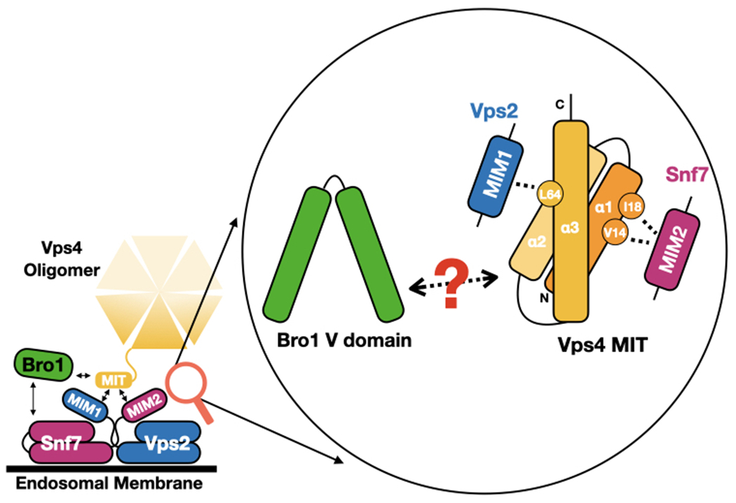
Bro1 V domain interacts with MIT domain of Vps4 via an unknown surface which is independent of MIM1/2. The MIT domain is a single bundle of three alpha-helices (α1, α2, α3). MIM2 interacts with the MIT via the groove between α1 and α3, while MIM1 interacts via the groove between α2 and α3.
Speculations and Implications: V domain stimulation and ESCRT-III “remodeling”
ESCRT-III is a heterooligomeric polymer comprised of four core subunits: 1) Vps20/CHMP6 is required for nucleation, 2) Snf7/CHMP4 is the major stoichiometric component, 3) Vps2/CHMP2 and Vps24/CHMP3 recruit Vps4 [17]. ESCRT-III subunits are capable of assembling into fibrils on membranes [2] and can remodel membranes in vitro [46,47]. Other ESCRT-III-like auxiliary subunits can be incorporated into ESCRT-III polymers, presumably to alter its function. These include Did2/CHMP1 and Ist1/CHMP8, which form their own subcomplex that is recruited late in the process of ESCRT-III polymerization and may help to change the shape of the polymer to facilitate scission [46,48]. Vps60/CHMP5 is another ESCRT-III-like protein required for efficient MVB sorting in yeast and proper function of signal transduction pathways in mammalian cells [49–51]. Recently, it was found that CHMP5 and Vps60 bind directly to the V domains of HD-PTP and Bro1, respectively [5]. Together, these results show that these two Bro1-family proteins have 3 distinct ways to connect to – and possibly alter the configuration or function of – ESCRT-III via their ability bind directly via the BOD (Snf7/CHMP4) and the V domain (Vps60/CHMP5) or indirectly via Vps4.
Precisely how ESCRT-III changes during ILV formation and what aspect(s) Bro1 family members may control remain to be resolved. However, a model in which ESCRT-III is controlled only at the level of assembly or disassembly is inadequate. Although we now appreciate details about ESCRT-III on yeast endosomes (such as ESCRT-III recruitment kinetics, average dwell-time, number of Snf7, Vps24 or Vps4 [52], nanometer scale structure of ESCRT-III polymers composed of either endogenous or chimeric subunits [2] and even the minimal features of ESCRT-III polymers [53], the lack of structural details of ESCRT-III assemblies with varying compositions at a resolution capable of identifying one subunit from another preclude predictions about what remodeling looks like. Recently ESCRT-III polymers were proposed to form parallel arrays, or co-polymers, consisting of a Snf7 polymer backbone laterally associated with a Vps2/Vps24 heteropolymer [46,53]. A co-polymer model fits well with various models for how vesicle scission is completed [2,3] by offering several possibilities of remodeling to regulate polymer dynamics, including A) rearranging ESCRT-III subunits, B) altering co-polymer composition, and C) changing subunit states (Figure 2). Altering the composition of different subunit ratios would require Vps4 to manipulate or remove subunits, which is consistent with the roles of AAA ATPases [54]. Vps4 is able to generate force and pull unfolded substrates through its central pore [55]. The first two scenarios are supported by the idea that rearranging subunits in or changing compositions of a co-polymer would alter the biophysical properties (rigidity, curvature, and/or torsion) of the polymer [48]. Such changes would subsequently produce different filament shapes, thereby driving membrane remodeling.
Figure 2.
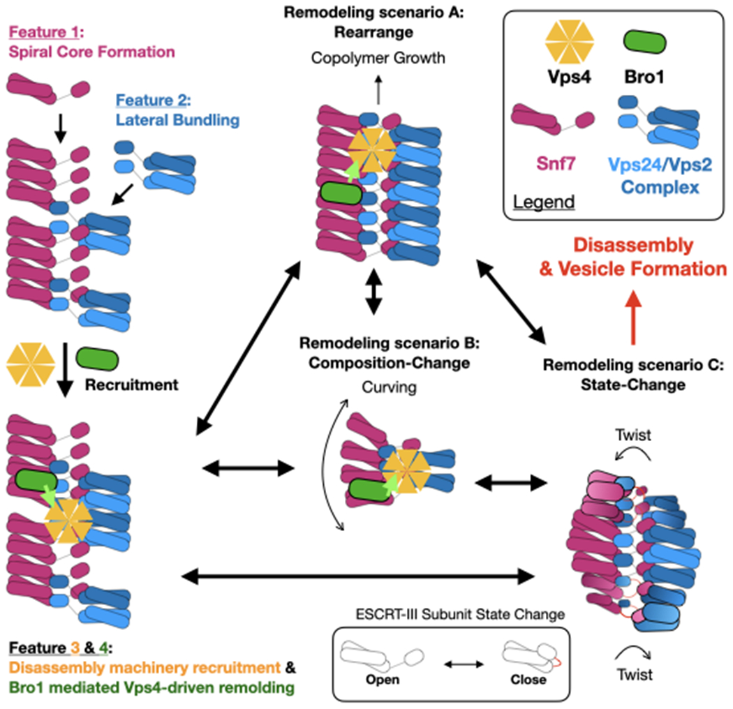
Proposed model for Bro1-regulated Vps4-driven ESCRT-III co-polymer remodeling. The ESCRT-III co-polymer contains four features. During ESCRT-III assembly, Snf7 forms the spiral core/backbone (Feature 1), while the Vps24/Vps2 complex binds to the spiral core laterally (Feature 2). Subsequently Bro1 and Vps4 are recruited to drive remodeling (Feature 3 & 4). In remodeling scenario C, Vps4 and Bro1 are omitted for better visualization of ESCRT-III polymer twisting. The neon green arrow indicates Bro1 stimulation of Vps4 ATPase activity.
An obvious consequence of a single stranded polymer transitioning into a double-stranded co-polymer would be an increase in rigidity. Altering the rigidity and curvature of ESCRT-III polymers is likely to provide dynamic conformations that would enable ESCRT-III to coordinate vesicle biogenesis with cargo incorporation. Pure lipid bilayers remain flat, and energy is required to overcome the tension within the membrane to generate membrane curvature [56]. As the curvature of a membrane increases, the energy required to bend it increases [57]. Thus, we might expect that during ILV formation an ESCRT-III filament that can change its physical properties such as rigidity to adapt to the changing membrane curvature might be better at facilitating the process. The formation of double-stranded and even three-stranded ESCRT-III filaments have been reported [46,58–62].
One could imagine that the difference in subunit numbers between the two associated polymers would produce curvature; alternatively, curvature could also be produced by combining two filaments of different rigidity (flexibility) [48], which could be caused by a difference in subunit state [63–65]. One could also imagine that a ribbon bends more easily along its face than its edges, a property that could impact function. Additionally, ESCRT-III polymers, dependent upon subunit composition, have been proposed to have distinct membrane interacting surfaces [61,66,67]. It may thus be possible that in addition to curve, the co-polymer ribbon would twist to generate torsion in order to maintain/align membrane interacting surfaces [48]. These observations, together with the fact that Snf7 polymer can store elastic energy [68,69], suggest a possibility that the formation of different co-polymer shapes on the membrane would generate and retain mechanical energies to contribute to membrane sculpting. The key mechanical feature these speculations offer is that the ESCRT-III polymer would be remodeled to transition through a series of subunit compositions and arrangements during vesicle formation rather than simply forming and disassembling a uniform polymer.
ESCRT-III subunits transition between open and closed conformation, with the open state facilitating inter-subunit interactions (i.e. polymerization) and membrane association [70–72]. ESCRT-III-driven membrane remodeling would require Vps4 ATP hydrolysis to alter the state of individual subunits to modify inter-/intra-unit contacts to change polymer shapes. Overexpression of the Bro1 domain stabilized membrane-associated ESCRT-III and delayed budding kinetics [11], suggesting that without V domain-mediated stimulation, Vps4 is unable to regulate/remodel ESCRT-III dynamics and the license for ESCRT-III-driven ILV formation is lost. Alternatively, Bro1 domain could also participate in regulating membrane-associated ESCRT-III dynamics by limiting Vps4 access to ESCRT-III subunits [42] or relieving Snf7 autoinhibition by blocking C-terminal tail from interacting with the helical body [73,74]. These concepts highlight the idea that polymer inter-/intra-subunit contacts regulate polymer conformation, dependent on Vps4-mediated remodeling.
Together, these remodeling possibilities require Vps4 to: 1) alter distinct ESCRT-III inter-subunit contacts in an ATP-dependent manner [72], and 2) rearrange/alter ESCRT-III co-polymer by exchanging individual subunits [54,75,76]. Therefore, it is possible that within this model Bro1 could impact either of these Vps4 functions to achieve coordination of cargo sorting and vesicle formation.
Conclusions
Despite significant advances in our understanding of ESCRT functions, fundamental aspects of how cargo sorting and vesicle formation are coordinated remain elusive. Here we propose a model wherein Bro1 coordinates the cargo sorting and membrane remodeling activities of ESCRTs via regulating Vps4-dependent ESCRT-III remodeling. This is achieved through interactions with ESCRT-III, Vps4, and Ub. Key is the ability of Bro1 to integrate its activity as both a receptor for Ub and a controller of ESCRT-III through remodeling and/or polymerization by Vps4. At the same time, Bro1 also coordinates the recruitment of deubiquitinating enzymes that recycle Ub from Ub-cargo at the right time and place to avoid escape of cargo from the forming ILV while ensuring cellular Ub levels. Bro1 sits at the nexus of these processes by interacting with proteins that execute these events. The observation that breaking this nexus disconnects cargo sorting from ILV formation highlights the pivotal role Bro1 plays in orchestrating these events. Bro1 acts as an ‘Ub-sensor’ for Vps4, and further work is required to determine what molecular changes and physiological tuning such a circuit provides. One possibility is that the V domain acts as an Ub-sensor and together with Vps4 maintains ESCRT-III in a state that facilitates cargo transfer and/or prevents cargo escape after Ub removal from cargo. Another possibility is that Ub-binding by Bro1 helps coordinate removal of Ub from cargo only when cargo has reached a sorting state mature enough to endure removal of its MVB sorting signal (Ub), so that MVB sorting can proceed without depleting Ub stores through lysosomal degradation. The molecular connections between the BOD with Snf7/CHMP4 and the V domain with the Vps4 MIT are conserved in human ALIX and HD-PTP as well as the yeast Bro1-family member, Rim20 [5,13]. For MVB sorting, ALIX plays a key role in sorting particular cargo into ILVs that go on to be secreted as exosomes [18] as well as viral budding [27–29]. HD-PTP plays a role very much like Bro1 in promoting the sorting of Ub-cargoes into ILVs that undergo delivery and degradation in lysosomes [16,21]. How these molecular interactions impact these and other pathways will be important to determine as will be the molecular details that explain the functional differences between Bro1 family members (e.g. ALIX and HDPTP).
Acknowledgements
We would like to thank the researchers who contributed to this field, and apologize to those who are not cited here, due to space constraints. We would like to acknowledge Dr. Brian Davies for his thoughtful discussions. The authors declare no competing financial interests.
Funding Sources
This research was supported, in whole or in part, by National Institutes of Health Grant R01 GM116826 (to D.J.K.) and R01 GM58202 (to R.C.P.). CT was supported by the Mayo Clinic Sidney Luckman Family Predoctoral Fellowship and the Mayo Clinic Graduate School of Biomedical Sciences. The authors declare that they have no conflicts of interest with the contents of this article.
References
- [1].Vietri M, Radulovic M, & Stenmark H (2020). The many functions of ESCRTs. Nature Reviews Molecular Cell Biology, 21(1), 25–42. doi: 10.1038/s41580-019-0177-4 [DOI] [PubMed] [Google Scholar]
- [2].Schoneberg J, Lee IH, Iwasa JH, & Hurley JH (2017). Reverse-topology membrane scission by the ESCRT proteins. Nature Reviews Molecular Cell Biology, 18(1), 5–17. doi: 10.1038/nrm.2016.121 [DOI] [PMC free article] [PubMed] [Google Scholar]
- [3].Hanson PI, & Cashikar A (2012). Multivesicular Body Morphogenesis. Annual Review of Cell and Developmental Biology, 28(1), 337–362. doi: 10.1146/annurev-cellbio-092910-154152 [DOI] [PubMed] [Google Scholar]
- [4].Pashkova N, Gakhar L, Winistorfer Stanley C., Sunshine Anna B., Rich M, Dunham Maitreya J., … Piper Robert C. (2013). The Yeast Alix Homolog Bro1 Functions as a Ubiquitin Receptor for Protein Sorting into Multivesicular Endosomes. Developmental Cell, 25(5), 520–533. doi: 10.1016/j.devcel.2013.04.007 [DOI] [PMC free article] [PubMed] [Google Scholar]
- [5].Pashkova N, Yu L, Schnicker NJ, Tseng C-C, Gakhar L, Katzmann DJ, & Piper RC (2021). Interactions of ubiquitin and CHMP5 with the V domain of HD-PTP reveals role for regulation of Vps4 ATPase. Molecular Biology of the Cell, mbc.E21-04-0219. doi: 10.1091/mbc.E21-04-0219 [DOI] [PMC free article] [PubMed] [Google Scholar]
- [6].Tang S, Buchkovich NJ, Henne WM, Banjade S, Kim YJ, & Emr SD (2016). ESCRT-III activation by parallel action of ESCRT-I/II and ESCRT-0/Bro1 during MVB biogenesis. Elife, 5, e15507. doi: 10.7554/eLife.15507 [DOI] [PMC free article] [PubMed] [Google Scholar]
- [7].Keren-Kaplan T, Attali I, Estrin M, Kuo LS, Farkash E, Jerabek-Willemsen M, … Prag G (2013). Structure-based in silico identification of ubiquitin-binding domains provides insights into the ALIX-V:ubiquitin complex and retrovirus budding. The EMBO Journal, 32(4), 538–551. doi: 10.1038/emboj.2013.4 [DOI] [PMC free article] [PubMed] [Google Scholar]
- [8].Vincent O, Rainbow L, Tilburn J, Arst HN Jr., & Peñalva MA (2003). YPXL/I is a protein interaction motif recognized by aspergillus PalA and its human homologue, AIP1/Alix. Mol Cell Biol, 23(5), 1647–1655. doi: 10.1128/mcb.23.5.1647-1655.2003 [DOI] [PMC free article] [PubMed] [Google Scholar]
- [9].Kimura Y, Tanigawa M, Kawawaki J, Takagi K, Mizushima T, Maeda T, & Tanaka K (2015). Conserved Mode of Interaction between Yeast Bro1 Family V Domains and YP(X)nL Motif-Containing Target Proteins. Eukaryot Cell, 14(10), 976–982. doi: 10.1128/ec.00091-15 [DOI] [PMC free article] [PubMed] [Google Scholar]
- [10].Kim J, Sitaraman S, Hierro A, Beach BM, Odorizzi G, & Hurley JH (2005). Structural Basis for Endosomal Targeting by the Bro1 Domain. Developmental Cell, 8(6), 937–947. doi: 10.1016/j.devcel.2005.04.001 [DOI] [PMC free article] [PubMed] [Google Scholar]
- [11].Wemmer M, Azmi I, West M, Davies B, Katzmann D, & Odorizzi G (2011). Bro1 binding to Snf7 regulates ESCRT-III membrane scission activity in yeast. J Cell Biol, 192(2), 295–306. doi: 10.1083/jcb.201007018 [DOI] [PMC free article] [PubMed] [Google Scholar]
- [12].McCullough J, Fisher RD, Whitby FG, Sundquist WI, & Hill CP (2008). ALIX-CHMP4 interactions in the human ESCRT pathway. Proceedings of the National Academy of Sciences, 105(22), 7687. doi: 10.1073/pnas.0801567105 [DOI] [PMC free article] [PubMed] [Google Scholar]
- [13].Tseng C-C, Dean S, Davies BA, Azmi IF, Pashkova N, Payne JA, … Katzmann DJ (2021). Bro1 stimulates Vps4 to promote intralumenal vesicle formation during multivesicular body biogenesis. Journal of Cell Biology, 220(8). doi: 10.1083/jcb.202102070 [DOI] [PMC free article] [PubMed] [Google Scholar]
- [14].Luhtala N, & Odorizzi G (2004). Bro1 coordinates deubiquitination in the multivesicular body pathway by recruiting Doa4 to endosomes. The Journal of Cell Biology, 166(5), 717. doi: 10.1083/jcb.200403139 [DOI] [PMC free article] [PubMed] [Google Scholar]
- [15].Kalinowska K, Nagel M-K, Goodman K, Cuyas L, Anzenberger F, Alkofer A, … Isono E (2015). Arabidopsis ALIX is required for the endosomal localization of the deubiquitinating enzyme AMSH3. Proceedings of the National Academy of Sciences, 112(40), E5543. doi: 10.1073/pnas.1510516112 [DOI] [PMC free article] [PubMed] [Google Scholar]
- [16].Ali N, Zhang L, Taylor S, Mironov A, Urbé S, & Woodman P (2013). Recruitment of UBPY and ESCRT Exchange Drive HD-PTP-Dependent Sorting of EGFR to the MVB. Current Biology, 23(6), 453–461. doi: 10.1016/j.cub.2013.02.033 [DOI] [PubMed] [Google Scholar]
- [17].McCullough J, Frost A, & Sundquist WI (2018). Structures, Functions, and Dynamics of ESCRT-III/Vps4 Membrane Remodeling and Fission Complexes. Annual Review of Cell and Developmental Biology, 34(1), 85–109. doi: 10.1146/annurev-cellbio-100616-060600 [DOI] [PMC free article] [PubMed] [Google Scholar]
- [18].Baietti MF, Zhang Z, Mortier E, Melchior A, Degeest G, Geeraerts A, … David G (2012). Syndecan–syntenin–ALIX regulates the biogenesis of exosomes. Nature Cell Biology, 14(7), 677–685. doi: 10.1038/ncb2502 [DOI] [PubMed] [Google Scholar]
- [19].Strack B, Calistri A, Craig S, Popova E, & Göttlinger HG (2003). AIP1/ALIX Is a Binding Partner for HIV-1 p6 and EIAV p9 Functioning in Virus Budding. Cell, 114(6), 689–699. doi: 10.1016/S0092-8674(03)00653-6 [DOI] [PubMed] [Google Scholar]
- [20].MacDonald C, Shields SB, Williams CA, Winistorfer S, & Piper RC (2020). A Cycle of Ubiquitination Regulates Adaptor Function of the Nedd4-Family Ubiquitin Ligase Rsp5. Curr Biol, 30(3), 465–479 e465. doi: 10.1016/j.cub.2019.11.086 [DOI] [PMC free article] [PubMed] [Google Scholar]
- [21].Doyotte A, Mironov A, McKenzie E, & Woodman P (2008). The Bro1-related protein HD-PTP/PTPN23 is required for endosomal cargo sorting and multivesicular body morphogenesis. Proc Natl Acad Sci U S A, 105(17), 6308–6313. doi: 10.1073/pnas.0707601105 [DOI] [PMC free article] [PubMed] [Google Scholar]
- [22].Dowlatshahi Dara P., Sandrin V, Vivona S, Shaler Thomas A., Kaiser Stephen E., Melandri F, … Kopito Ron R. (2012). ALIX Is a Lys63-Specific Polyubiquitin Binding Protein that Functions in Retrovirus Budding. Developmental Cell, 23(6), 1247–1254. doi: 10.1016/j.devcel.2012.10.023 [DOI] [PMC free article] [PubMed] [Google Scholar]
- [23].Alfred V, & Vaccari T (2016). When membranes need an ESCRT: endosomal sorting and membrane remodelling in health and disease. Swiss Med Wkly, 146, w14347. doi: 10.4414/smw.2016.14347 [DOI] [PubMed] [Google Scholar]
- [24].Michelet X, Djeddi A, & Legouis R (2010). Developmental and cellular functions of the ESCRT machinery in pluricellular organisms. Biology of the Cell, 102(3), 191–202. doi: 10.1042/BC20090145 [DOI] [PubMed] [Google Scholar]
- [25].Gingras MC, Kazan JM, & Pause A (2017). Role of ESCRT component HD-PTP/PTPN23 in cancer. Biochem Soc Trans, 45(3), 845–854. doi: 10.1042/bst20160332 [DOI] [PubMed] [Google Scholar]
- [26].Gingras MC, Kharitidi D, Chénard V, Uetani N, Bouchard M, Tremblay ML, & Pause A (2009). Expression analysis and essential role of the putative tyrosine phosphatase His-domain-containing protein tyrosine phosphatase (HD-PTP). Int J Dev Biol, 53(7), 1069–1074. doi: 10.1387/ijdb.082820mg [DOI] [PubMed] [Google Scholar]
- [27].Fisher RD, Chung H-Y, Zhai Q, Robinson H, Sundquist WI, & Hill CP (2007). Structural and Biochemical Studies of ALIX/AIP1 and Its Role in Retrovirus Budding. Cell, 128(5), 841–852. doi: 10.1016/j.cell.2007.01.035 [DOI] [PubMed] [Google Scholar]
- [28].Fujii K, Munshi UM, Ablan SD, Demirov DG, Soheilian F, Nagashima K, … Freed EO (2009). Functional role of Alix in HIV-1 replication. Virology, 391(2), 284–292. doi: 10.1016/j.virol.2009.06.016 [DOI] [PMC free article] [PubMed] [Google Scholar]
- [29].Morita E, Sandrin V, McCullough J, Katsuyama A, Baci Hamilton I, & Sundquist WI (2011). ESCRT-III protein requirements for HIV-1 budding. Cell Host & Microbe, 9(3), 235–242. doi: 10.1016/j.chom.2011.02.004 [DOI] [PMC free article] [PubMed] [Google Scholar]
- [30].Laporte MH, Chatellard C, Vauchez V, Hemming FJ, Deloulme J-C, Vossier F, … Sadoul R (2017). Alix is required during development for normal growth of the mouse brain. Scientific Reports, 7(1), 44767. doi: 10.1038/srep44767 [DOI] [PMC free article] [PubMed] [Google Scholar]
- [31].Carlton JG, Agromayor M, & Martin-Serrano J (2008). Differential requirements for Alix and ESCRT-III in cytokinesis and HIV-1 release. Proceedings of the National Academy of Sciences, 105(30), 10541. doi: 10.1073/pnas.0802008105 [DOI] [PMC free article] [PubMed] [Google Scholar]
- [32].Alazami AM, Patel N, Shamseldin HE, Anazi S, Al-Dosari MS, Alzahrani F, … Alkuraya FS (2015). Accelerating novel candidate gene discovery in neurogenetic disorders via whole-exome sequencing of prescreened multiplex consanguineous families. Cell Rep, 10(2), 148–161. doi: 10.1016/j.celrep.2014.12.015 [DOI] [PubMed] [Google Scholar]
- [33].Bend R, Cohen L, Carter MT, Lyons MJ, Niyazov D, Mikati MA, … Gonzaga-Jauregui C (2020). Phenotype and mutation expansion of the PTPN23 associated disorder characterized by neurodevelopmental delay and structural brain abnormalities. Eur J Hum Genet, 28(1), 76–87. doi: 10.1038/s41431-019-0487-1 [DOI] [PMC free article] [PubMed] [Google Scholar]
- [34].Smigiel R, Landsberg G, Schilling M, Rydzanicz M, Pollak A, Walczak A, … Ploski R (2018). Developmental epileptic encephalopathy with hypomyelination and brain atrophy associated with PTPN23 variants affecting the assembly of UsnRNPs. Eur J Hum Genet, 26(10), 1502–1511. doi: 10.1038/s41431-018-0179-2 [DOI] [PMC free article] [PubMed] [Google Scholar]
- [35].Sowada N, Hashem MO, Yilmaz R, Hamad M, Kakar N, Thiele H, … Borck G (2017). Mutations of PTPN23 in developmental and epileptic encephalopathy. Hum Genet, 136(11-12), 1455–1461. doi: 10.1007/s00439-017-1850-3 [DOI] [PubMed] [Google Scholar]
- [36].Trujillano D, Bertoli-Avella AM, Kumar Kandaswamy K, Weiss ME, Köster J, Marais A, … Abou Jamra R (2017). Clinical exome sequencing: results from 2819 samples reflecting 1000 families. Eur J Hum Genet, 25(2), 176–182. doi: 10.1038/ejhg.2016.146 [DOI] [PMC free article] [PubMed] [Google Scholar]
- [37].Lahaie S, Morales D, Bagci H, Hamoud N, Castonguay C-E, Kazan JM, … Kania A (2019). The endosomal sorting adaptor HD-PTP is required for ephrin-B:EphB signalling in cellular collapse and spinal motor axon guidance. Scientific Reports, 9(1), 11945. doi: 10.1038/s41598-019-48421-9 [DOI] [PMC free article] [PubMed] [Google Scholar]
- [38].Bulgari D, Jha A, Deitcher David L, & Levitan Edwin S (2018). Myopic (HD-PTP, PTPN23) selectively regulates synaptic neuropeptide release. Proceedings of the National Academy of Sciences, 115(7), 1617–1622. doi: 10.1073/pnas.1716801115 [DOI] [PMC free article] [PubMed] [Google Scholar]
- [39].MacDonald C, Buchkovich NJ, Stringer DK, Emr SD, & Piper RC (2012). Cargo ubiquitination is essential for multivesicular body intralumenal vesicle formation. EMBO reports, 13(4), 331. doi: 10.1038/embor.2012.18 [DOI] [PMC free article] [PubMed] [Google Scholar]
- [40].Stringer DK, & Piper RC (2011). A single ubiquitin is sufficient for cargo protein entry into MVBs in the absence of ESCRT ubiquitination. The Journal of Cell Biology, 192(2), 229. doi: 10.1083/jcb.201008121 [DOI] [PMC free article] [PubMed] [Google Scholar]
- [41].Azmi I, Davies B, Dimaano C, Payne J, Eckert D, Babst M, & Katzmann DJ (2006). Recycling of ESCRTs by the AAA-ATPase Vps4 is regulated by a conserved VSL region in Vta1. J Cell Biol, 172(5), 705–717. doi: 10.1083/jcb.200508166 [DOI] [PMC free article] [PubMed] [Google Scholar]
- [42].Azmi IF, Davies BA, Xiao J, Babst M, Xu Z, & Katzmann DJ (2008). ESCRT-III Family Members Stimulate Vps4 ATPase Activity Directly or via Vta1. Developmental Cell, 14(1), 50–61. doi: 10.1016/j.devcel.2007.10.021 [DOI] [PubMed] [Google Scholar]
- [43].Babst M, Katzmann DJ, Estepa-Sabal EJ, Meerloo T, & Emr SD (2002). Escrt-III: An endosome-associated heterooligomeric protein complex required for mvb sorting. Developmental Cell, 3(2), 271–282. doi: 10.1016/S1534-5807(02)00220-4 [DOI] [PubMed] [Google Scholar]
- [44].Nickerson DP, West M, Henry R, & Odorizzi G (2010). Regulators of Vps4 ATPase Activity at Endosomes Differentially Influence the Size and Rate of Formation of Intralumenal Vesicles. Molecular Biology of the Cell, 21(6), 1023–1032. doi: 10.1091/mbc.e09-09-0776 [DOI] [PMC free article] [PubMed] [Google Scholar]
- [45].Vajjhala PR, Catchpoole E, Nguyen CH, Kistler C, & Munn AL (2007). Vps4 regulates a subset of protein interactions at the multivesicular endosome. The FEBS Journal, 274(8), 1894–1907. doi: 10.1111/j.1742-4658.2007.05736.x [DOI] [PubMed] [Google Scholar]
- [46].Pfitzner A-K, Mercier V, Jiang X, Moser von Filseck J, Baum B, Šarić A, & Roux A (2020). An ESCRT-III Polymerization Sequence Drives Membrane Deformation and Fission. Cell, 182(5), 1140–1155.e1118. doi: 10.1016/j.cell.2020.07.021 [DOI] [PMC free article] [PubMed] [Google Scholar]
- [47].Wollert T, Wunder C, Lippincott-Schwartz J, & Hurley JH (2009). Membrane scission by the ESCRT-III complex. Nature, 458(7235), 172–177. doi: 10.1038/nature07836 [DOI] [PMC free article] [PubMed] [Google Scholar]
- [48].Pfitzner A-K, Moser von Filseck J, & Roux A (2021). Principles of membrane remodeling by dynamic ESCRT-III polymers. Trends in Cell Biology. doi: 10.1016/j.tcb.2021.04.005 [DOI] [PubMed] [Google Scholar]
- [49].Adoro S, Park KH, Bettigole SE, Lis R, Shin HR, Seo H, … Glimcher LH (2017). Post-translational control of T cell development by the ESCRT protein CHMP5. Nat Immunol, 18(7), 780–790. doi: 10.1038/ni.3764 [DOI] [PubMed] [Google Scholar]
- [50].Shim JH, Xiao C, Hayden MS, Lee KY, Trombetta ES, Pypaert M, … Ghosh S (2006). CHMP5 is essential for late endosome function and down-regulation of receptor signaling during mouse embryogenesis. J Cell Biol, 172(7), 1045–1056. doi: 10.1083/jcb.200509041 [DOI] [PMC free article] [PubMed] [Google Scholar]
- [51].Shiflett SL, Ward DM, Huynh D, Vaughn MB, Simmons JC, & Kaplan J (2004). Characterization of Vta1p, a class E Vps protein in Saccharomyces cerevisiae. J Biol Chem, 279(12), 10982–10990. doi: 10.1074/jbc.M312669200 [DOI] [PubMed] [Google Scholar]
- [52].Adell MAY, Migliano SM, Upadhyayula S, Bykov YS, Sprenger S, Pakdel M, … Teis D (2017). Recruitment dynamics of ESCRT-III and Vps4 to endosomes and implications for reverse membrane budding. Elife, 6. doi: 10.7554/eLife.31652 [DOI] [PMC free article] [PubMed] [Google Scholar]
- [53].Banjade S, Shah YH, Tang S, & Emr SD (2021). Design principles of the ESCRT-III Vps24-Vps2 module. Elife, 10, e67709. doi: 10.7554/eLife.67709 [DOI] [PMC free article] [PubMed] [Google Scholar]
- [54].Schöneberg J, Pavlin MR, Yan S, Righini M, Lee I-H, Carlson L-A, … Hummer G (2018). ATP-dependent force generation and membrane scission by ESCRT-III and Vps4. Science, 362(6421), 1423–1428. [DOI] [PMC free article] [PubMed] [Google Scholar]
- [55].Remec Pavlin M, & Hurley JH (2020). The ESCRTs – converging on mechanism. Journal of Cell Science, 133(18), jcs240333. doi: 10.1242/jcs.240333 [DOI] [PMC free article] [PubMed] [Google Scholar]
- [56].Jarsch IK, Daste F, & Gallop JL (2016). Membrane curvature in cell biology: An integration of molecular mechanisms. Journal of Cell Biology, 214(4), 375–387. doi: 10.1083/jcb.201604003 [DOI] [PMC free article] [PubMed] [Google Scholar]
- [57].Helfrich P, & Jakobsson E (1990). Calculation of deformation energies and conformations in lipid membranes containing gramicidin channels. Biophys J, 57(5), 1075–1084. doi: 10.1016/s0006-3495(90)82625-4 [DOI] [PMC free article] [PubMed] [Google Scholar]
- [58].Banjade S, Tang S, Shah YH, & Emr SD (2019). Electrostatic lateral interactions drive ESCRT-III heteropolymer assembly. Elife, 8, e46207. doi: 10.7554/eLife.46207 [DOI] [PMC free article] [PubMed] [Google Scholar]
- [59].Bertin A, de Franceschi N, de la Mora E, Maity S, Alqabandi M, Miguet N, … Bassereau P (2020). Human ESCRT-III polymers assemble on positively curved membranes and induce helical membrane tube formation. Nature Communications, 11(1), 2663. doi: 10.1038/s41467-020-16368-5 [DOI] [PMC free article] [PubMed] [Google Scholar]
- [60].McCullough J, Clippinger AK, Talledge N, Skowyra ML, Saunders MG, Naismith TV, … Frost A (2015). Structure and membrane remodeling activity of ESCRT-III helical polymers. Science, 350(6267), 1548–1551. doi: 10.1126/science.aad8305 [DOI] [PMC free article] [PubMed] [Google Scholar]
- [61].Mierzwa BE, Chiaruttini N, Redondo-Morata L, Moser von Filseck J, König J, Larios J, … Gerlich DW (2017). Dynamic subunit turnover in ESCRT-III assemblies is regulated by Vps4 to mediate membrane remodelling during cytokinesis. Nature Cell Biology, 19(7), 787–798. doi: 10.1038/ncb3559 [DOI] [PMC free article] [PubMed] [Google Scholar]
- [62].Nguyen HC, Talledge N, McCullough J, Sharma A, Moss FR 3rd, Iwasa JH, … Frost A (2020). Membrane constriction and thinning by sequential ESCRT-III polymerization. Nat Struct Mol Biol, 27(4), 392–399. doi: 10.1038/s41594-020-0404-x [DOI] [PMC free article] [PubMed] [Google Scholar]
- [63].Gupta TK, Klumpe S, Gries K, Heinz S, Wietrzynski W, Ohnishi N, … Rast A (2021). Structural basis for VIPP1 oligomerization and maintenance of thylakoid membrane integrity. Cell. [DOI] [PubMed] [Google Scholar]
- [64].Junglas B, Huber ST, Heidler T, Schlösser L, Mann D, Hennig R, … Sachse C (2021). PspA adopts an ESCRT-III-like fold and remodels bacterial membranes. Cell. doi: 10.1016/j.cell.2021.05.042 [DOI] [PubMed] [Google Scholar]
- [65].Liu J, Tassinari M, Souza DP, Naskar S, Noel JK, Bohuszewicz O, … Low HH (2021). Bacterial Vipp1 and PspA are members of the ancient ESCRT-III membrane-remodeling superfamily. Cell, 184(14), 3660–3673.e3618. doi: 10.1016/j.cell.2021.05.041 [DOI] [PMC free article] [PubMed] [Google Scholar]
- [66].Chiaruttini N, & Roux A (2017). Dynamic and elastic shape transitions in curved ESCRT-III filaments. Current Opinion in Cell Biology, 47, 126–135. doi: 10.1016/j.ceb.2017.07.002 [DOI] [PubMed] [Google Scholar]
- [67].Moser von Filseck J, Barberi L, Talledge N, Johnson IE, Frost A, Lenz M, & Roux A (2020). Anisotropic ESCRT-III architecture governs helical membrane tube formation. Nature Communications, 11(1), 1516. doi: 10.1038/s41467-020-15327-4 [DOI] [PMC free article] [PubMed] [Google Scholar]
- [68].Chiaruttini N, Redondo-Morata L, Colom A, Humbert F, Lenz M, Scheuring S, & Roux A (2015). Relaxation of Loaded ESCRT-III Spiral Springs Drives Membrane Deformation. Cell, 163(4), 866–879. doi: 10.1016/j.cell.2015.10.017 [DOI] [PMC free article] [PubMed] [Google Scholar]
- [69].Lenz M, Crow DJG, & Joanny J-F (2009). Membrane Buckling Induced by Curved Filaments. Physical Review Letters, 103(3), 038101. doi: 10.1103/PhysRevLett.103.038101 [DOI] [PubMed] [Google Scholar]
- [70].Hanson PI, Roth R, Lin Y, & Heuser JE (2008). Plasma membrane deformation by circular arrays of ESCRT-III protein filaments. The Journal of Cell Biology, 180(2), 389–402. doi: 10.1083/jcb.200707031 [DOI] [PMC free article] [PubMed] [Google Scholar]
- [71].Shim S, Kimpler LA, & Hanson PI (2007). Structure/function analysis of four core ESCRT-III proteins reveals common regulatory role for extreme C-terminal domain. Traffic, 8(8), 1068–1079. doi: 10.1111/j.1600-0854.2007.00584.x [DOI] [PubMed] [Google Scholar]
- [72].Adell MA, & Teis D (2011). Assembly and disassembly of the ESCRT-III membrane scission complex. FEBS Lett, 585(20), 3191–3196. doi: 10.1016/j.febslet.2011.09.001 [DOI] [PMC free article] [PubMed] [Google Scholar]
- [73].Teis D, Saksena S, & Emr SD (2008). Ordered Assembly of the ESCRT-III Complex on Endosomes Is Required to Sequester Cargo during MVB Formation. Developmental Cell, 15(4), 578–589. doi: 10.1016/j.devcel.2008.08.013 [DOI] [PubMed] [Google Scholar]
- [74].Tang S, Henne WM, Borbat PP, Buchkovich NJ, Freed JH, Mao Y, … Emr SD (2015). Structural basis for activation, assembly and membrane binding of ESCRT-III Snf7 filaments. Elife, 4, e12548. doi: 10.7554/eLife.12548 [DOI] [PMC free article] [PubMed] [Google Scholar]
- [75].Monroe N, Han H, Shen PS, Sundquist WI, & Hill CP (2017). Structural basis of protein translocation by the Vps4-Vta1 AAA ATPase. Elife, 6, e24487. doi: 10.7554/eLife.24487 [DOI] [PMC free article] [PubMed] [Google Scholar]
- [76].Yang B, Stjepanovic G, Shen Q, Martin A, & Hurley JH (2015). Vps4 disassembles an ESCRT-III filament by global unfolding and processive translocation. Nature Structural & Molecular Biology, 22(6), 492–498. doi: 10.1038/nsmb.3015 [DOI] [PMC free article] [PubMed] [Google Scholar]
- [77].Matsuo H, Chevallier J, Mayran N, Le Blanc I, Ferguson C, Fauré J, … Gruenberg J (2004). Role of LBPA and Alix in multivesicular liposome formation and endosome organization. Science, 303(5657), 531–534. doi: 10.1126/science.1092425 [DOI] [PubMed] [Google Scholar]
- [78].Von Schwedler UK, Stuchell M, Müller B, Ward DM, Chung H-Y, Morita E, … Cimbora DM (2003). The protein network of HIV budding. Cell, 114(6), 701–713. [DOI] [PubMed] [Google Scholar]
- [79].Morita E, Sandrin V, Chung H-Y, Morham SG, Gygi SP, Rodesch CK, & Sundquist WI (2007). Human ESCRT and ALIX proteins interact with proteins of the midbody and function in cytokinesis. The EMBO Journal, 26(19), 4215–4227. doi: 10.1038/sj.emboj.7601850 [DOI] [PMC free article] [PubMed] [Google Scholar]
- [80].Sun S, Zhou X, Corvera J, Gallick GE, Lin SH, & Kuang J (2015). ALG-2 activates the MVB sorting function of ALIX through relieving its intramolecular interaction. Cell Discov, 1, 15018. doi: 10.1038/celldisc.2015.18 [DOI] [PMC free article] [PubMed] [Google Scholar]
- [81].Ichioka F, Takaya E, Suzuki H, Kajigaya S, Buchman VL, Shibata H, & Maki M (2007). HD-PTP and Alix share some membrane-traffic related proteins that interact with their Bro1 domains or proline-rich regions. Archives of Biochemistry and Biophysics, 457(2), 142–149. doi: 10.1016/j.abb.2006.11.008 [DOI] [PubMed] [Google Scholar]
- [82].Stefani F, Zhang L, Taylor S, Donovan J, Rollinson S, Doyotte A, … Woodman P (2011). UBAP1 is a component of an endosome-specific ESCRT-I complex that is essential for MVB sorting. Curr Biol, 21(14), 1245–1250. doi: 10.1016/j.cub.2011.06.028 [DOI] [PubMed] [Google Scholar]
- [83].Odorizzi G, Katzmann DJ, Babst M, Audhya A, & Emr SD (2003). Bro1 is an endosome-associated protein that functions in the MVB pathway in Saccharomyces cerevisiae. J Cell Sci, 116(Pt 10), 1893–1903. doi: 10.1242/jcs.00395 [DOI] [PubMed] [Google Scholar]
- [84].Kimura Y, Yashiroda H, Kudo T, Koitabashi S, Murata S, Kakizuka A, & Tanaka K (2009). An Inhibitor of a Deubiquitinating Enzyme Regulates Ubiquitin Homeostasis. Cell, 137(3), 549–559. doi: 10.1016/j.cell.2009.02.028 [DOI] [PubMed] [Google Scholar]
- [85].Nikko E, & André B (2007). Split-Ubiquitin Two-Hybrid Assay To Analyze Protein-Protein Interactions at the Endosome: Application to Saccharomyces cerevisiae Bro1 Interacting with ESCRT Complexes, the Doa4 Ubiquitin Hydrolase, and the Rsp5 Ubiquitin Ligase. Eukaryotic Cell, 6(8), 1266. doi: 10.1128/EC.00024-07 [DOI] [PMC free article] [PubMed] [Google Scholar]