PLOS ONE logoLink to PLOS ONE
. 2022 Oct 17;17(10):e0274938. doi: 10.1371/journal.pone.0274938

Diagnostic performances of pepsinogens and gastrin-17 for atrophic gastritis and gastric cancer in Mongolian subjects

Ganchimeg Dondov 1,2, Dashmaa Amarbayasgalan 3, Batbold Batsaikhan 1, Tegshjargal Badamjav 1, Batchimeg Batbaatar 4, Baljinnyam Tuvdenjamts 4, Nasanjargal Tumurbat 1, Bayar Davaa 1, Erkhembulgan Purevdorj 1, Bayarmaa Nyamaa 2, Tulgaa Lonjid 1,*
Editor: Muhammad Tarek Abdel Ghafar5
PMCID: PMC9576095  PMID: 36251649

Abstract

In Mongolia, gastric cancer morbidity and mortality are high, and more than 80 percent of cases are diagnosed at an advanced stage. This study aimed to evaluate pepsinogens (PGIs) and gastrin-17 (G-17) levels and to determine the diagnostic performances for gastric cancer and chronic atrophic gastritis among Mongolian individuals. We enrolled a total of 120 subjects, including gastric cancer (40), atrophic gastritis (40), and healthy control (40), matched by age (±2) and sex. Pepsinogen I (PGI), Pepsinogen II (PGII), G-17, and H. pylori IgG levels were measured using GastroPanel ELISA kit (Biohit, Helsinki, Finland). Also, PGI to PGII ratio (PGR) was calculated. For atrophic gastritis, when the optimal cut-off value of PGI was ≤75.07 ng/ml, the sensitivity and specificity were 75% and 50%, respectively; when the optimal cut-off value of PGR was ≤6.25, sensitivity and specificity were 85% and 44.7%, respectively. For gastric cancer, when the optimal cut-off value of PGI was ≤35.25 ng/ml, the sensitivity and specificity were 47.2% and 86.8%, respectively; when the optimal cut-off value of PGR was ≤5.27, sensitivity and specificity were 75% and 60.5%, respectively. Combinations of biomarkers with risk factors could improve diagnostic accuracy (AUC for atrophic gastritis 74.8, 95% CI 64.0–85.7, p<0.001; AUC for gastric cancer 75.5, 95% CI 64.2–86.8, p<0.001). PGI, PGR biomarkers combined with the risk of age, family history of gastric cancer, and previous gastric disease could not be an alternative test for upper endoscopy but might be a supportive method which is identifying individuals at medium- and high risk of gastric cancer and precancerous lesions who may need upper endoscopy.

Introduction

Gastric cancer is still a major health problem worldwide, despite a dramatic reduction in incidence and mortality rates [1]. In Mongolia, gastric cancer is ranking second after liver cancer and it is increasing in the last decade [2]. According to data of the International Agency for Research on Cancer, Mongolia had the highest rate (100’000:32.5) of gastric cancer and leads to mortality (100’000:24.6) of gastric cancer [3]. Many epidemiological studies have revealed a strong association between H. pylori infection and gastric cancer. Previous studies have shown that the prevalence of H. pylori infection is high among the population of Mongolia [4]. Besides, over 80% of gastric cancer cases are diagnosed in the late stage in our country [5]. But a screening program has not introduced to decrease the gastric cancer rate. According to the study, early detection of gastric cancer could reduce death related gastric cancer by 30–65% [6]. In our country, we performed a gastroduodenoscopy for screening and histological evaluation to diagnose gastric cancer. These methods are an effective diagnostic modality for gastric diseases; however, invasive and cause discomfort, making it an undesirable procedure for patients. Thus, there is a demand to introduce a non-invasive, easy-to-use early detection method for screening to general the population. Gastric cancer is the end of a long and multistep process, including atrophic gastritis, intestinal metaplasia, low- and high-grade dysplasia [7]. Accordingly, we considered that atrophy is the key condition of gastric cancer and monitoring atrophic gastritis is a preventive measure against gastric cancer. In some developed countries, H. pylori IgG, pepsinogens (PGs), and gastrin-17 (G-17) have been studied as non-invasive serological evaluation of gastric cancer and precancerous gastric lesions and have been suggested a variety of cut-off values [811]. Human PGs, which are protein-digestive enzymes secreted as proenzymes by chief cells, classified as pepsinogen I (PGI) and pepsinogen II (PGII). PGs may function as a marker of the functional and morphologic status of the gastric mucosa [12,13]. Gastrins are synthesized by endocrine G cells of the stomach and stimulates the secretion of gastric acid by the parietal cells. Gastric acid is necessary for the conversion of inactive pepsinogen to active pepsin. G-17 and G-34 are classical gastrins, G-17 is more prevalent in antral mucosa and G-34 predominates in duodenum. Also, gastrins allow proliferation, inhibit apoptosis and support migration of gastric epithelial cells [14]. Based on these physiologies, the loss of glands in atrophy would decrease PGs and G-17 levels. Therefore, we aimed to evaluate serum PGIs and G-17 levels and to determine the diagnostic performances for gastric cancer and chronic atrophic gastritis among Mongolian individuals.

Materials and methods

Study subjects

This study enrolled a total of 120 subjects who attended the gastrointestinal endoscopy at the National Center of Cancer of Mongolia between January 2019 and October 2019. There were 40 gastric cancer patients enrolled before surgery and other therapies. Besides, there were 40 chronic atrophic gastritis patients and 40 healthy controls enrolled. Gastric cancer and chronic atrophic gastritis were confirmed by gastroscopy and pathological examination; healthy controls were without obvious disease by basic tests, and healthy mucosa by gastroscopy. Subjects of three groups were matched by age (±2) and sex. Exclusion criteria were followed: age <18, pregnancy, recent use of proton pump inhibitor or H2 receptor blockers, history of H. pylorieradication within three months, history of gastric surgery or malignancy of cancers. After the exclusion of 6 subjects, 36 with gastric cancer, 40 with chronic atrophic gastritis, and 38 healthy subjects were included. Table 1 summarizes the baseline characteristics of the study population. This study was performed by the Helsinki Declaration and all subjects signed informed consent to participate in this study, which was approved by the Ethics Review Committee of the Ministry of Health of Mongolia on August 24, 2018 (Approval №67).

Table 1. Baseline characteristics of the study population.

Variables Total (n = 114) Healthy control, (n = 38) Atrophic gastritis (n = 40) Gastric cancer (n = 36) р value
Age, mean (SD) * 59.98 (10.88) 59.87 (11.62) 58.73 (10.79) 61.50 (10.26) -
Gender, male (%) * 60 (52.6) 20 (52.6) 21 (52.5) 19 (52.8) -
Family history of gastric cancer (%) 24 (21.1) 7 (18.4) 4 (10.0) 13 (36.1) 0.018
Previous history of gastrointestinal diseases (%) 35 (30.7) 6 (15.8) 10 (25.0) 19 (52.8) 0.002
H. pylori IgG >30EIU (%) 67 (58.8) 24 (63.2) 22 (55.0) 21 (58.3) 0.764
PGI ng/ml, median (min, max) 59.38 (4.23, 223.94) 74.32 (14.65, 223.94) 56.52 (4.23, 209.28) 46.94 (6.52, 212.67) 0.067
PGII ng/ml, median (min, max) 13.11 (3.73, 41.77) 13.32 (6.24, 39.48) 11.39 (4.20, 40.75) 16.60 (3.73, 41.77) 0.084
PGR, median (min, max) 4.80 (0.58, 13.37) 5.77 (1.71, 12.87) 5.03 (0.60,13.73) 3.76 (0.58, 8.71) 0.004
G-17 pmol/l, median (min, max) 12.36 (0.11, 78.69) 13.99 (0.80, 78.69) 10.64 (0.96–45.80) 13.41 (0.11, 77.49) 0.091

*Age and gender were matched between study groups.

PGI, pepsinogen I; PGII, pepsinogen II; PGR, pepsinogen I to pepsinogen II ratio; G-17, gastrin-17.

Gastrointestinal endoscopy

Gastrointestinal endoscopy was performed at the National Cancer Center of Mongolia with accordance national standard MNS5747-1:2007 using endoscope EVIS Exera III. After the 10-hour fast, simethicone solution was used to improve the visibility of the mucosa, followed by 10% lydocaine spray. Endoscopies were initially performed using white light. Subsequently, narrow-band imaging was activated, if any further evaluation was required. At least four biopsies were obtained from the corpus, antrum, and lesions detected macroscopically and by narrow-band imaging. The biopsies were transferred into 4% formalin buffer and sent to the Department of Pathology for pathological examination. The diagnosis was confirmed based on the gastroscopy and pathological examinations by a single expert, respectively.

Measurement of serum biomarkers using GastroPanel

PGI, PGII, G-17, and H. pylori IgG levels were measured using GastroPanel enzyme-linked immunosorbent assay kit (Biohit, Helsinki, Finland). To obtain more accurate analysis results, the biomarkers concentration was used for the average value of the results of a triplicate analysis repeated twice. The fasting blood samples were collected into an EDTA tube from all subjects. The blood samples were centrifuged at 2000 rpm for 10 minutes, and the supernatant was stored at -70°C freezer until testing. Before the assay, the samples were diluted with diluent buffer for the assays following the manufacturer’s package insert: 1:5 for G-17, 1:20 for PGI and PGII, and 1:400 for H. pyloriIgG. The plasma concentrations of PGI, PGII, G-17, and H. pylori IgG were determined by following protocol. First, the blank solutions, calibrators, controls and diluted samples were pipetted into microplate wells at a volume of 100 μl. Each individuals sample were pipetted into 3 microplate wells. The microplates were incubated at room temperature for 60 minutes with shaking at 750 rpm. Microplate strips were automatically washed three times with 350 μl of diluted buffer and gently tapped on a clean towel. Subsequently, 100 μl of specific conjugate solutions were pipetted into each microplate wells and incubated at room temperature for 60 minutes with shaking at 750 rpm. Microplate strips were automatically washed using a BIOBASE-EL10A reader three times with 350 μl of diluted buffer and gently tapped on a clean towel. After that, 100 μl of substrate solutions were pipetted into each microplate wells and incubated for 30 minutes at ambient temperature avoiding exposure to light. Finally, 100 μl of stop solutions were pipetted into microplate wells. The absorbance of the microplate wells was measured at 450 nm using a BIOBASE-EL10A microplate reader (Biobase Biodustry, Shandong, China). Also, PGI to PGII ratio (PGR) was calculated.

Statistical analysis

All statistical analyses were performed by SPSS version 26.0 (SPSS, Chicago, IL, USA). Categorical variables were presented as numbers and proportions and differences were assessed using the Chi-square test. Plasma levels of biomarkers were presented as medians and differences assessed using Kruskal-Wallis Test. The diagnostic accuracy and cut-off values were assessed by ROC curves and the Youden index, with evaluations of sensitivity, specificity, positive predictive value, negative predictive value, positive likelihood ratio, and negative likelihood ratio. Additionally, we evaluated all subjects by giving one point to each of the age ≤40, positive family history of gastric cancer, positive previous disease history, PGI ≤75.07 ng/ml, PGR ≤6.25, or two point to each of PGI ≤35.25 ng/ml, and PGR ≤5.27, with score ranging from 0 to 7 to predict gastric cancer and atrophic gastritis risks. Differences with p<0.05 considered to be statistically significant.

Results

Subjects and biomarkers levels

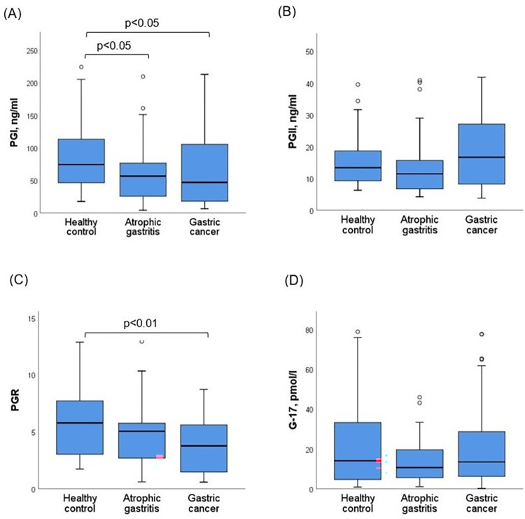
Baseline characteristics and levels of PGI, PGII, G-17 and PGR of study subjects are presented in Table 1. Median age of the subjects was 62 (min 27, max 80), 52.6% (n = 60) were male. Proportions of family history of gastric cancer and previous history of gastric disease were significantly higher in the gastric cancer group compared with atrophic gastritis and healthy control groups. Biopsy of gastric cancer patients yielded the following findings: adenocarcinoma 75.0% (n = 27), mucinous carcinoma 11.1% (n = 4), poorly cohesive 8.3% (n = 3), tubular carcinoma 2.8% (n = 1), papillary carcinoma 2.8% (n = 1). The median of PGI was 74.32 ng/ml (14.65 to 223.94) for healthy controls, 56.52 ng/ml (4.23 to 209.28) for atrophic gastritis and 46.94 ng/ml (6.52 to 212.67) for gastric cancer patients. The PGI level was significantly decreased in gastric cancer and atrophic gastritis groups as compared to the healthy control (p<0.05, p<0.05) (Fig 1A). The median of PGII was 13.32 ng/ml (6.24 to 39.48) for healthy controls, 11.39 ng/ml (4.20 to 40.75) for atrophic gastritis and 16.60 ng/ml (3.73 to 41.77) for gastric cancer patients. And the median of G-17 was 13.99 pmol/l (0.80 to 78.89) for healthy controls, 11.39 pmol/l (0.96 to 45.80) for atrophic gastritis and 13.41 pmol/l (0.11 to 77.49) for gastric cancer patients. There were no significant differences in the PGII and G-17 levels between study groups (Fig 1B and 1D). The median of PGR was 5.77 (1.71 to 12.87), 5.03 (0.60 to 13.73), and 3.76 (0.58 to 8.71) for healthy controls, atrophic gastritis, and gastric cancer patients, respectively. The PGR was significantly lower in the gastric cancer group compared with the healthy control (p<0.01) (Fig 1C).

Fig 1.

Fig 1

Comparisons of (A) PGI, (B) PGII, (C) PGR, (D) G-17 biomarkers levels between healthy control and patients with atrophic gastritis and gastric cancer. The graph indicates the medians and the boxes of 25%-75% quartiles. PGI, pepsinogen I; PGII, pepsinogen II; PGR, pepsinogen I to pepsinogen II ratio; G-17, gastrin-17. H. pylori was positive in 67 (58.8%) subjects according to H. pylori IgG assay and there was no difference between study groups. We estimated PGI, PGII, PGR and G-17 levels between H. pylori IgG negative and positive groups. The median of PGI was 46.11 (4.22 to 188.07) and 70.26 (7.53 to 223.94), the median of PGII was 8.40 (3.73 to 41.77) and 15.78 (4.43 to 40.75) for H. pylori IgG negative and positive subjects, respectively. The PGI and PGII levels were higher in H. pylori positive subjects than in H. pylori negative subjects (p<0.01), while there was no difference in PGR and G-17 levels between them. The median of PGR was 4.62 (0.60 to 13.37) and 4.85 (0.58 to 12.86), the median of G-17 was 10.52 (0.11 to 77.49) and 13.69 (1.02 to 78.69) for H. pylori IgG negative and positive subjects, respectively.

Diagnostic performance of the biomarkers for atrophic gastritis

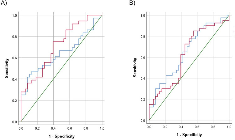
The corresponding ROC curves of PGI and PGR were developed to predict atrophic gastritis, and AUC were 65.2 (95% CI 53.0–77.3, p<0.05) and 62.7 (95% CI 50.1–75.3, p<0.05), respectively (Fig 2A). The results calculated diagnostic values of PGI, PGR, G-17 and combinations of biomarkers for atrophic gastritis have summarized in Table 2. When the optimal cut-off value of PGI was ≤75.07 ng/ml, the sensitivity and specificity were 75% and 50%, respectively. Also, when the optimal cut-off value of PGR was ≤6.25, sensitivity and specificity were 85% and 44.7%, respectively. Because the AUC was relatively lower, we developed ROC curves for combinations of biomarkers. But AUC of PGI and/or PGR combination was not significant to predict atrophic gastritis. However, level of G-17 was not different among study groups, AUC of G-17 (cut-off value ≤23.42) combined with PGI and/or PGR was 70.3 (95% CI 58.4–82.1, p<0.01) with 80% sensitivity and 60.5% specificity (Table 2).

Fig 2.

Fig 2

Receiver Operating Characteristic (ROC) curves for diagnosing gastric cancer (A) and atrophic gastritis (B). The blue line indicates PGI and the red line indicates PGR. A green line is reference line. PGI, pepsinogen I; PGR, pepsinogen I to pepsinogen II ratio.

Table 2. Diagnostic performance of biomarkers for detection of gastric cancer and atrophic gastritis.

Biomarker Outcome* Cut-off Sens % Spec % PPV % NPV % LR+ LR- AUC (95% CI) p value
PGI AG ≤75.07 75.0 50.0 61.2 65.5 1.5 0.7 65.2 (53.0–77.3) 0.021
GC ≤35.25 47.2 86.8 77.3 63.5 3.6 0.3 64.3 (51.3–77.2) 0.035
PGR AG ≤6.25 85.0 44.7 61.8 73.9 1.5 0.7 62.7 (50.1–75.3) 0.048
GC ≤5.27 75.0 60.5 58.7 67.9 1.9 0.5 71.6 (69.6–82.8) 0.002
G-17 AG ≤23.42 85.0 39.5 59.6 71.4 1.4 0.7 57.3 (44.3–70.3) 0.267
PGI and/or PGR AG PGI≤75.07
PGR≤6.25
95.0 26.3 57.5 83.3 1.3 0.8 60.7 (48.0–73.3) 0.105
GC PGI≤35.25
PGR≤5.27
77.7 60.5 65.1 74.2 2.0 0.5 69.2 (56.9–81.4) 0.005
G-17 and PGI and/or PGR AG G-17≤23.42
PGI≤75.07
PGR≤6.25
80.0 60.5 68.1 74.2 2.0 0.5 70.3 (58.4–52.1) 0.002

*Outcome of AG was analyzed between healthy controls (n = 38) and atrophic gastritis (n = 40) subjects; outcome of GC was analyzed between healthy controls (n = 38) and gastric cancer (n = 36) subjects.

AG, atrophic gastritis; GC, gastric cancer; PGI, pepsinogen I; PGR, pepsinogen I to pepsinogen II ratio; G-17, gastrin-17; Sens, sensitivity; spec, specificity; PPV, positive predictive value; NPV, negative predictive value; LR+, positive likelihood ratio, LR-, negative likelihood ratio, AUC, area under the curve.

Diagnostic performance of the biomarkers for gastric cancer

The corresponding ROC curves of PGI and PGR were developed to predict gastric cancer, and AUC were 64.3 (95% CI 51.3–77.2, p<0.05) and 71.6 (95% CI 69.6–82.8, p<0.01), respectively (Fig 2B). The results calculated diagnostic values of PGI, PGR and combinations of biomarkers for gastric cancer have presented in Table 2. When the optimal cut-off value of PGI was ≤35.25 ng/ml, the sensitivity and specificity were 47.2% and 86.8%, respectively. When the optimal cut-off value of PGR was ≤5.27, sensitivity and specificity were 75% and 60.5%, respectively. Also, we developed ROC curves for combinations of biomarkers, to increase AUC. The AUC of PGI and/or PGR combination was 69.2 (95% CI 56.9–81.4, p<0.01), with sensitivity of 77.7% and specificity of 60.5% (Table 2).

Scoring system to predict risk of atrophic gastritis and gastric cancer

In addition, we evaluated all subjects by giving one point to each of the age ≤40, positive family history of gastric cancer, positive previous gastric disease history, PGI ≤75.07 ng/ml, PGR ≤6.25, or two point to each of PGI ≤35.25 ng/ml, and PGR ≤5.27, with score ranging from 0 to 7. As score increased, the risk of atrophic gastritis or gastric cancer increased. Scores 0 to 2, 3 to 4, 5 to 7 were classified into three categories, corresponding to low-, medium-, high-risk, respectively. According to classification, 29 (25.4%) subjects were classified into the low-risk category, 40 (35.1%) subjects into medium-risk category, and 45 (39.5%) subjects into high-risk category. For the atrophic gastritis patients, 17 (42.5%) were classified into medium-risk category (OR 4.49, 95% CI 1.38–14.58) and 17 (42.5%) were classified into high-risk category (OR 7.69, 95% CI 2.16–27.43). Whereas, 11 (30.6%) patients with gastric cancer were classified into medium-risk category (OR 4.35, 95% CI 1.13–16.85), 21 (58.3%) were classified into high-risk category (OR 14.25, 95% CI 3.60–56.43) (Table 3).

Table 3. Prevalence rate and risks of atrophic gastritis and gastric cancer by risk category.

Score category Healthy control
n (%)
Atrophic gastritis Gastric cancer
n (%) OR (95% CI) n (%) OR (95% CI)
Low-risk 19 (50.0%) 6 (15.0%) ref 4 (11.1%) ref
Medium-risk 12 (31.6%) 17 (42.5%) 4.49 (1.38–14.58) 11 (30.6%) 4.35 (1.13–16.85)
High-risk 7 (18.4%) 17 (42.5%) 7.69 (2.16–27.43) 21 (58.3%) 14.25 (3.60–56.43)

Discussion

In Mongolia, gastric cancer morbidity and mortality are high, and more than 80 percent of cases are diagnosed at an advanced stage. So, it’s recommended to have a gastrointestinal endoscopy annually for individuals who are over 40 years. But the endoscopic and histological examinations are invasive and unpleasant for individuals. There is a demand to introduce a non-invasive, easy-to-use early detection methods for screening to the general population. The purpose of this case-control study was to evaluate PGI, PGII, G-17 level and PGR and determine diagnostic performances for atrophic gastritis and gastric cancer compared to healthy controls. Because some studies showed that male gender was associated with higher PGI than female [15,16] and positive correlation between age and PGI or PGII [17], we matched our study subjects by age (±2) and sex.

According to the results, atrophic gastritis and gastric cancer patients were associated with a low level of PGI and PGR. Previous studies have shown that the low level of PGI, PGR with H. pylori examination can predict gastric cancer and precancerous lesions with a variety of cut-off values [4,8,18,19]. In our study, the optimal cut-off value of PGI was ≤75.07 ng/ml with 75% sensitivity, 50% specificity and ≤35.25 ng/ml with 47.2% sensitivity, 86.8% specificity for atrophic gastritis and gastric cancer, respectively. These findings were approximate to other studies which have been suggested a PGI cut off ≈70 ng/ml and ≈30 ng/ml for atrophic gastritis and gastric cancer, respectively [8,13,18]. PGI sensitivity for gastric cancer (47.2%) in our result is consistent with previous studies noted that sensitivity is lower (36.8%-62.3%) than the assessment of gastric atrophy [2022]. In contrast, PGR has better sensitivity for the assessment of atrophic gastritis and gastric cancer in this study (85% and 75.0%) and previous studies (73.5–87.1%) [20,22]. PGR cut-off values (≤6.25 for atrophic gastritis, ≤5.27 for gastric cancer), were quite higher in our result than some studies suggested [8,18]. But a similar outcome had reported in a study by Cao Q et al (2007), for the best discrimination of atrophic gastritis, the cut-off values of PGI and PGR were 82.3 microg/L and 6.05, respectively [19].

Previous study revealed that high level of serum G-17 (>15 pmol/L) was significantly associated with increased risk of atrophic gastritis in healthy population. But the progression of stomach diseases, the diagnostic strength of serum G-17 for atrophic gastritis declined in more advanced situations, such as gastric cancer [23]. Our finding showed that the AUCs of the models with G-17 for atrophic gastritis was higher than without G-17; however, G-17 level had not difference between study groups. Therefore, we recommend including G-17 in the screening of precancerous lesions and prediction models for accurate risk stratification of gastric cancer.

The age of patients, positive family history of gastric cancer, and positive previous gastric disease history are known as risk factors for gastric cancer from our study. So, we combined these risk factors with biomarkers to increase diagnostic accuracy. Our finding highlighted the combinations of biomarkers with risk factors could improve diagnostic accuracy (AUC for atrophic gastritis 74.8, 95% CI 64.0–85.7, p<0.001; AUC for gastric cancer 75.5, 95% CI 64.2–86.8, p<0.001). But, the sensitivity and specificity of biomarkers in our finding indicated that these non-invasive biomarkers cannot be perfect alternative methods to endoscopic examination for the diagnosis of gastric cancer and precancerous lesions. However, these are might be valuable screening markers for the high-risk population, who may need upper endoscopy. Cai Q et al (2019) comprised seven variables, including age, sex, PGR, G-17 level, H. pylori infection, pickled food and fried food, with scores from 0 to 25 to stratify high-risk population in China. According their results, the observed prevalence rates of gastric cancer in the derivation cohort at low-risk (≤11), medium-risk (12–16) or high-risk (17–25) group were 1.2%, 4.4% and 12.3%, respectively (p<0.001) [24]. In this study, we created a risk prediction scoring system with a score ranging from 0 to 7, based on variables age, family history of gastric cancer, prior disease history, PGI and PGR levels. Our findings revealed that medium-risk (3–4 score) or high-risk (5–7 score) categories have more prevalence of patients with atrophic gastritis and gastric cancer. So, we recommended that patients who are classified into medium-risk or high-risk category have to investigate further examination, such as upper endoscopy.

In this study, H. pylori IgG was not associated with atrophic gastritis and gastric cancer. This finding can be explained by the high prevalence of H. pylori infection among the population of our country [25]. Moreover, our data showed that the PGI and PGII levels were higher in H. pylori positive subjects, while there was no difference in PGR. The PGI and PGII levels were markedly increased in H. pylori infection, in contrast PGI level was declined in atrophic mucosal change and cancer [26]. Based on this findings, PGR might be more valuable biomarker to distinguish gastritis.

Different population-based screening strategies currently being adopted successfully in Korea, Japan, and high incidence regions of China and Taiwan for gastric cancer [27]. Mongolia leads to the morbidity and mortality of gastric cancer and 80% of gastric cancer cases are diagnosed in the late stage [5]. Unfortunately, a screening program has not introduced to decrease the gastric cancer rate. Therefore, there is an urgent need to introduce effective screening methods to decreasing gastric cancer incidence and mortality. Recently, endoscopy is a predicting method for gastric cancer and precancerous lesions in our country. The sensitivity and specificity of endoscopic screening are varying in different countries. For example, in countries such as South Korea and Japan, where the incidence of gastric cancer is high, the sensitivity of the endoscopy is more than 80% [28,29]. The use of endoscopic screening method for gastric cancer has several limitations in our country, such as insufficient patient enrollment due to invasiveness, poor supply of endoscopic devices for all regions, and deficiency well-trained endoscopist to meet the increased demand. Therefore, these biomarkers, combined with the risk of age, family history, and previous gastric disease might be considered supportive method for the mass screening of gastric cancer and precancerous lesions in our country.

Our study has several limitations. First, due to the small number of subjects, subjects with atrophic gastritis and gastric cancer have not classified into different clinical classifications. Previous studies reported that a low level of serum PGI and PGR more related to corpus atrophy and diffuse type gastric cancer [4,30]. Therefore, a study with the large number of subjects is needed. Second, H. pylori infection evaluated only using antibody assay. According to some studies, serum PGII and PGR level is associated with H. pylori infection status and its eradication [27,31].

In conclusion, PGI, and PGR biomarkers could not be an alternative test for upper endoscopy but might be a supportive method. The scoring system, based on PGI, PGR, risk of age, family history of gastric cancer and previous history of gastric disease could identify individuals with medium- and high-risk gastric cancer and precancerous lesions who may need upper endoscopy.

Acknowledgments

We would like to thank National Cancer Center of Mongolia, especially for Dr Amarbat Baatarnum, Dr Serjbayar Ganbold, Dr Chinzorig Munkhjargal, and Dr Erkhembayar Enkhbat for enrolling their patients in this study.

Data Availability

All CSV data and README files are available from the Dryad database (https://doi.org/10.5061/dryad.6wwpzgn1t).

Funding Statement

Tulgaa Lonjid and Erkhembulgan Purevdorj received grant and supervised study.The current study was funded by Science and Technology Foundation, Ulaanbaatar, Mongolia (Grant No: SHUSs2018/15). URL: http://stf.mn/en/. The funders had no role in study design, data collection and analysis, decision to publish, or preparation of the manuscript.

References

  • 1.Balakrishnan M, George R, Sharma A, Graham DY. Changing Trends in Stomach Cancer Throughout the World. Current gastroenterology reports. 2017;19(8):36. Epub 2017/07/22. doi: 10.1007/s11894-017-0575-8 ; PubMed Central PMCID: PMC6918953. [DOI] [PMC free article] [PubMed] [Google Scholar]
  • 2.Lonjid T, Sambuu T, Tumurbat N, Banzragch U, Dondov G, Badamjav T, et al. Incidence of Stomach and Esophageal Cancers in Mongolia: Data from 2009 to 2018. Euroasian journal of hepato-gastroenterology. 2020;10(1):16–21. Epub 2020/08/04. doi: 10.5005/jp-journals-10018-1313 ; PubMed Central PMCID: PMC7376595. [DOI] [PMC free article] [PubMed] [Google Scholar]
  • 3.WHO. [cited 2020 05.20]. Available from: https://gco.iarc.fr/today/home.
  • 4.Gantuya B, Oyuntsetseg K, Bolor D, Erdene-Ochir Y, Sanduijav R, Davaadorj D, et al. Evaluation of serum markers for gastric cancer and its precursor diseases among high incidence and mortality rate of gastric cancer area. Gastric cancer: official journal of the International Gastric Cancer Association and the Japanese Gastric Cancer Association. 2019;22(1):104–12. Epub 2018/06/24. doi: 10.1007/s10120-018-0844-8 . [DOI] [PubMed] [Google Scholar]
  • 5.Center of Health Development MoH, Mongolia. Non-communicable diseases. Ulaanbaatar, Mongolia: 2019. [Google Scholar]
  • 6.Hamashima C, Ogoshi K, Okamoto M, Shabana M, Kishimoto T, Fukao A. A community-based, case-control study evaluating mortality reduction from gastric cancer by endoscopic screening in Japan. PloS one. 2013;8(11):e79088. doi: 10.1371/journal.pone.0079088 ; PubMed Central PMCID: PMC3827316. [DOI] [PMC free article] [PubMed] [Google Scholar]
  • 7.Correa P, Piazuelo MB. The gastric precancerous cascade. Journal of digestive diseases. 2012;13(1):2–9. doi: 10.1111/j.1751-2980.2011.00550.x ; PubMed Central PMCID: PMC3404600. [DOI] [PMC free article] [PubMed] [Google Scholar]
  • 8.Tu H, Sun L, Dong X, Gong Y, Xu Q, Jing J, et al. A Serological Biopsy Using Five Stomach-Specific Circulating Biomarkers for Gastric Cancer Risk Assessment: A Multi-Phase Study. The American journal of gastroenterology. 2017;112(5):704–15. doi: 10.1038/ajg.2017.55 . [DOI] [PubMed] [Google Scholar]
  • 9.Mattar R, Marques SB, Ribeiro IB, Visconti TAC, Funari M, EGH DEM. Diagnostic Accuracy of Gastropanel(R) for Atrophic Gastritis in Brazilian Subjects and the Effect of Proton Pump Inhibitors. Arquivos de gastroenterologia. 2020;57(2):154–60. doi: . [DOI] [PubMed] [Google Scholar]
  • 10.Chapelle N, Petryszyn P, Blin J, Leroy M, Le Berre-Scoul C, Jirka I, et al. A panel of stomach-specific biomarkers (GastroPanel(R)) for the diagnosis of atrophic gastritis: A prospective, multicenter study in a low gastric cancer incidence area. Helicobacter. 2020;25(5):e12727. doi: 10.1111/hel.12727 . [DOI] [PubMed] [Google Scholar]
  • 11.Yoshida T, Kato J, Inoue I, Yoshimura N, Deguchi H, Mukoubayashi C, et al. Cancer development based on chronic active gastritis and resulting gastric atrophy as assessed by serum levels of pepsinogen and Helicobacter pylori antibody titer. International journal of cancer. 2014;134(6):1445–57. doi: 10.1002/ijc.28470 . [DOI] [PubMed] [Google Scholar]
  • 12.Samloff IM, Varis K, Ihamaki T, Siurala M, Rotter JI. Relationships among serum pepsinogen I, serum pepsinogen II, and gastric mucosal histology. A study in relatives of patients with pernicious anemia. Gastroenterology. 1982;83(1 Pt 2):204–9. Epub 1982/07/01. . [PubMed] [Google Scholar]
  • 13.Kim YJ, Chung WC. Is serum pepsinogen testing necessary in populationbased screening for gastric cancer? The Korean journal of internal medicine. 2020;35(3):544–6. Epub 2020/05/12. doi: 10.3904/kjim.2020.139 ; PubMed Central PMCID: PMC7214357. [DOI] [PMC free article] [PubMed] [Google Scholar]
  • 14.Copps J, Murphy RF, Lovas S. The production and role of gastrin-17 and gastrin-17-gly in gastrointestinal cancers. Protein Pept Lett. 2009;16(12):1504–18. Epub 2009/12/17. doi: 10.2174/092986609789839269 ; PubMed Central PMCID: PMC2872940. [DOI] [PMC free article] [PubMed] [Google Scholar]
  • 15.Mezmale L, Isajevs S, Bogdanova I, Polaka I, Krigere A, Rudzite D, et al. Prevalence of Atrophic Gastritis in Kazakhstan and the Accuracy of Pepsinogen Tests to Detect Gastric Mucosal Atrophy. Asian Pacific journal of cancer prevention: APJCP. 2019;20(12):3825–9. Epub 2019/12/25. doi: 10.31557/APJCP.2019.20.12.3825 ; PubMed Central PMCID: PMC7173362. [DOI] [PMC free article] [PubMed] [Google Scholar]
  • 16.Kim HY, Kim N, Kang JM, Park YS, Lee DH, Kim YR, et al. Clinical meaning of pepsinogen test and Helicobacter pylori serology in the health check-up population in Korea. European journal of gastroenterology & hepatology. 2009;21(6):606–12. Epub 2009/03/19. doi: 10.1097/MEG.0b013e3283086757 . [DOI] [PubMed] [Google Scholar]
  • 17.Miftahussurur M, Waskito LA, Aftab H, Vilaichone RK, Subsomwong P, Nusi IA, et al. Serum pepsinogens as a gastric cancer and gastritis biomarker in South and Southeast Asian populations. PloS one. 2020;15(4):e0230064. Epub 2020/04/10. doi: 10.1371/journal.pone.0230064 ; PubMed Central PMCID: PMC7145115. [DOI] [PMC free article] [PubMed] [Google Scholar]
  • 18.Miki K. Gastric cancer screening by combined assay for serum anti-Helicobacter pylori IgG antibody and serum pepsinogen levels—"ABC method". Proceedings of the Japan Academy Series B, Physical and biological sciences. 2011;87(7):405–14. doi: 10.2183/pjab.87.405 ; PubMed Central PMCID: PMC3171284. [DOI] [PMC free article] [PubMed] [Google Scholar]
  • 19.Cao Q, Ran Z, Xiao S. Screening of atrophic gastritis and gastric cancer by serum pepsinogen, gastrin-17 and Helicobacter pylori immunoglobulin G antibodies. Journal of digestive diseases. 2007;8:15–22. doi: 10.1111/j.1443-9573.2007.00271.x [DOI] [PubMed] [Google Scholar]
  • 20.Bornschein J, Selgrad M, Wex T, Kuester D, Malfertheiner P. Serological assessment of gastric mucosal atrophy in gastric cancer. BMC gastroenterology. 2012;12:10. Epub 2012/02/01. doi: 10.1186/1471-230X-12-10 ; PubMed Central PMCID: PMC3280182. [DOI] [PMC free article] [PubMed] [Google Scholar]
  • 21.Mizuno S, Kobayashi M, Tomita S, Miki I, Masuda A, Onoyama M, et al. Validation of the pepsinogen test method for gastric cancer screening using a follow-up study. Gastric cancer: official journal of the International Gastric Cancer Association and the Japanese Gastric Cancer Association. 2009;12(3):158–63. Epub 2009/11/06. doi: 10.1007/s10120-009-0522-y . [DOI] [PubMed] [Google Scholar]
  • 22.Kang JM, Kim N, Yoo JY, Park YS, Lee DH, Kim HY, et al. The role of serum pepsinogen and gastrin test for the detection of gastric cancer in Korea. Helicobacter. 2008;13(2):146–56. Epub 2008/03/07. doi: 10.1111/j.1523-5378.2008.00592.x . [DOI] [PubMed] [Google Scholar]
  • 23.Wang R, Chen X-Z. Prevalence of atrophic gastritis in southwest China and predictive strength of serum gastrin-17: A cross-sectional study (SIGES). Scientific Reports. 2020;10(1):4523. doi: 10.1038/s41598-020-61472-7 [DOI] [PMC free article] [PubMed] [Google Scholar]
  • 24.Cai Q, Zhu C, Yuan Y, Feng Q, Feng Y, Hao Y, et al. Development and validation of a prediction rule for estimating gastric cancer risk in the Chinese high-risk population: a nationwide multicentre study. Gut. 2019;68(9):1576–87. Epub 2019/03/31. doi: 10.1136/gutjnl-2018-317556 ; PubMed Central PMCID: PMC6709770. [DOI] [PMC free article] [PubMed] [Google Scholar]
  • 25.Byambajav TO, Bira N, Choijamts G, Davaadorj D, Gantuya B, Sarantuya T, et al. Initial Trials With Susceptibility-Based and Empiric Anti-H. pylori Therapies in Mongolia. Frontiers in pharmacology. 2019;10:394. Epub 2019/05/02. doi: 10.3389/fphar.2019.00394 ; PubMed Central PMCID: PMC6476916. [DOI] [PMC free article] [PubMed] [Google Scholar]
  • 26.Kim N, Jung HC. The role of serum pepsinogen in the detection of gastric cancer. Gut and liver. 2010;4(3):307–19. Epub 09/24. doi: 10.5009/gnl.2010.4.3.307 . [DOI] [PMC free article] [PubMed] [Google Scholar]
  • 27.Baek SM, Kim N, Kwon YJ, Lee HS, Kim HY, Lee J, et al. Role of Serum Pepsinogen II and Helicobacter pylori Status in the Detection of Diffuse-Type Early Gastric Cancer in Young Individuals in South Korea. Gut and liver. 2020;14(4):439–49. doi: 10.5009/gnl19091 ; PubMed Central PMCID: PMC7366145. [DOI] [PMC free article] [PubMed] [Google Scholar]
  • 28.Hamashima C, Okamoto M, Shabana M, Osaki Y, Kishimoto T. Sensitivity of endoscopic screening for gastric cancer by the incidence method. International journal of cancer. 2013;133(3):653–9. doi: 10.1002/ijc.28065 . [DOI] [PubMed] [Google Scholar]
  • 29.Choi KS, Suh M. Screening for gastric cancer: the usefulness of endoscopy. Clinical endoscopy. 2014;47(6):490–6. doi: 10.5946/ce.2014.47.6.490 ; PubMed Central PMCID: PMC4260095. [DOI] [PMC free article] [PubMed] [Google Scholar]
  • 30.Song HJ, Jang SJ, Yun SC, Park YS, Kim MJ, Lee SM, et al. Low Levels of Pepsinogen I and Pepsinogen I/II Ratio are Valuable Serologic Markers for Predicting Extensive Gastric Corpus Atrophy in Patients Undergoing Endoscopic Mucosectomy. Gut and liver. 2010;4(4):475–80. doi: 10.5009/gnl.2010.4.4.475 ; PubMed Central PMCID: PMC3021602. [DOI] [PMC free article] [PubMed] [Google Scholar]
  • 31.Massarrat S, Haj-Sheykholeslami A, Mohamadkhani A, Zendehdel N, Aliasgari A, Rakhshani N, et al. Pepsinogen II can be a potential surrogate marker of morphological changes in corpus before and after H. pylori eradication. BioMed research international. 2014;2014:481607. doi: 10.1155/2014/481607 ; PubMed Central PMCID: PMC4083213. [DOI] [PMC free article] [PubMed] [Google Scholar]

Decision Letter 0

Muhammad Tarek Abdel Ghafar

5 Jan 2022

PONE-D-21-19578Diagnostic performances of serum Pepsinogens and Gastrin-17 for atrophic gastritis and gastric cancer in Mongolian subjectsPLOS ONE

Dear Dr. Lonjid,

Thank you for submitting your manuscript to PLOS ONE. After careful consideration, we feel that it has merit but does not fully meet PLOS ONE’s publication criteria as it currently stands. Therefore, we invite you to submit a revised version of the manuscript that addresses the points raised during the review process.

Please submit your revised manuscript by Feb 19 2022 11:59PM. If you will need more time than this to complete your revisions, please reply to this message or contact the journal office at plosone@plos.org. When you're ready to submit your revision, log on to https://www.editorialmanager.com/pone/ and select the 'Submissions Needing Revision' folder to locate your manuscript file.

Please include the following items when submitting your revised manuscript:

  • A rebuttal letter that responds to each point raised by the academic editor and reviewer(s). You should upload this letter as a separate file labeled 'Response to Reviewers'.

  • A marked-up copy of your manuscript that highlights changes made to the original version. You should upload this as a separate file labeled 'Revised Manuscript with Track Changes'.

  • An unmarked version of your revised paper without tracked changes. You should upload this as a separate file labeled 'Manuscript'.

If you would like to make changes to your financial disclosure, please include your updated statement in your cover letter. Guidelines for resubmitting your figure files are available below the reviewer comments at the end of this letter.

If applicable, we recommend that you deposit your laboratory protocols in protocols.io to enhance the reproducibility of your results. Protocols.io assigns your protocol its own identifier (DOI) so that it can be cited independently in the future. For instructions see: https://journals.plos.org/plosone/s/submission-guidelines#loc-laboratory-protocols. Additionally, PLOS ONE offers an option for publishing peer-reviewed Lab Protocol articles, which describe protocols hosted on protocols.io. Read more information on sharing protocols at https://plos.org/protocols?utm_medium=editorial-email&utm_source=authorletters&utm_campaign=protocols.

We look forward to receiving your revised manuscript.

Kind regards,

Muhammad Tarek Abdel Ghafar, M.D

Academic Editor

PLOS ONE

Journal Requirements:

When submitting your revision, we need you to address these additional requirements.

1. Please ensure that your manuscript meets PLOS ONE's style requirements, including those for file naming. The PLOS ONE style templates can be found at 

https://journals.plos.org/plosone/s/file?id=wjVg/PLOSOne_formatting_sample_main_body.pdf and 

https://journals.plos.org/plosone/s/file?id=ba62/PLOSOne_formatting_sample_title_authors_affiliations.pdf.

2. We note that you have stated that you will provide repository information for your data at acceptance. Should your manuscript be accepted for publication, we will hold it until you provide the relevant accession numbers or DOIs necessary to access your data. If you wish to make changes to your Data Availability statement, please describe these changes in your cover letter and we will update your Data Availability statement to reflect the information you provide.

Additional Editor Comments:

The sample size appears to be small. Please add a justification for the sample size used in this study.

[Note: HTML markup is below. Please do not edit.]

Reviewers' comments:

Reviewer's Responses to Questions

Comments to the Author

1. Is the manuscript technically sound, and do the data support the conclusions?

The manuscript must describe a technically sound piece of scientific research with data that supports the conclusions. Experiments must have been conducted rigorously, with appropriate controls, replication, and sample sizes. The conclusions must be drawn appropriately based on the data presented.

Reviewer #1: Yes

Reviewer #2: Yes

Reviewer #3: Partly

Reviewer #4: No

**********

2. Has the statistical analysis been performed appropriately and rigorously?

Reviewer #1: Yes

Reviewer #2: Yes

Reviewer #3: No

Reviewer #4: No

**********

3. Have the authors made all data underlying the findings in their manuscript fully available?

The PLOS Data policy requires authors to make all data underlying the findings described in their manuscript fully available without restriction, with rare exception (please refer to the Data Availability Statement in the manuscript PDF file). The data should be provided as part of the manuscript or its supporting information, or deposited to a public repository. For example, in addition to summary statistics, the data points behind means, medians and variance measures should be available. If there are restrictions on publicly sharing data—e.g. participant privacy or use of data from a third party—those must be specified.

Reviewer #1: Yes

Reviewer #2: Yes

Reviewer #3: No

Reviewer #4: No

**********

4. Is the manuscript presented in an intelligible fashion and written in standard English?

PLOS ONE does not copyedit accepted manuscripts, so the language in submitted articles must be clear, correct, and unambiguous. Any typographical or grammatical errors should be corrected at revision, so please note any specific errors here.

Reviewer #1: Yes

Reviewer #2: Yes

Reviewer #3: Yes

Reviewer #4: No

**********

5. Review Comments to the Author

Please use the space provided to explain your answers to the questions above. You may also include additional comments for the author, including concerns about dual publication, research ethics, or publication ethics. (Please upload your review as an attachment if it exceeds 20,000 characters)

Reviewer #1: This manuscript entitles "Diagnostic performances of serum pepsinogens and Gastrin-17 for atrophic gastritis and gastric cancer in Mongolian subjects" by Dondov G et. al have assessed the efficacy of serum pepsinogens and gastrin-17 levels as biomarkers for gastric cancer in Mongolia. It is a well-reported phenomenon that serum pepsinogens and Gastrin-17 exhibit the potential to be used as diagnostic markers for gastric cancer. Given the discomfort associated with endoscopy, serum markers can pave the way for an easy diagnosis of the disease. This article provide novel insights about the levels of pepsinogens and gastric-17 in Mongolian population which has not been studied yet. The only serious drawback or limitation of the study is its small population size, which the authors have mentioned.

I would recommend this paper for publication.

Reviewer #2: The topic is important and the manuscript is well-structured.

-Please provide a reference for the score used to combine the biomarkers with other risk factors.

-The sensitivity and specificity is relatively low, even after applying the score. I think this is not sufficient to reach this conclusion.

Reviewer #3: This is an interesting paper in some regards. The authors selection of a matched case study was good, the study arms are balanced and well controlled.

Unfortunately, there are a number of issues with the study results and interpretation, listed below:

1. The data plotted in Figure 1 demonstrate a very substantial overlap between the control population and the two disease populations, Atrophic Gastritis and Gastric Cancer. The graph of the PGR biomarker (pepsinogen ratios) shows a believable trend towards decrease with advancing disease. This is known from the literature. The data presented here and in Table 2, although showing a "significant" statistical difference in the populations, demonstrate that the serum assay cannot distinguish the benign from malignant disease. The data in both the figure and table demonstrate a very low clinical sensitivity for separation of the two disease populations from the healthy population.

2. The authors proposed a combined biomarker using 4 scoring factors. This is an interesting approach. However, not enough data was provided on the application of the 4 factor biomarker score on this study. A Table and Figure covering the population data set for all patients for the proposed marker should be included.

3. There is a Figure 2 in the manuscript, showing ROC plots for the two biomarkers selected for analysis, PGI (serum pepsinogen I) and PGR (serum pepsinogen I-II ratios). The plots appear to show a somewhat better performance of the markers when comparing gastric cancer to normal vs gastric atrophy to normal. The tabular analysis demonstrates that the serum marker levels will not distinguish the two diseases. Therefor the authors need to provide a cogent argument on how the serum assay would actually be useful in the clinic and what would be the benefit of a low sensitivity biomarker assay that cannot distinguish benign from malignant disease; the authors point out the lack of available endoscopy facilities in Mongolia but also point out that only pathology workup can distinguish benign from cancerous lesions; does this mean that endoscopy is unlikely to be useful or will endoscopy replace the serum assay in most circumstances due to better capability of separating normal from diseased (benign plus cancer) populations.

Other items to be addressed:

Line 80; Solution is misspelled

Line 96; change wording to "following the manufacturer's package insert"

Line 105; automatically washed, this probably means with a plate washer; please specify including model used

Lines 138-144; demonstrate that there is not an adequate rationale for including the PG-II and G17 measurements in the data analyses

Line 162-164; given that there is no measurable difference across the populations for PG-II and G17, there inclusion to make the data more statistically significant is not justified

Table 2; there are two columns labelled as positive likelihood ratio. presumable one should be labelled negative; this reviewer does not understand what the authors mean by a positive likelihood ratio. is this just that the biomarker is positive or is this a probability of disease. if the latter, is it restricted to cancer or is it either benign or cancer.

Line 225; the high background incidence of H pylori in Mongolia is a critical factor in understanding this study; this sentence should be moved to the introduction.

Lines 252-272; this paragraph is unnecessarily long and redundant, and should be reworked.

Line 280 is unsupported by the data presented, and yet is likely the most important outcome of this study if true.

Reviewer #4: Review: PONE-D-21-19578

The authors reported the levels of PG1, PGR, Gastrin-17 as diagnostic markers for gastric cancer in the

Mongolian subjects. The authors used the GastroPanel (ELISA, BioHit) to measure the levels of these

three molecules and H. pylori IgG. GastroPanel has been extensively used to identify healthy gastric

mucosa, H. pylori infection, anacidic stomach, atrophic gastritis and guide risk patients to gastroscopy.

These four molecules have been extensively studied in the Asian population due to high prevalence of

gastric cancer in this geography. The authors reported the H. pylori positive (58.8%) in the study groups

but did not incorporate H. pylori results as a risk factor for chronic atrophic gastritis or gastric cancer. H.

pylori IgG, age, gender, and smoking status have been reported as risk factors for gastric cancer. The

authors did not cite either old or recent publications describing these four molecules for gastric cancer,

for examples:

1) Cao et al. (2007) in Journal of Digestive Disease, “Screening of atrophic gastritis and gastric cancer by

serum pepsinogen, gastrin-17 and Helicobacter pylori immunoglobulin G antibodies”, where H. pylori

IgG results were also incorporated in the analyses with PGI and PGR. These authors reported the

significant increase in G-17 in the gastric cancer group.

2) Reviewed publications, Kim N. & Jung C. (2010) Gut Liver “The role of serum pepsinogen in the

detection of gastric cancer”.

3) Wang & Chen (2020) Scientific Reports, “Prevalence of atrophic gastritis in southwest China and

predictive strength of serum gastrin-17: A cross-sectional study (SIGES)”

4) Shen et al. (2021) Gastroenterology Research and Practice “The diagnostic value of serum gastin-17

and pepsinogen for gastric cancer screening in eastern China”.

A few highlights for authors to consider:

Line 80, “simethicone silution” should be “solution”.

Line 83, the authors described the collection of biopsies, but nowhere in the manuscript described the

results from these samples.

Line 92 of the manuscript “The fasting blood samples were collected in an EDTA tube from all subjects.”

The presence of EDTA as anticoagulant in the tube is for plasma collection, not serum, as the authors

used “serum” throughout the manuscript.

Line 224, “In this study, H. pylori IgG was not associated with atrophic gastritis and gastric cancer”.

Nowhere in the manuscript the authors described the analyses for PGI, PGR, and G-17 relative to H.

pylori IgG positive and negative subgroups.

**********

6. PLOS authors have the option to publish the peer review history of their article (what does this mean?). If published, this will include your full peer review and any attached files.

If you choose “no”, your identity will remain anonymous but your review may still be made public.

Do you want your identity to be public for this peer review? For information about this choice, including consent withdrawal, please see our Privacy Policy.

Reviewer #1: No

Reviewer #2: Yes: Dina M. El-Guindy

Reviewer #3: No

Reviewer #4: No

[NOTE: If reviewer comments were submitted as an attachment file, they will be attached to this email and accessible via the submission site. Please log into your account, locate the manuscript record, and check for the action link "View Attachments". If this link does not appear, there are no attachment files.]

While revising your submission, please upload your figure files to the Preflight Analysis and Conversion Engine (PACE) digital diagnostic tool, https://pacev2.apexcovantage.com/. PACE helps ensure that figures meet PLOS requirements. To use PACE, you must first register as a user. Registration is free. Then, login and navigate to the UPLOAD tab, where you will find detailed instructions on how to use the tool. If you encounter any issues or have any questions when using PACE, please email PLOS at figures@plos.org. Please note that Supporting Information files do not need this step.

PLoS One. 2022 Oct 17;17(10):e0274938. doi: 10.1371/journal.pone.0274938.r002

Author response to Decision Letter 0


29 Mar 2022

We would like to appreciate for editor. We are grateful your consideration of our manuscript and for giving us the opportunity to improve it. Also, we thank to the reviewers for their careful reading of our manuscript. Reviewers comments and suggestions were very helpful and valuable in improving the article. Updated cover letter and two versions of the manuscript are attached, one where all the changes have been highlighted blue texts and another one without any highlights.

We hope that our revision to the manuscript will facilitate the decision to publish our article in your journal.

Thank you again consideration.

Attachment

Submitted filename: Response to Rewiewers.docx

Decision Letter 1

Muhammad Tarek Abdel Ghafar

30 May 2022

PONE-D-21-19578R1Diagnostic performances of Pepsinogens and Gastrin-17 for atrophic gastritis and gastric cancer in Mongolian subjectsPLOS ONE

Dear Dr. Lonjid,

Thank you for submitting your manuscript to PLOS ONE. After careful consideration, we feel that it has merit but does not fully meet PLOS ONE’s publication criteria as it currently stands. Therefore, we invite you to submit a revised version of the manuscript that addresses the points raised during the review process.

Please submit your revised manuscript by Jul 14 2022 11:59PM. If you will need more time than this to complete your revisions, please reply to this message or contact the journal office at plosone@plos.org. When you're ready to submit your revision, log on to https://www.editorialmanager.com/pone/ and select the 'Submissions Needing Revision' folder to locate your manuscript file.

Please include the following items when submitting your revised manuscript:

  • A rebuttal letter that responds to each point raised by the academic editor and reviewer(s). You should upload this letter as a separate file labeled 'Response to Reviewers'.

  • A marked-up copy of your manuscript that highlights changes made to the original version. You should upload this as a separate file labeled 'Revised Manuscript with Track Changes'.

  • An unmarked version of your revised paper without tracked changes. You should upload this as a separate file labeled 'Manuscript'.

If you would like to make changes to your financial disclosure, please include your updated statement in your cover letter. Guidelines for resubmitting your figure files are available below the reviewer comments at the end of this letter.

If applicable, we recommend that you deposit your laboratory protocols in protocols.io to enhance the reproducibility of your results. Protocols.io assigns your protocol its own identifier (DOI) so that it can be cited independently in the future. For instructions see: https://journals.plos.org/plosone/s/submission-guidelines#loc-laboratory-protocols. Additionally, PLOS ONE offers an option for publishing peer-reviewed Lab Protocol articles, which describe protocols hosted on protocols.io. Read more information on sharing protocols at https://plos.org/protocols?utm_medium=editorial-email&utm_source=authorletters&utm_campaign=protocols.

We look forward to receiving your revised manuscript.

Kind regards,

Muhammad Tarek Abdel Ghafar, M.D

Academic Editor

PLOS ONE

[Note: HTML markup is below. Please do not edit.]

Reviewers' comments:

Reviewer's Responses to Questions

Comments to the Author

1. If the authors have adequately addressed your comments raised in a previous round of review and you feel that this manuscript is now acceptable for publication, you may indicate that here to bypass the “Comments to the Author” section, enter your conflict of interest statement in the “Confidential to Editor” section, and submit your "Accept" recommendation.

Reviewer #2: All comments have been addressed

Reviewer #3: All comments have been addressed

**********

2. Is the manuscript technically sound, and do the data support the conclusions?

The manuscript must describe a technically sound piece of scientific research with data that supports the conclusions. Experiments must have been conducted rigorously, with appropriate controls, replication, and sample sizes. The conclusions must be drawn appropriately based on the data presented.

Reviewer #2: Yes

Reviewer #3: Yes

**********

3. Has the statistical analysis been performed appropriately and rigorously?

Reviewer #2: Yes

Reviewer #3: Yes

**********

4. Have the authors made all data underlying the findings in their manuscript fully available?

The PLOS Data policy requires authors to make all data underlying the findings described in their manuscript fully available without restriction, with rare exception (please refer to the Data Availability Statement in the manuscript PDF file). The data should be provided as part of the manuscript or its supporting information, or deposited to a public repository. For example, in addition to summary statistics, the data points behind means, medians and variance measures should be available. If there are restrictions on publicly sharing data—e.g. participant privacy or use of data from a third party—those must be specified.

Reviewer #2: Yes

Reviewer #3: Yes

**********

5. Is the manuscript presented in an intelligible fashion and written in standard English?

PLOS ONE does not copyedit accepted manuscripts, so the language in submitted articles must be clear, correct, and unambiguous. Any typographical or grammatical errors should be corrected at revision, so please note any specific errors here.

Reviewer #2: Yes

Reviewer #3: Yes

**********

6. Review Comments to the Author

Please use the space provided to explain your answers to the questions above. You may also include additional comments for the author, including concerns about dual publication, research ethics, or publication ethics. (Please upload your review as an attachment if it exceeds 20,000 characters)

Reviewer #2: Please provide a reference for similar scoring.

Why the score variables are different regarding AG and GC?

The score used need to be clearly mentioned in the methods section.

The aim and title need to be modified as the authors didn't evaluated pepsinogen and gastrin 17 alone.

Reviewer #3: (No Response)

**********

7. PLOS authors have the option to publish the peer review history of their article (what does this mean?). If published, this will include your full peer review and any attached files.

If you choose “no”, your identity will remain anonymous but your review may still be made public.

Do you want your identity to be public for this peer review? For information about this choice, including consent withdrawal, please see our Privacy Policy.

Reviewer #2: No

Reviewer #3: No

[NOTE: If reviewer comments were submitted as an attachment file, they will be attached to this email and accessible via the submission site. Please log into your account, locate the manuscript record, and check for the action link "View Attachments". If this link does not appear, there are no attachment files.]

While revising your submission, please upload your figure files to the Preflight Analysis and Conversion Engine (PACE) digital diagnostic tool, https://pacev2.apexcovantage.com/. PACE helps ensure that figures meet PLOS requirements. To use PACE, you must first register as a user. Registration is free. Then, login and navigate to the UPLOAD tab, where you will find detailed instructions on how to use the tool. If you encounter any issues or have any questions when using PACE, please email PLOS at figures@plos.org. Please note that Supporting Information files do not need this step.

PLoS One. 2022 Oct 17;17(10):e0274938. doi: 10.1371/journal.pone.0274938.r004

Author response to Decision Letter 1


9 Jul 2022

Reviewer 2’s comments:

Comment 1: Please provide a reference for similar scoring. Why the score variables are different regarding AG and GC? The score used need to be clearly mentioned in the methods section.

Response: We strongly agree with your comment that score variables are different. So, we conducted new analysis and created new scoring system combined with atrophic gastritis and gastric cancer. We have added our results under additional subtitle “Scoring system to predict risk of atrophic gastritis and gastric cancer” (Line 206-222). Cai Q et al (2019) comprised seven variables, including age, sex, PGR, G-17 level, H. pylori infection, pickled food and fried food, with scores from 0 to 25 to stratify high-risk population in China (ref 24). Thank you for your valuable

Comment 2: The score used need to be clearly mentioned in the methods section.

Response: According to your comment, we have mentioned about the scoring system in the method section (Line 120-124). Thank you for pointing out this.

Attachment

Submitted filename: Response to Rewiewers_second.docx

Decision Letter 2

Muhammad Tarek Abdel Ghafar

12 Jul 2022

PONE-D-21-19578R2Diagnostic performances of Pepsinogens and Gastrin-17 for atrophic gastritis and gastric cancer in Mongolian subjectsPLOS ONE

Dear Dr. Lonjid,

Thank you for submitting your manuscript to PLOS ONE. After careful consideration, we feel that it has merit but does not fully meet PLOS ONE’s publication criteria as it currently stands. Therefore, we invite you to submit a revised version of the manuscript that addresses the points raised during the review process.

Please submit your revised manuscript by Aug 26 2022 11:59PM. If you will need more time than this to complete your revisions, please reply to this message or contact the journal office at plosone@plos.org. When you're ready to submit your revision, log on to https://www.editorialmanager.com/pone/ and select the 'Submissions Needing Revision' folder to locate your manuscript file.

Please include the following items when submitting your revised manuscript:

  • A rebuttal letter that responds to each point raised by the academic editor and reviewer(s). You should upload this letter as a separate file labeled 'Response to Reviewers'.

  • A marked-up copy of your manuscript that highlights changes made to the original version. You should upload this as a separate file labeled 'Revised Manuscript with Track Changes'.

  • An unmarked version of your revised paper without tracked changes. You should upload this as a separate file labeled 'Manuscript'.

If you would like to make changes to your financial disclosure, please include your updated statement in your cover letter. Guidelines for resubmitting your figure files are available below the reviewer comments at the end of this letter.

If applicable, we recommend that you deposit your laboratory protocols in protocols.io to enhance the reproducibility of your results. Protocols.io assigns your protocol its own identifier (DOI) so that it can be cited independently in the future. For instructions see: https://journals.plos.org/plosone/s/submission-guidelines#loc-laboratory-protocols. Additionally, PLOS ONE offers an option for publishing peer-reviewed Lab Protocol articles, which describe protocols hosted on protocols.io. Read more information on sharing protocols at https://plos.org/protocols?utm_medium=editorial-email&utm_source=authorletters&utm_campaign=protocols.

We look forward to receiving your revised manuscript.

Kind regards,

Muhammad Tarek Abdel Ghafar, M.D

Academic Editor

PLOS ONE

Additional Editor Comments (if provided):

Please resubmit the revised manuscript and point-by-point response files while deleting any redundant files from the previous revisions.

[Note: HTML markup is below. Please do not edit.]

Reviewers' comments:

[NOTE: If reviewer comments were submitted as an attachment file, they will be attached to this email and accessible via the submission site. Please log into your account, locate the manuscript record, and check for the action link "View Attachments". If this link does not appear, there are no attachment files.]

While revising your submission, please upload your figure files to the Preflight Analysis and Conversion Engine (PACE) digital diagnostic tool, https://pacev2.apexcovantage.com/. PACE helps ensure that figures meet PLOS requirements. To use PACE, you must first register as a user. Registration is free. Then, login and navigate to the UPLOAD tab, where you will find detailed instructions on how to use the tool. If you encounter any issues or have any questions when using PACE, please email PLOS at figures@plos.org. Please note that Supporting Information files do not need this step.

PLoS One. 2022 Oct 17;17(10):e0274938. doi: 10.1371/journal.pone.0274938.r006

Author response to Decision Letter 2


17 Jul 2022

We have deleted previous manuscript and response files and resubmitted the new revised manuscript and response to reviewer's files. Also, we have uploaded and checked our figure files to the Preflight Analysis and Conversion Engine (PACE) digital diagnostic tool to PLOS requirements. Thank you for your suggestions.

Attachment

Submitted filename: Response to Rewiewers.docx

Decision Letter 3

Muhammad Tarek Abdel Ghafar

1 Aug 2022

PONE-D-21-19578R3Diagnostic performances of Pepsinogens and Gastrin-17 for atrophic gastritis and gastric cancer in Mongolian subjectsPLOS ONE

Dear Dr. Lonjid,

Thank you for submitting your manuscript to PLOS ONE. After careful consideration, we feel that it has merit but does not fully meet PLOS ONE’s publication criteria as it currently stands. Therefore, we invite you to submit a revised version of the manuscript that addresses the points raised during the review process.

Please submit your revised manuscript by Sep 15 2022 11:59PM. If you will need more time than this to complete your revisions, please reply to this message or contact the journal office at plosone@plos.org. When you're ready to submit your revision, log on to https://www.editorialmanager.com/pone/ and select the 'Submissions Needing Revision' folder to locate your manuscript file.

Please include the following items when submitting your revised manuscript:

  • A rebuttal letter that responds to each point raised by the academic editor and reviewer(s). You should upload this letter as a separate file labeled 'Response to Reviewers'.

  • A marked-up copy of your manuscript that highlights changes made to the original version. You should upload this as a separate file labeled 'Revised Manuscript with Track Changes'.

  • An unmarked version of your revised paper without tracked changes. You should upload this as a separate file labeled 'Manuscript'.

If you would like to make changes to your financial disclosure, please include your updated statement in your cover letter. Guidelines for resubmitting your figure files are available below the reviewer comments at the end of this letter.

If applicable, we recommend that you deposit your laboratory protocols in protocols.io to enhance the reproducibility of your results. Protocols.io assigns your protocol its own identifier (DOI) so that it can be cited independently in the future. For instructions see: https://journals.plos.org/plosone/s/submission-guidelines#loc-laboratory-protocols. Additionally, PLOS ONE offers an option for publishing peer-reviewed Lab Protocol articles, which describe protocols hosted on protocols.io. Read more information on sharing protocols at https://plos.org/protocols?utm_medium=editorial-email&utm_source=authorletters&utm_campaign=protocols.

We look forward to receiving your revised manuscript.

Kind regards,

Muhammad Tarek Abdel Ghafar, M.D

Academic Editor

PLOS ONE

Journal Requirements:

Please review your reference list to ensure that it is complete and correct. If you have cited papers that have been retracted, please include the rationale for doing so in the manuscript text, or remove these references and replace them with relevant current references. Any changes to the reference list should be mentioned in the rebuttal letter that accompanies your revised manuscript. If you need to cite a retracted article, indicate the article’s retracted status in the References list and also include a citation and full reference for the retraction notice.

[Note: HTML markup is below. Please do not edit.]

Reviewers' comments:

Reviewer's Responses to Questions

Comments to the Author

1. If the authors have adequately addressed your comments raised in a previous round of review and you feel that this manuscript is now acceptable for publication, you may indicate that here to bypass the “Comments to the Author” section, enter your conflict of interest statement in the “Confidential to Editor” section, and submit your "Accept" recommendation.

Reviewer #2: (No Response)

**********

2. Is the manuscript technically sound, and do the data support the conclusions?

The manuscript must describe a technically sound piece of scientific research with data that supports the conclusions. Experiments must have been conducted rigorously, with appropriate controls, replication, and sample sizes. The conclusions must be drawn appropriately based on the data presented.

Reviewer #2: Partly

**********

3. Has the statistical analysis been performed appropriately and rigorously?

Reviewer #2: Yes

**********

4. Have the authors made all data underlying the findings in their manuscript fully available?

The PLOS Data policy requires authors to make all data underlying the findings described in their manuscript fully available without restriction, with rare exception (please refer to the Data Availability Statement in the manuscript PDF file). The data should be provided as part of the manuscript or its supporting information, or deposited to a public repository. For example, in addition to summary statistics, the data points behind means, medians and variance measures should be available. If there are restrictions on publicly sharing data—e.g. participant privacy or use of data from a third party—those must be specified.

Reviewer #2: Yes

**********

5. Is the manuscript presented in an intelligible fashion and written in standard English?

PLOS ONE does not copyedit accepted manuscripts, so the language in submitted articles must be clear, correct, and unambiguous. Any typographical or grammatical errors should be corrected at revision, so please note any specific errors here.

Reviewer #2: Yes

**********

6. Review Comments to the Author

Please use the space provided to explain your answers to the questions above. You may also include additional comments for the author, including concerns about dual publication, research ethics, or publication ethics. (Please upload your review as an attachment if it exceeds 20,000 characters)

Reviewer #2: The authors didn't provide a reference for the used score.

Have the authors proposed this score?

If so, then the conclusion needs to be modified as they are not evaluating a well established score.

**********

7. PLOS authors have the option to publish the peer review history of their article (what does this mean?). If published, this will include your full peer review and any attached files.

If you choose “no”, your identity will remain anonymous but your review may still be made public.

Do you want your identity to be public for this peer review? For information about this choice, including consent withdrawal, please see our Privacy Policy.

Reviewer #2: Yes: Dina M. El-Guindy

**********

[NOTE: If reviewer comments were submitted as an attachment file, they will be attached to this email and accessible via the submission site. Please log into your account, locate the manuscript record, and check for the action link "View Attachments". If this link does not appear, there are no attachment files.]

While revising your submission, please upload your figure files to the Preflight Analysis and Conversion Engine (PACE) digital diagnostic tool, https://pacev2.apexcovantage.com/. PACE helps ensure that figures meet PLOS requirements. To use PACE, you must first register as a user. Registration is free. Then, login and navigate to the UPLOAD tab, where you will find detailed instructions on how to use the tool. If you encounter any issues or have any questions when using PACE, please email PLOS at figures@plos.org. Please note that Supporting Information files do not need this step.

PLoS One. 2022 Oct 17;17(10):e0274938. doi: 10.1371/journal.pone.0274938.r008

Author response to Decision Letter 3


30 Aug 2022

Dear Editor

Thank you for giving us the opportunity to submit a revised draft of the manuscript “Diagnostic performances of serum Pepsinogens and Gastrin-17 for atrophic gastritis and gastric cancer in Mongolian subjects” for publication in the PLOS ONE. We appreciate the time and effort that you and reviewers dedicated to providing feedback on our manuscript and are grateful for the generous comments on and valuable improvements to our paper. Following reviewer’s suggestion, we have made a minor revisions to the manuscript. Those changes are highlighted within the manuscript. Changes to the manuscript are shown in blue.

We hope the revised version is now suitable for publication and look forward to hearing from you.

Here, we have answered comment below.

Reviewer 2’s comments:

Comment: The authors didn't provide a reference for the used score. Have the authors proposed this score? If so, then the conclusion needs to be modified as they are not evaluating a well established score.

Response: We strongly agree with your comment. We have added reference score of medium- and high risk in round bracket to make it more clear (Line 274-276). Also, we have modified our conclusion according to your comment that conclusion needs to be proposed scoring system (Line 309-313). We considered that our scoring system could identify individuals who may need upper endoscopy. The use of endoscopic screening method for gastric cancer has several limitations in our country, such as insufficient patient enrollment due to invasiveness, poor supply of endoscopic devices for all regions, and deficiency well-trained endoscopists to meet the increased demand. Therefore, these biomarkers, combined with the risk of age, family history, and previous gastric disease might be considered supportive method for the mass screening of gastric cancer and precancerous lesions in our country. Thank you for your useful comments. We believe that our manuscript are improving with your valuable suggestions.

We look forward to hearing from you and responding to any further questions and comments you may have.

Attachment

Submitted filename: Response to Reviewers.docx

Decision Letter 4

Muhammad Tarek Abdel Ghafar

8 Sep 2022

Diagnostic performances of Pepsinogens and Gastrin-17 for atrophic gastritis and gastric cancer in Mongolian subjects

PONE-D-21-19578R4

Dear Dr. Lonjid,

We’re pleased to inform you that your manuscript has been judged scientifically suitable for publication and will be formally accepted for publication once it meets all outstanding technical requirements.

Within one week, you’ll receive an e-mail detailing the required amendments. When these have been addressed, you’ll receive a formal acceptance letter and your manuscript will be scheduled for publication.

An invoice for payment will follow shortly after the formal acceptance. To ensure an efficient process, please log into Editorial Manager at http://www.editorialmanager.com/pone/, click the 'Update My Information' link at the top of the page, and double check that your user information is up-to-date. If you have any billing related questions, please contact our Author Billing department directly at authorbilling@plos.org.

If your institution or institutions have a press office, please notify them about your upcoming paper to help maximize its impact. If they’ll be preparing press materials, please inform our press team as soon as possible -- no later than 48 hours after receiving the formal acceptance. Your manuscript will remain under strict press embargo until 2 pm Eastern Time on the date of publication. For more information, please contact onepress@plos.org.

Kind regards,

Muhammad Tarek Abdel Ghafar, M.D

Academic Editor

PLOS ONE

Additional Editor Comments (optional):

Reviewers' comments:

Reviewer's Responses to Questions

Comments to the Author

1. If the authors have adequately addressed your comments raised in a previous round of review and you feel that this manuscript is now acceptable for publication, you may indicate that here to bypass the “Comments to the Author” section, enter your conflict of interest statement in the “Confidential to Editor” section, and submit your "Accept" recommendation.

Reviewer #2: All comments have been addressed

**********

2. Is the manuscript technically sound, and do the data support the conclusions?

The manuscript must describe a technically sound piece of scientific research with data that supports the conclusions. Experiments must have been conducted rigorously, with appropriate controls, replication, and sample sizes. The conclusions must be drawn appropriately based on the data presented.

Reviewer #2: Yes

**********

3. Has the statistical analysis been performed appropriately and rigorously?

Reviewer #2: Yes

**********

4. Have the authors made all data underlying the findings in their manuscript fully available?

The PLOS Data policy requires authors to make all data underlying the findings described in their manuscript fully available without restriction, with rare exception (please refer to the Data Availability Statement in the manuscript PDF file). The data should be provided as part of the manuscript or its supporting information, or deposited to a public repository. For example, in addition to summary statistics, the data points behind means, medians and variance measures should be available. If there are restrictions on publicly sharing data—e.g. participant privacy or use of data from a third party—those must be specified.

Reviewer #2: Yes

**********

5. Is the manuscript presented in an intelligible fashion and written in standard English?

PLOS ONE does not copyedit accepted manuscripts, so the language in submitted articles must be clear, correct, and unambiguous. Any typographical or grammatical errors should be corrected at revision, so please note any specific errors here.

Reviewer #2: Yes

**********

6. Review Comments to the Author

Please use the space provided to explain your answers to the questions above. You may also include additional comments for the author, including concerns about dual publication, research ethics, or publication ethics. (Please upload your review as an attachment if it exceeds 20,000 characters)

Reviewer #2: (No Response)

**********

7. PLOS authors have the option to publish the peer review history of their article (what does this mean?). If published, this will include your full peer review and any attached files.

If you choose “no”, your identity will remain anonymous but your review may still be made public.

Do you want your identity to be public for this peer review? For information about this choice, including consent withdrawal, please see our Privacy Policy.

Reviewer #2: No

**********

Acceptance letter

Muhammad Tarek Abdel Ghafar

6 Oct 2022

PONE-D-21-19578R4

Diagnostic performances of Pepsinogens and Gastrin-17 for atrophic gastritis and gastric cancer in Mongolian subjects

Dear Dr. Lonjid:

I'm pleased to inform you that your manuscript has been deemed suitable for publication in PLOS ONE. Congratulations! Your manuscript is now with our production department.

If your institution or institutions have a press office, please let them know about your upcoming paper now to help maximize its impact. If they'll be preparing press materials, please inform our press team within the next 48 hours. Your manuscript will remain under strict press embargo until 2 pm Eastern Time on the date of publication. For more information please contact onepress@plos.org.

If we can help with anything else, please email us at plosone@plos.org.

Thank you for submitting your work to PLOS ONE and supporting open access.

Kind regards,

PLOS ONE Editorial Office Staff

on behalf of

Prof Muhammad Tarek Abdel Ghafar

Academic Editor

PLOS ONE

Associated Data

    This section collects any data citations, data availability statements, or supplementary materials included in this article.

    Supplementary Materials

    Attachment

    Submitted filename: Response to Rewiewers.docx

    Attachment

    Submitted filename: Response to Rewiewers_second.docx

    Attachment

    Submitted filename: Response to Rewiewers.docx

    Attachment

    Submitted filename: Response to Reviewers.docx

    Data Availability Statement

    All CSV data and README files are available from the Dryad database (https://doi.org/10.5061/dryad.6wwpzgn1t).


    Articles from PLoS ONE are provided here courtesy of PLOS

    RESOURCES