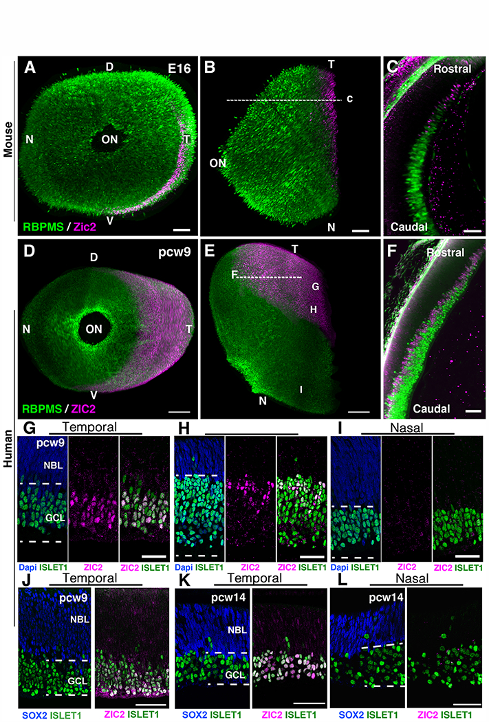
Fig. 3. Zic2 is expressed in the temporal retina of mammal embryos.

(A to C) Whole-mount immunohistochemistry of an E16 mouse eye labeled with the pan ganglion cell marker RBPMS and the ipsilateral ganglion cell marker Zic2. (A) frontal view and (B) top view. (C) Optical section at the level indicated by the dashed line in (C). (D to F), 3D light-sheet fluorescence microscopy of pcw9 human embryonic eye cleared using EyeDISCO and labeled for RBPMS and ZIC2. (D) frontal view and (E) top view. (F) Optical section at the level indicated by the dashed line in (C). G, H, I indicate the approximate positions of images in panels G-I. (G to I) retinal cryosections of a pcw9 human embryo eye labeled with ZIC2 and ISLET1 at 3 different levels: temporal and close to retinal outer limit (G), temporomedial (H) and nasal (I). ZIC2 cells are in the ganglion cell layer (GCL) and co-express ISLET1. They are absent from the neuroblastic layer (NBL). (J) retinal cryosection of a pcw9 human embryo eye labeled with ZIC2 and SOX2. ZIC2 is absent from the NBL which contains SOX2+ progenitors. (K and L) retinal cryosections of a pcw14 human embryo eye labeled with ZIC2, SOX2 and ISLET1. ZIC2 is only present in ISLET1+ ganglion cells in the temporal retina and absent from SOX2+ progenitors (K). Abbreviations: D, dorsal ; V, ventral ; N, nasal ; T, temporal ; ON, optic nerve. Scale bars are 70 μm in (A and B) and 20 μm in (C) and 300 μm in (D and E) and 50 μm in (F to L).
