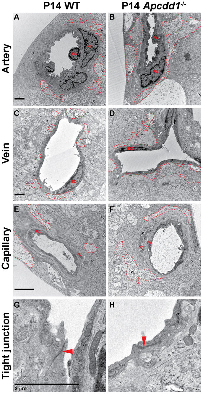
Fig. 7.

Apcdd1−/− retinal vessels have more extensive astrocyte endfeet coverage at the electron microscopy level compared with wild type. (A-F) TEM analyses of astrocyte endfeet coverage (red outlines) around the WT (A,C,E) and Apcdd1−/− (B,D,F) retinal superficial blood vessels show more-extensive endfeet coverage of arteries and veins in Apcdd1−/− retinas. (G,H) Ultrastructural analysis of endothelial tight junctions (red arrowheads) shows no obvious difference between WT and Apcdd1−/− retinas.
