. 2022 Sep 7;11:e63327. doi: 10.7554/eLife.63327

Figure 5. Recombination of complementing viruses after serial passaging.

(A) PCR strategy to detect gene arrays with outward-facing primers. (B) Representative PCR products amplified with outward-facing primers for VC-R2 and three independent HGT3 passages. (C) Recombination of complementing viruses resulted in tandem arrays of the H4L locus as determined by long-read sequencing.

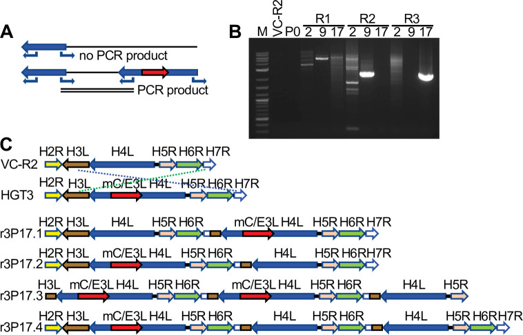
Figure 5—source data 1. PCR products amplified with outward-facing primers for VC-R2 and three independent HGT3 passages.

Figure 5.

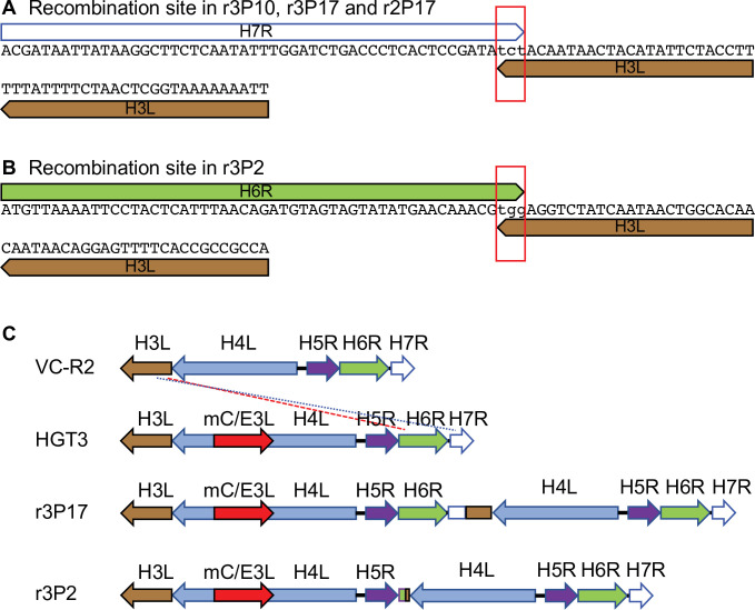
Figure 5—figure supplement 1. Recombination sites in passaged HGT3.

Figure 5—figure supplement 1.

(A) Recombination site in HGT3 between H7R and H3L identified in passage 10 of replicate 3 and passage 17 of replicate 2 by PCR and Sanger sequencing, as well as in passage 17 of replicate 3 by PacBio sequencing. The red rectangle shows three shared nucleotides between the genes at the breakpoint. (B) Recombination site in HGT3 between H6R and H3L identified in passage 2 of replicate 3 by PCR and Sanger sequencing. The red rectangle shows three shared nucleotides between the genes at the breakpoint. (C) Schematic representation of recombined loci in parental VC-R2 and HGT3, and recombined loci in r3P17 (blue dotted line) and r3P2 (red dashed line).