Abstract
Described herein is a morbidly obese 57-year-old man with an aneurysm involving the tubular portion of the aorta. Examination of the wall of the operatively resected aneurysm disclosed classic findings of aortic syphilis, a condition that clearly has not disappeared. If there is an aneurysm involving the tubular portion of the ascending aorta in the absence of aortic dissection or involvement of the sinuses of Valsalva, the most likely diagnosis is aortic syphilis. In these circumstances, the serologic test for syphilis is often negative.
Keywords: Aortic aneurysm, aortic operation, aortic syphilis
The patient, either a man or a woman, is >50 years of age, usually asymptomatic, may or may not have a precordial murmur, and possesses a normal-sized heart, but the area of the ascending aorta on the chest radiograph is enlarged. An echocardiogram and/or computed tomographic image shows the tubular portion of the aorta to be dilated without evidence of aortic dissection, the sinuses of Valsalva to be of normal size, and focal calcific deposits may or may not be present in the aneurysmal wall. The serologic test for syphilis may or may not be reactive. This scenario is the characteristic picture of the patient with aortic syphilis.1–3 Such a case is described herein.
CASE DESCRIPTION
A 57-year-old asymptomatic, obese (body mass index 40.5 kg/m2) woman on routine checkup was found to have a soft precordial murmur; a chest radiograph and an echocardiogram showed a dilated ascending aorta (involving the tubular portion only). These findings led to her referral to Baylor University Medical Center’s Aortic Clinic, where a computed tomographic image confirmed the fusiform aneurysm involving the tubular portion of the ascending aorta, its maximal diameter being 5.1 cm. The left ventricular ejection fraction was 55% to 60%, and the left ventricular cavity was of normal size. The electrocardiogram was normal. A serologic test for syphilis was negative. The aortic aneurysm was replaced with a 30-mm graft. Her postoperative course was uneventful. The operatively excised aorta is shown in Figures 1 and 2, and photomicrographs of portions of the aortic media are shown in Figure 3.
Figure 1.
(a) Diagram of the aortic aneurysm before and after operative therapy. (b) View of the dilated ascending aorta before and after its replacement.
Figure 2.
View of the aortogram showing the (a, b) dilated tubular portion of the aorta. The sinus of Valsalva (SV) is of normal size. AA indicates ascending aorta; DTA, descending thoracic aorta. (c) Opened aorta showing the extensive fibrous and calcific deposits on the intimal surface. The circumference of the aorta is 16 cm and, when intact, the diameter is 5.1 cm. (d) Photomicrograph of the wall of the fusiform aneurysm showing large collections of cholesterol clefts in the intima (I) and destroyed part of the media (M). The adventitia (A) is thickened by fibrous tissue within which are collections of lymphocytes and plasmacytes (small arrows). Movat stain, ×20.
Figure 3.
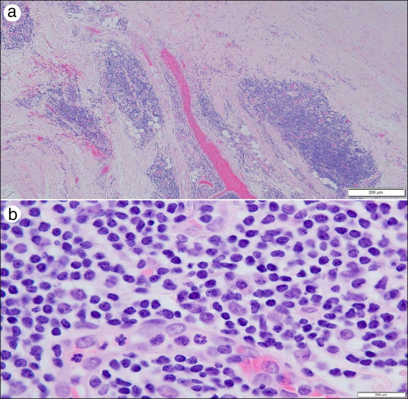
Photomicrographs of portions of the aortic media showing (a) large clumps of lymphocytes and plasmacytes and (b) a close-up of cells in the clumps. Hematoxylin-eosin stains, × 20 (a) and ×1000 (b).
DISCUSSION
Aortic syphilis has not disappeared.1–4 It remains a major cause of aneurysm of the tubular portion of the aorta.4 The process begins at the sinotubular junction, thus sparing the walls of the sinuses of Valsalva. Although on occasion the syphilitic process may involve one or more arteries arising from the arch and also the descending thoracic aorta, the process never involves the abdominal aorta (because there are no vaso vasora in this portion of the aorta). The wall of the aneurysm is 100% involved by the syphilitic process or nearly so. The aneurysm, thus, is fusiform, but on occasion one or more saccular aneurysms may arise from the wall of the fusiform aneurysm.
The histologic features of the aneurysmal wall are characteristic. All three layers of the wall are involved in the process. The adventitia is thickened by fibrous tissue containing focal collections of lymphocytes and plasmacytes. The walls of the vaso vasora are thicker than normal and their lumens are narrowed. (The latter appears to be responsible for the focal scarring of the media and the destruction of many of its elastic fibers. Sometimes the inflammatory process in the adventitia extends into the media, which is not thickened by the process.) The intima is thickened mainly by fibrous tissue, often with focal calcific deposits. The consequence of this process is that the wall of the aorta is thicker than normal, but because of the disappearance of many elastic fibers in the media, the wall is weaker than normal, leading to the aneurysmal dilation. The presence of the scar tissue in the media essentially prevents the occurrence of aortic dissection in these patients.5
The main reason to diagnose syphilis as the cause of an aortic aneurysm is to prevent its massive expansion and rupture and to prompt the use of antibiotics to retard or prevent the occurrence of neurological syphilis. The unusual feature of the present case is the disposition in the aortic intima of huge quantities of calcific deposits.
References
- 1.Roberts WC, Ko JM, Vowels TJ.. Natural history of syphilitic aortitis. Am J Cardiol. 2009;104(11):1578–1587. doi: 10.1016/j.amjcard.2009.07.031. [DOI] [PubMed] [Google Scholar]
- 2.Roberts WC, Bose R, Ko JM, Henry AC, Hamman BL.. Identifying cardiovascular syphilis at operation. Am J Cardiol. 2009;104(11):1588–1594. doi: 10.1016/j.amjcard.2009.06.071. [DOI] [PubMed] [Google Scholar]
- 3.Roberts WC, Barbin CM, Weissenborn MR, Ko JM, Henry AC.. Syphilis as a cause of thoracic aortic aneurysm. Am J Cardiol. 2015;116(8):1298–1303. doi: 10.1016/j.amjcard.2015.07.030. [DOI] [PubMed] [Google Scholar]
- 4.Roberts WC, Moore AJ, Roberts CS.. Syphilitic aortitis: still a current common cause of aneurysm of the tubular portion of ascending aorta. Cardiovasc Pathol. 2020;46:107175. doi: 10.1016/j.carpath.2019.107175. [DOI] [PubMed] [Google Scholar]
- 5.Roberts WC, Roberts CS.. Combined cardiovascular syphilis and type A acute aortic dissection. Am J Cardiol. 2022;168:159–162. doi: 10.1016/j.amjcard.2021.10.040. [DOI] [PubMed] [Google Scholar]


