



Abstract
The current treatment of osteomyelitis includes systemic antibiotic therapy and a debridement procedure of the formed biofilm and necrotic tissue. Moreover, cements and three-dimensional scaffolds are used both for the delivery of therapeutic agents and as fillers for bone defects. The aim of our research was to test, on cellular cultures, the biocompatibility of a previously synthesized microporous biocomposite containing hydroxyapatite and a collagen matrix including a therapeutic agent (ciprofloxacin and gentamicin). The scaffold was obtained by direct mineralization namely co-precipitation of hydroxyapatite on a collagen matrix.
Keywords: Biocomposite, collagen-hydroxyapatite, biocompatibility
Introduction
The treatment of bone infections continues to be a challenge due to low penetrability of systemic administered antibiotics in the bone tissue [1].
Bacteria adheres to the bone matrix and the orthopedic implant and eludes the defense mechanism of the host by forming a biofilm or by developing an extremely slow growth rate [1].
Literature proposes incorporating traditional antibiotics in biodegradable composites in order to avoid bacterial colonization during surgical interventions. These devices present several advantages that benefit both the health system and the patient by optimizing the cost generated by long periods of hospitalization, repeated surgical interventions, the cost of pharmacologic treatment, reducing the risks of reinfection and improving compliance [2].
The latest approach in osteomyelitis management involves a single surgical intervention and the use of these local antibiotic carriers in order to ensure a therapeutic local concentration and prevent further toxicity [2].
A collagen-hydroxyapatite (Col-HA) biocomposite was first mentioned in the literature fifteen years ago. In this time frame, research was able to establish the advantages and limitations of the biocomposite and it is currently directed into the optimization of this biomaterial [2,3].
Both materials namely hydroxyapatite (HA) and collagen (Col) played a key role in orthopedic surgery and implantology. Although they each may have several limitations, when used together they exhibit remarkable properties. Early research followed the development of a composite scaffold containing a polymer (to ensure flexibility) and a ceramic material (to ensure strength and rigidity) in order to use it as connective tissue replacement or a filler for bone defects [3].
Currently, research focuses on using bioactive scaffolds in bone tissue engineering or as a targeted delivery system for drugs [3].
Hydroxyapatite ensures both an increased loading and a prolonged release of the drug due to its porosity. Furthermore, these properties are essential for the efficacy of the delivery system [4].
Moreover, adding hydroxyapatite to collagen increases the compression of the collagen matrix and provides a larger and more abrasive adhesion surface. This improves bioactivity and cell proliferation [4].
Collagen acts as a delivery support for therapeutic agents due to its ability to expand without disintegrating and to incorporate hydrophobic materials. In addition, collagen shows an adjustable degradation rate which allows a controlled release of the drug [4].
The aim of this study is to asses cell response to a previously synthesized Col-HA bioactive scaffold containing either ciprofloxacin (Cipro) or gentamicin (Genta). Hydroxyapatite was synthesized from its precursors, namely calcium hydroxide and diammonium phosphate, using a method adapted to its application i.e. hydroxyapatite co precipitation in collagen matrix. This ensures a better control over particle size, morphology, ion substitution and to avoid possible impurities. Furthermore, the method is reliable, economic and fast [5,6].
Hydroxyapatite may have several applications namely to repair and replace the deteriorated hard tissue and as a drug delivery system that promotes tissue growth. Regarding calcium phosphates, HA has the lowest degradation rate and a higher stability in aqueous media at a 4.2-8 pH [7].
Collagen is a polymer that has the ability to influence the nucleation, growth and assembly of HA crystals in its fiber network.
Ciprofloxacin is a broad-spectrum antibiotic and shows a good penetrability through both soft and hard tissues. Furthermore, it is effective against beta- lactamase producing strains [8,9].
Gentamicin is used for both the prophylaxis and treatment of orthopedic infections. Moreover, it is soluble in water, heat stable and it may prevent bacteria adherence the days following implantation. Gentamicin encapsulated into [poly (methyl methacrylate)] (PMMA) pearls is already used for the local treatment of bone infections; although the main drawback is that PMMA is not biodegradable and it requires a second intervention for its removal [10].
Here, we show that Col-HA scaffolds obtained by co-precipitation for antibiotic drug delivery to bone tissue are biocompatible in a mesenchymal stem cell tissue culture model. Further research is necessary to develop this biomaterial for use in osteomyelitis treatment applications.
Materials and Methods
Materials
Paraformaldehyde, HMDS, Triton X-100 and 25% glutaraldehyde were purchased from SIGMA (Table 1). Phosphate buffered saline (PBS) was purchased from Santa Cruz.
Table 1.
Producers/providers for the media, kits and reagents used
|
Media |
Producer/provider |
|
DMEM 4,5g-L |
Gibco Invitrogen |
|
Kits and reagents |
|
|
Phalloidin Alexa Fluor 488 conjugate |
Molecular Probes, Invitrogen |
|
CellTiter 96® AQueous One Solution Cell Proliferation Assay |
Promega, Dextercom SRL, Bucureşti, Romania |
|
NBT-BCIP (nitro blue tetrazolium chloride-/5-bromo-4-chloro-3-indolylphosphate). |
Roche |
|
Alizarin Red |
Fluka, REDOX, Bucuresti, Romania |
|
Prolong Gold Antifade mountant with DAPI |
Invitrogen, Medist, Bucuresti, Romania |
Methods
Synthesis of the Antibiotic Doped Col-HA Scaffolds
We preferred to synthesize hydroxyapatite in the laboratory from precursors (calcium hydroxide-Ca (OH)2 and dibasic ammonium phosphate-(NH4)2HPO4 ) by a method adapted to the application (co-precipitation of hydroxyapatite in the collagen matrix), in order to have a better control over the particle size, morphology, ion substitution and synthesis parameters and to avoid possible impurities. It is also a reliable, fast and economical method [11].
The principle of the in situ co-precipitation method (described in detail in our previous articles) is the crystallization of hydroxyapatite in solution in the presence of a natural polymer (collagen), a process that facilitates a high degree of interaction between inorganic and organic components of the composite. Ca2+ binds to the HA substrate and the-COO- groups on the collagen structure, which subsequently give better mechanical properties to the final biocomposite [6,12].
To optimize the molecular bonds between the constituents, the composite material was chemically crosslinked.
An important parameter that can be easily calibrated by our synthesis method is the pH (maintained in the pH range=7.5-8 with 1M HCl solution) which can influence both the interaction between the polymer and the inorganic ions and the diffusion rate and its physical and chemical properties.
After synthesis the biomaterial was dehydrated by lyophilization.
The biopunch device no. 6 was used resulting in discs with a diameter of 5-6mm and a variable mass (8-9mg).
Morphology of Col-HA Scaffolds
The samples were fixed in 4% paraformaldehyde (PFA) for 20 minutes at room temperature. After three washes in PBS, the samples were sequentially immersed in 70%, 90% and 100% ethanol for 15 minutes twice for each concentration in order to dehydrate the cells. The samples were air dried.
Then, they were metallized (coated with a 60nm thick film of gold particles for 60 sec) in order to obtain electrical conductivity and they were fixed on a support (Al) using carbon conductive tape. A high-resolution scanning electron microscope, FEI Inspect F50, at 30KeV was used to evaluate the morphology of the samples.
Biocompatibility of Col-HA Biomaterials
Isolation of Mesenchymal Stem Cells
Mesenchymal stem cells were isolated from the bone marrow extracted from patients who underwent orthopedic implant surgery at the orthopedics department of the Emergency Hospital of Craiova. The cell line was subsequently obtained at the Institute of Biochemistry of the Romanian Academy following established protocols [12,13].
Biomaterial Sterilization
The UV sterilization method was applied with a three-minute exposure into the laminar flow hood (Holten LaminAir, Thermo).
Cell Proliferation Assay Using the MTS Test
CellTiter 96® AQueous One Solution Cell Proliferation Assay (MTS) Promega kit was used to evaluate cell proliferation at 450nm. After being cultured, both the materials and the adherent cells were moved into new 48 well plates and incubated together with 300 µl fresh medium that contained MTS reagent. 90 minutes later, the supernatant was added in duplicate into a well plate to perform a spectroscopic analysis using a Mithras spectrophotometer (Berthold).
Cell Adhesion Evaluation by Fluorescence Microscopy
The cells were fixed with 4% paraformaldehyde (PFA) for a period of 10min and then permeabilized with a 0.2% Triton-X-100 solution at room temperature for 3 minutes. The cells were firstly incubated with primary antibodies and then with Alexa Fluor 594 conjugated secondary antibodies for 30 minutes each. After each stage the samples were washed with PBS. Actin filaments were labeled with phalloidin Alexa Fluor 488 conjugate 1: 100. Furthermore, nuclei were labeled with Hoechst at a 1: 10000 dilution of a stock solution of 10 mg / ml. The samples were mounted in a Prolong Gold Antifade Invitrogen mounting medium and then visualized under the fluorescence microscope. The images were acquired using TissueFAXSiPlus image cytometry reader (TissueGnostics, Austria) and then processed in Power Point.
Evaluation of the Enzymatic Activity of Active Alkaline Phosphatase (ALP) in Differentiated Osteoblasts
The test was performed using the colorimetric technique using a Roche kit that contains BCIP (5-Bromo-4-chloro-3- indolyl phosphate), which serves as a substrate for ALP and NBT (nitro blue tetrazolium), which acts as an oxidant.
Results
Morphology of Col-HA Scaffolds
All samples analyzed showed collagen fibers with a folded lamellar appearance (Figure 1). HA appears as larger agglomerated particles both inside and on the lamellar surface of collagen. Following the addition of the inorganic phase, the volume of macro pores in the collagen matrix decreases. Furthermore, smaller pores, only a few microns (1.6-1.9µm) may also be observed. The addition of drugs slightly changed the morphology of the biomaterial. The antibiotic may reside inside the pores namely gentamicin or be adsorbed on the HA surface such as ciprofloxacin, which seems to have a special affinity for the Ca atoms in the HA structure.
Figure 1.


SEM images for the scaffolds tested: A. Col-HA, B. Col-HA-Gentamicin, C. Col-HA-Ciprofloxacin
Biocompatibility of Col-HA Biomaterials
The MTS test on mesenchymal stem cells was used to evaluate the cytotoxicity of the tested materials after being cultivated for 48 hours.
The results showed that osteogenic cells cultivated on the Col-HA scaffolds maintain their mitochondrial metabolism. Furthermore, the cells are relatively uniformly distributed on the surface of the material (Figure 2A).
Figure 2.

Cytotoxicity evaluation of Col-HA scaffolds both with and without antibiotics (ciprofloxacin and gentamicin) using the MTS test. A. Sample images after the MTS reaction and supernatant removal. B.Graphic representation of the absorbance signal obtained following the MTS reaction
By comparison, the number of viable cells identified on scaffolds containing antibiotics such as Col-HA-Cipro and Col-HA-Genta was lower (Figure 2A).
These observations are also confirmed by the spectroscopic analysis of the media containing the MTS substrate which shows a decrease in the number of active metabolic cells (viable) for the antibiotic containing scaffolds as compared to the Col-HA scaffold (Figure 2B).
Another test conducted consisted of a complementary analysis of cell viability using fluorescence microscopy. This test was performed on the MSCs cultured on Col-HA scaffolds. Following the labelling with calcein and ethidium homodimer (EthD-1) it was observed that most cells retained their viability (Figure 3, top panel and bottom left); only a small fraction of the cell nuclei were labelled with EthD-1 (Figure 3, bottom right, magenta staining obtained due to the overlapping of EthD-1 with Hoechst).
Figure 3.

Mesenchymal stem cells viability after being cultured together with the Col-HA scaffold. Calcein (green) and ethidium homodimer-1 (red) were used to stain the viable cells and the dead cells, respectively inside the cellular culture. Hoechst was used to stain the nuclei. The upper image represents an overlap of images taken on 3 fluorescence channels (calcein/FITC+EthD-1/TxRed+Hoechst/DAPI). Below the first image are two decomposed images: left-calcein / FITC and right-EthD-1/TxRed+Hoechst/DAPI). Scale=100μm
This test confirms that the measured MTS signal is determined by the majority of the viable cells that grew on the scaffold. The cells tend to slide into the pores of the material and then they grow along the collagen fibers which support cell adhesion.
The viability test was also repeated on cells cultivated together with each of the three scaffolds in DMEM expansion medium (Figure 4).
Figure 4.

Viability test performed on mesenchymal stem cells stained with calcein and co-cultivated with the scaffolds. After three days the samples were evaluated using the fluorescence microscope. Cell nuclei were identified by Hoechst labelling on the DAPI channel (left)
Fluorescence microscopy analysis shows that the cells do not lose their viability. Furthermore, we estimate that the decrease in MTS signal may be determined by a decrease in cell proliferation or mitochondrial metabolism and not a result of cell apoptosis. A high cell occupancy was evidenced by DAPI staining for all three scaffolds.
In order to evaluate the capacity of the material to support osteogenic differentiation of MSC, we performed comparative tests in expansion medium (DMEM) and osteoinduction medium (OIM).
The fluorescence microscopy assay on cells stained with phalloidin and Hoechst and cultured for 14 days on DMEM or OIM media shows a rather equal distribution of the cells inside the scaffolds (Figure 5).
Figure 5.

Cell adhesion analysis after staining MSC with AlexaFluor488 phalloidin which labels actin cytoskeleton (green); cell nuclei were stained with Hoechst (blue). The images were obtained using the TissueFAXSiPlus system which automatically scans the entire surface of the samples and then renders a virtual reconstruction of the sample (left); images on the right side represent selected detailed visual fields
Under osteogenic conditions, the number of cells is slightly decreased, which we anticipated because during differentiation cells decrease their proliferation rate.
Samples grown in parallel under the same conditions were fixed, dehydrated, and analysed under a scanning microscope to assess the interaction between cells and scaffolds (Figure 6).
Figure 6.

SEM images of the MSC morphology 14 days after cell culture in osteoinduction medium compared to expansion medium
Cells may be detected on the surface of the materials tested under expansion conditions (DMEM) or osteoinduction (OIM) when compared to the Col-HA simple scaffold. No significant morphological differences were observed between the variants with and without antibiotic.
From a functional point of view, an analysis was performed to outline the active alkaline phosphatase (ALP), an osteoblast marker, by incubating cells with their enzymatic substrate (NBT-BCIP). The results showed that cells efficiently infiltrate Col-HA simple scaffolds and those containing ciprofloxacin but far fewer cells with active ALP were identified in Col-HA-Genta samples under osteoinduction conditions (Figure 7-OIM).
Figure 7.
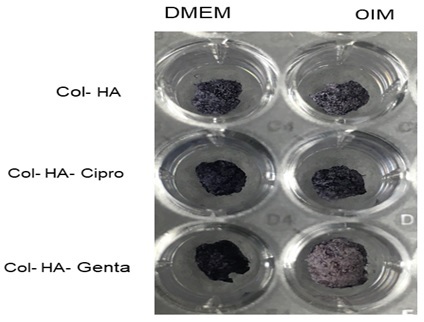
Analysis of the alkaline phosphatase enzymatic activity after 14 days of cultivating MSC in osteoinduction medium (OIM) compared to expansion medium (DMEM) by incubating samples with NBT-BCIP substrate (the result of the colorimetric reaction is highlighted in purple)
Interestingly, both antibiotics were able to increase ALP activity even under expansion conditions (DMEM), in the absence of osteoinductive factors (Figure 7-DMEM).
The latter test advances the Col-HA-Cipro formula for further development to be used in bone therapies or interventions.
Discussion
The principle of the in-situ co-precipitation method (inspired by natural mineralization processes) resides in the crystallization of hydroxyapatite in a solution in the presence of a natural polymer (collagen). This process facilitates a high degree of interaction between inorganic and organic components of the composite (Ca 2+ ions attached to HA substrate and the-COO- groups on the collagen structure) which subsequently give better mechanical properties to the final biocomposite. To optimize the molecular bonds between the constituents, the composite material was chemically crosslinked [11,15].
The Col-HA scaffolds were doped with antibiotics by absorption. This was determined by the “swelling” properties of the biocomposite resulted from combining the two materials [16].
An adsorption process truly occurs for ciprofloxacin due to a chemical reaction that takes place between the surface of the biocomposite and the adsorbent. Therefore, new interactions are possible between the Ca atoms in the HA structure and the O atoms in the Cipro structure [17].
The amount of antibiotic (Cipro and Genta) encapsulated in the composite was assessed by high performance liquid chromatography (HPLC). The encapsulation efficiency, for both antibiotics was significant (18.027% Cipro and 15.92% Genta). This percentage resembles the results obtained using other methods (e.g.: addition of antibiotic during the synthesis phase of HA by wet precipitation). The high binding rate of the antibiotic may be a result of the porosity of the biocomposite substrate, determined by collagen. [6]
Lyophilization was the method chosen to dehydrate the material. Furthermore, lyophilization is recommended for obtaining porous structures because a succession of freezing and sublimation phases leads to pores (ice crystals formed after sublimation will create gaps). This is very important for our material because, later, it is easily doped with antibiotics [18].
Porosity is an important parameter for a bone scaffold, as it ensures an increased fluid absorption. Therefore, all the necessary ions are absorbed from the environment, in order to regenerate damaged bone faster. This could also be associated with an improved cell adhesion, potentially increased osteoconductivity, and implicitly it may determine the good results these materials show when in vitro cytotoxicity tests are performed [19,20].
Conclusions
Biocompatibility tests have shown that human mesenchymal cells can infiltrate and remain viable after culture on both the Col-HA scaffolds and the antibiotic-doped substrates (ciprofloxacin and gentamicin).
The material has proven biomimetic properties that allow cell adhesion and proliferation. Therefore, we consider it qualifies for further development for applications in implantology.
The results suggest that the biocomposite may be eligible for a possible use as an antibiotic delivery system in order to treat bone infections that occur as a complication following surgery in orthopedic departments.
This approach, as a local drug delivery system, offers a number of advantages over standard therapy such as increasing local antibiotic concentration, reducing drug toxicity and showing a better bioavailability of the drug. Furthermore, it offers a new perspective on the long-term treatment of osteomyelitis and patient compliance.
Conflict of interests
None to declare.
Acknowledgments
Acknowledgments
The authors would like to thank Dr. Livia Sima in the Department of Molecular and Cellular Biology at the Institute of Biochemistry of the Romanian Academy, Bucharest, for support in testing the biocompatibility of the material, adaptation of testing methods, discussions and valuable advices.
References
- 1.Macha IJ, Cazalbou S, Ben-Nissan B, Harvey KL, Milthorpe B. Marine structure derived calcium phosphate-polymer biocomposites for local antibiotic delivery. Mar Drugs. 2015;13(1):666–680. doi: 10.3390/md13010666. [DOI] [PMC free article] [PubMed] [Google Scholar]
- 2.Ferguson J, Alexander M, Bruce S, O'Connell M, Beecroft S, McNally M. A retrospective cohort study comparing clinical outcomes and healthcare resource utilisation in patients undergoing surgery for osteomyelitis in England: a case for reorganising orthopaedic infection services. J Bone Jt Infect. 2021;28;6(5):151–163. doi: 10.5194/jbji-6-151-2021. [DOI] [PMC free article] [PubMed] [Google Scholar]
- 3.Kołodziejska B, Kaflak A, Kolmas J. Biologically Inspired Collagen/Apatite Composite Biomaterials for Potential Use in Bone Tissue Regeneration-A Review. Materials (Basel) 2020;13(7):1748–1748. doi: 10.3390/ma13071748. [DOI] [PMC free article] [PubMed] [Google Scholar]
- 4.Thirumalai J, editor. Hydroxyapatite-Advances in Composite Nanomaterials,Biomedical Applications and Its Technological Facets. Rijeka: InTech; 2018. pp. 109–109. [Google Scholar]
- 5.Ciocilteu MV, Mocanu AG, Mocanu A, Ducu C, Nicolaescu OE, Manda VC, Turcu-Stiolica A, Nicolicescu C, Melinte R, Balasoiu M, Croitoru O, Neamtu J. Hydroxyapatite-ciprofloxacin delivery system: Synthesis, characterisation and antibacterial activity. Acta Pharm. 2018;68(2):129–144. doi: 10.2478/acph-2018-0011. [DOI] [PubMed] [Google Scholar]
- 6.Ciocîlteu MV, Filip (Ionescu) OL, Manda CV, Nicolaescu OE, Neacşu IA, Ficai A, Buzatu IM, Nicolicescu C, Croitoru O, Neamţu J. Physico-chemical characterization and antibacterial activity of a controlled collagen-hydroxyapatite-ciprofloxacin release system. Farmacia. 2020;68(6):1055–1061. [Google Scholar]
- 7.Kumar A, Kargozar S, Baino F, Han SS. Additive Manufacturing Methods for Producing Hydroxyapatite and Hydroxyapatite-Based Composite Scaffolds: A Review. Frontiers in Materials. 2019;6:313–313. [Google Scholar]
- 8.Stevanović M, Djošić M, Janković A, Nešović K, Kojić V, Stojanović J, Grujić S, Matić Bujagić I, Rhee KY, Mišković-Stanković V. Assessing the Bioactivity of Gentamicin-Preloaded Hydroxyapatite/Chitosan Composite Coating on Titanium Substrate. ACS Omega. 2020;5(25):15433–15445. doi: 10.1021/acsomega.0c01583. [DOI] [PMC free article] [PubMed] [Google Scholar]
- 9.Mocanu AG, Turcu-Stiolica A, Belu I, Ciocilteu MV, Manda VC, Nicolicescu C, Neamtu J. Quality by Design for Ciprofloxacin Encapsulation in PLGA. J. Revista de Chimie. 2018;69(6):1332–1338. [Google Scholar]
- 10.Jackson J, Lo J, Hsu E, Burt HM, Shademani A, Lange D. The Combined Use of Gentamicin and Silver Nitrate in Bone Cement for a Synergistic and Extended Antibiotic Action against Gram-Positive and Gram-Negative Bacteria. Materials (Basel) 2021;14(12):3413–3413. doi: 10.3390/ma14123413. [DOI] [PMC free article] [PubMed] [Google Scholar]
- 11.Ficai A, Andronescu E, Ghitulica C. Colagen/Hydroxyapatite Interaction in Composite Biomaterials. Materiale Plastice. 2009;46(1):11–15. [Google Scholar]
- 12.Ionescu (Filip) OL, Ciocîlteu MV, Manda CV, Neacsu IA, Ficai A, Amzoiu E, Turcu Stiolica A, Croitoru O, Neamtu J. Bone-Graft Delivery Systems of type PLGA-Gentamicin and Collagen- Hydroxyapatite- Gentamicine. Materiale Plastice. 2019;56(3):534–537. [Google Scholar]
- 13.Sima LE, Stan GE, Morosanu CO, Melinescu A, Ianculescu A, Melinte R, Neamtu J, Petrescu SM. Differentiation of Mesenchymal Stem Cells onto Highly Adherent Radio Frequency-Sputtered Carbonated Hydroxyapatite thin Films. J Biomed Mater Res A. 2010;95(4):1203–1214. doi: 10.1002/jbm.a.32947. [DOI] [PubMed] [Google Scholar]
- 14.Nicolaescu OE, Turcu-Stiolica A, Varut RM, Mocanu AG, Belu I, Sima LE, Neamtu J. Evaluation of Mesenchymal Stem Cells and Osteoblasts Adhesion and Proliferation in the Presence of HA- AL Biomaterials. Coatings. 2019;9(12):782–782. [Google Scholar]
- 15.Rahman MS, Rana MM, Spitzhorn LS, Akhtar N, Hasan MZ, Choudhury N, Fehm T, Czernuszka JT, Adjaye J, Asaduzzaman SM. Fabrication of biocompatible porous scaffolds based on hydroxyapatite/collagen/chitosan composite for restoration of defected maxillofacial mandible bone. Prog Biomater. 2019;8(3):137–154. doi: 10.1007/s40204-019-0113-x. [DOI] [PMC free article] [PubMed] [Google Scholar]
- 16.Mulazzi M, Campodoni E, Bassi G, Montesi M, Panseri S, Bonvicini F, Gentilomi GA, Tampieri A, Sandri M. Medicated Hydroxyapatite/Collagen Hybrid Scaffolds for Bone Regeneration and Local Antimicrobial Therapy to Prevent Bone Infections. Pharmaceutics. 2021;13(7):1090–1090. doi: 10.3390/pharmaceutics13071090. [DOI] [PMC free article] [PubMed] [Google Scholar]
- 17.Walmsley GG, McArdle A, Tevlin R, Momeni A, Atashroo D, Hu MS, Feroze AH, Wong VW, Lorenz PH, Longaker MT, Wan DC. Nanotechnology in bone tissue engineering. Nanomedicine. 2015;11(5):1253–1263. doi: 10.1016/j.nano.2015.02.013. [DOI] [PMC free article] [PubMed] [Google Scholar]
- 18.Vranceanu M, Saban R, Antoniac I, Albu M, Miculescu F. Development and novel porous collagen based biocomposite for bone tissue regeneration. UPB Scientific Bulletin Series B: Chemistry and Materials Science. 2012;74(3):145–156. [Google Scholar]
- 19.Dumitrescu CR, Neacsu IA, Surdu VA, Nicoara AI, Iordache F, Trusca R, Ciocan LT, Ficai A, Andronescu E. Nano-Hydroxyapatite vs. Xenografts: Synthesis, Characterization, and In Vitro Behavior. Nanomaterials (Basel) 2021;11(9):2289–2289. doi: 10.3390/nano11092289. [DOI] [PMC free article] [PubMed] [Google Scholar]
- 20.Neacsu IA, Serban AP, Nicoara AI, Trusca R, Ene VL, Iordache F. Biomimetic Composite Scaffold Based on Naturally Derived Biomaterials. Polymers (Basel) 2020;12(5):1161–1161. doi: 10.3390/polym12051161. [DOI] [PMC free article] [PubMed] [Google Scholar]