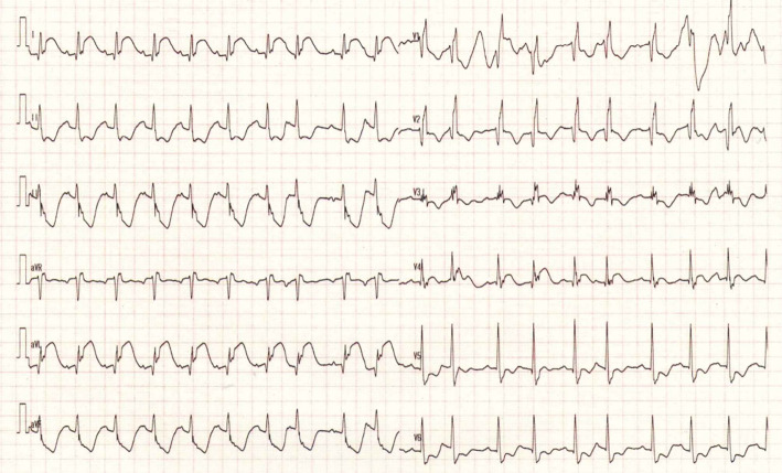
A 72-year-old woman without any significant medical history presented to the emergency department with pulseless electrical activity. A return of spontaneous circulation was obtained before arrival; 50 minutes had passed since the onset. Upon admission, her blood pressure was 199/107 mmHg, with sinus tachycardia. Lateral ST-segment elevation was documented (Picture 1). Echocardiography revealed left ventricle lateral wall hypokinesia. We used computed tomography (CT) to rule out aortic dissection.
Picture 1.
Suddenly, her blood pressure dropped after plain CT, and she experienced cardiac arrest. This resulted from a blow-out type free-wall rupture (Picture 2, 3). The patient could not be revived. Picture 2 shows the left ventricle with small pericardial effusions. Picture 3 shows the flow of contrast medium from the left ventricle into the pericardial space. Blow-out type free-wall ruptures occur abruptly, and are associated with a poor prognosis (1). Imaging studies that capture the exact moment of rupture are very rare.
Picture 2.

Picture 3.

The authors state that they have no Conflict of Interest (COI).
References
- 1. Matteucci M, Kowalewski M, De Bonis M, et al. Surgical treatment of post-infarction left ventricular free-wall rupture: a multicenter study. Ann Thorac Surg 112: 1186-1192, 2021. [DOI] [PubMed] [Google Scholar]


