Figure 1.
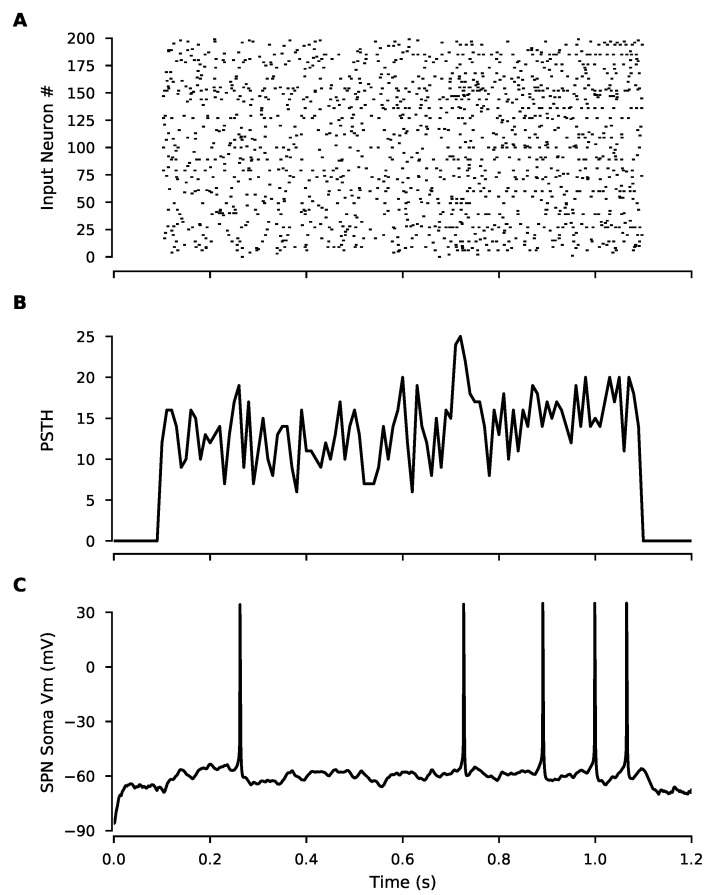
In vivo-like inputs constructed from cortical spike trains produce spiking in SPNs. (A) Raster plot shows spike times for each cortical input in the model, constructed from in vivo spike train recordings. (B) Peri-stimulus time histogram of the above raster plot (spike counts per 10 ms bin) (C) Somatic membrane potential of the SPN model showing spiking output induced by cortical input.

