Abstract
Mitochondria are important organelles responsible for energy production, redox homeostasis, oncogenic signaling, cell death, and apoptosis. Deregulated mitochondrial metabolism and biogenesis are often observed during cancer development and progression. Reports have described the crucial roles of mitochondria in urothelial carcinoma (UC), which is a major global health challenge. This review focuses on research advances in the role of mitochondria in UC. Here, we discuss the pathogenic roles of mitochondria in UC and update the mitochondria-targeted therapies. We aim to offer a better understanding of the mitochondria-modulated pathogenesis of UC and hope that this review will allow the development of novel mitochondria-targeted therapies.
Keywords: mitochondrial metabolism, redox homeostasis, apoptosis, tumorigenesis, urothelial carcinoma
1. Introduction
Urothelial carcinoma (UC) is a malignancy of the urinary system lining. The majority of UC cases (approximately 90–95%) arise in the urinary bladder. The remaining 5–10% are upper tract urothelial carcinomas (UTUCs), which refer to malignancies that originate from the renal calyceal system to the distal ureter [1,2]. Bladder cancer is indeed a major health threat, with an estimated 573,278 incident cases and 158,785 deaths worldwide in 2020 [3]. Although approximately 75% of newly diagnosed cases are non-muscle-invasive, 70% of tumors will recur, and 20% of the recurrences will progress to muscle-invasive disease that carries a high risk of tumor progression or metastasis. The 5-year survival rate of patients with metastatic disease is only 5% [4,5,6]. UTUC and bladder cancer are biologically similar and possess certain common risk factors, such as smoking and occupational exposure. However, they represent distinct entities owing to anatomical and practical differences [1,7,8,9]. The overall 5-year survival rate for UTUC is approximately 59–67% [10,11] and has been decreasing in recent years [12,13]. Depending on the type of UC and the stage of the disease, the mainstay of treatment includes surgery, radiation therapy, chemotherapy, and immunotherapy. Despite the great progress made in the diagnosis and treatment of UC, especially the rapid advances in immunotherapy, targeted therapy, and combinations [4,14,15,16], the high recurrence and mortality rates indicate that there are unmet needs in the management of UC. Revisiting the pathogenesis of UC may be a solution to the current bottleneck.
Mitochondria are important organelles responsible for energy production, redox homeostasis, oncogenic signaling, cell death, and apoptosis. Mitochondrial metabolism comprises pathways that generate adenosine triphosphate (ATP) and produce components necessary for macromolecule biosynthesis. It has become clear that mitochondrial metabolism plays an influential role in governing cell fate and function by controlling gene expression through the release of metabolites and reactive oxygen species (ROS) [17]. The compartmentalization of mitochondrial protein complexes and enzymes is essential for the maintenance of signaling pathways within the cell. The kidneys are second only to the heart in terms of mitochondrial abundance. In addition to their crucial roles in renal physiology, mitochondria have been recognized as key participants in kidney cancers [18,19,20]. Reduced mitochondrial DNA (mtDNA) content has been observed in renal cell carcinoma (RCC) [21]. In addition, impaired mitochondrial respiratory capacity has been observed in clear cell RCC [22]. Altered mitochondria-regulated apoptotic pathways have been reported in UTUC [23,24].
This review focuses on research advances in the role of mitochondria in UC. We aimed to offer a better understanding of the link between mitochondria and the pathogenesis of UC. We hope that this review will facilitate the development of novel mitochondria-targeted therapies for UC.
2. Roles of Mitochondria in UC
2.1. Alterations of mtDNA in UC
MtDNA mutations tend to be induced by oxidative damage, defects in nuclear genes in mtDNA stability and replication, altered nucleotide biosynthesis or transport, and exogenous sources (e.g., tobacco smoking, ionizing radiation, ozone, pesticides, and heavy metals) [25,26]. mtDNA mutations have been identified in 64% of bladder cancers and demonstrated in cancer tissue in the form of single-base deletions, point mutations, and insertions in the non-coding D-loop region or deletions in the coding regions of proteins involved in oxidative phosphorylation [27]. For example, among the mitochondrial genes cytochrome B, ATPase6, ND1, and the D310 region, G14905A, G8697A, C15452A, and A15607G polymorphisms were reported to be more frequent in UC patients than in controls [28]. The tumorigenic role of mtDNA mutations in UC was demonstrated for the 21-bp deletion in the cytochrome B (CYTB) gene. This mutation was found in urine samples and cancer tissues from patients with bladder cancer [27]. Overexpression of the 21-bp deletion mutation of the CYTB gene induces rapid cell cycle progression through upregulation of the nuclear factor-kappa B2 signaling pathway and eventually leads to tumor growth in vivo and in vitro [29]. Additionally, mtDNA mutations in the electron transport chain (ETC) have been reported. Mutations in the NADH dehydrogenase subunit 4 (ND4) gene have been identified in UTUC. Approximately 85% of mutated ND4 exists before the development of UTUC [30].
2.2. MtDNA Copy Number in UC
MtDNA copy number has been examined in bladder cancer and adjacent normal tissues using next-generation DNA sequencing. Compared with cells from normal tissues, bladder cancer cells were found to have lower mtDNA content. However, this reduction in mtDNA copy number was not accompanied by a reduction in mitochondrial gene expression. This discrepancy suggests that the expression of mitochondrial genes is not always correlated with mtDNA copy number and that mitochondrial activity may not be suppressed in bladder cancer [31].
2.3. Impact of Altered Expression of Mitochondrial Proteins on UC
Lon protease is an ATP-dependent serine protease in the mitochondrial matrix that is responsible for the degradation of abnormal proteins and maintenance of the mitochondrial genome. In cancers, Lon protease is essential for the proliferation and survival of cancer cells. Lon upregulation also contributes to metabolic reprogramming, facilitating the switch from respiratory to glycolytic metabolism in the cancer microenvironment [32]. In patients with bladder cancer, Lon protease expression is substantially higher in cancerous tissue than in non-cancerous tissue and is directly related to cancer grade and stage [33]. The mitochondrial GTPase mitofusin-2 (MFN2) is the key regulator of mitochondrial fusion at the outer mitochondrial membrane. Mitochondrial fusion/fission machinery plays a crucial role in mitochondrial quality control. Changes in mitochondrial fusion/fission machinery have been demonstrated in an increasing number of diseases including cancer [34,35]. The downregulation of MFN2 expression has been demonstrated in bladder cancer. In bladder cancer cell lines, MFN2 overexpression has been shown to inhibit cell proliferation by arresting the cell cycle and inducing apoptosis via caspase-3 [36]. Mitochondrial transcription factor A (TFAM) is a mitochondrial protein required for mtDNA stability, transcription, and replication [37,38]. TFAM expression was significantly enhanced in bladder cancer cells and directly related to cancer stage. In vitro studies have shown TFAM to induce bladder cancer cell proliferation, migration, and colony formation [39]. Leucine-rich pentatricopeptide repeat motif-containing protein (LRPPRC) is a multifunctional protein localized to the mitochondria, endoplasmic reticulum, outer and inner nuclear membranes, nucleoplasm, and cytoskeleton [40,41]. Besides being a prognostic factor, LRPPRC has recently been demonstrated to enhance tumorigenesis in bladder cancer [42].
The mitochondrial fusion/fission machinery is regulated by other genes. MicroRNA-98 (miR-98) regulates this fusion/fission machinery and affects mitochondrial membrane potential (MMP) in cancers. MiR-98 is known to be upregulated in bladder cancer cell lines and promote proliferation [43]. The role of miR-98 in chemoresistance depends on longevity assurance homolog 2 of yeast LAG1 (LASS2). LASS2 is a potent tumor suppressor that induces mitochondrial fusion and inhibits MMP. LASS2 consumption may lead to the proliferation and invasion of bladder cancer cells [44], and LASS2 negativity is associated with poor prognosis in bladder cancer [45].
2.4. Mitochondria Regulate Energy Metabolism in UC
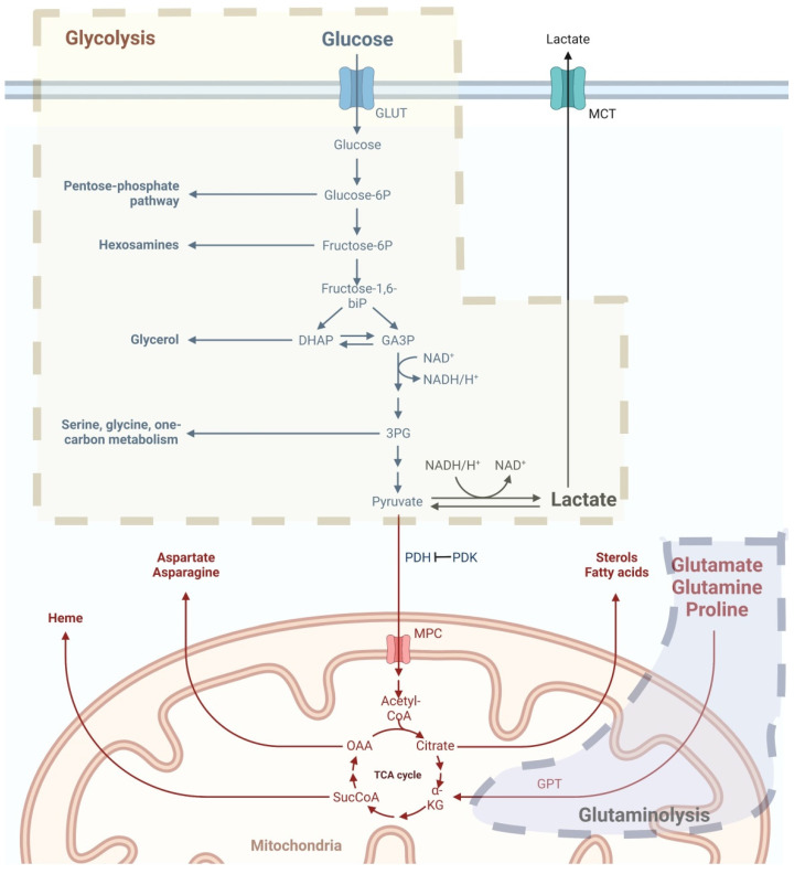
Mitochondria participate in the metabolic reprogramming of cancer cells (Figure 1). The Warburg effect, which was first reported by Otto Warburg in 1926, describes that tumor cells uptake substantial glucose and undergo glycolysis as an energy supplement, even with sufficient oxygen. Aerobic glycolysis results in increased production of cytosolic lactate [46]. Non-neoplastic cells produce energy by glucose oxidation via mitochondria, which oxidizes pyruvate to acetyl-co-enzyme-A under aerobic conditions. In this situation, pyruvate dehydrogenase (PDH) enables pyruvate to enter mitochondria. Carcinogenesis is preferred in hypoxic tissues because glucose consumption is low. Hypoxia-inducible factor (HIF) 1α is then activated together with upregulated glucose transporters (GLUTs) and pyruvate dehydrogenase kinase (PDK). The activation of PDK leads to the inhibition of PDH, and thus, the inhibition of glycolysis. In UC, PDK3 overexpression has recently been linked to poor oncological outcomes. Together with the overexpression of PDK3, these co-upregulated genes were associated with DNA repair and replication. These results suggest that PDK3 plays a crucial role in the development and proliferation of UC [47].
Figure 1.
Mitochondria regulate metabolic reprogramming in UC. The major pathways of metabolic reprogramming in UC are enhanced aerobic glycolysis and glutaminolysis. Aerobic glycolysis leads to increased cytosolic lactate production. Glutaminolysis supports cancer cells by providing energy and pools of TCA cycle intermediates for biosynthesis of proteins, lipids, and nucleotides Abbreviations: GLUT, glucose transporter; GPT, glutamate pyruvate transaminase; MCT, monocarboxylate transporter; OAA, oxaloacetate; PDH, pyruvate dehydrogenase; PDK, pyruvate dehydrogenase kinases; TCA, tricarboxylic acid. The figure was created with BioRender.com.
The mitochondrial matrix hosts the tricarboxylic acid (TCA) cycle. In UC, the mitochondrial TCA cycle produces reducing equivalents to fuel ETC and generate biosynthetic intermediates that are necessary for cell proliferation [48,49]. In addition to lactate, other substrates, including glutamine, are known to fuel the TCA cycle and participate in energy production when coupled with oxidative phosphorylations [50,51,52]. By interacting with heterogeneous nuclear ribonucleoprotein (hnRNP) I/L to upregulate glutamate pyruvate transaminase (GPT2) expression, long non-coding RNA urothelial cancer associated 1 (UCA1) has recently been demonstrated to promote glutamine-driven anaplerosis in bladder cancer [53].
2.5. Altered Mitochondrial ROS Production and ETC Activity in UC
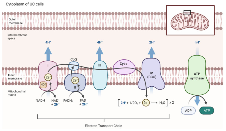
Redox homeostasis is a crucial mechanism in the progression and development of cancers [54]. Mitochondria generate ROS, which serve as toxic species for cellular macromolecules and regulate metabolic pathways [48]. Mitochondrial ROS are produced at the ETC by the leakage of electrons at the ubiquinone-binding sites of Complex I and Complex III [18,55]. Increased levels of ROS are related to increased metabolic activities and altered antioxidant capacities, which are often found in malignant conditions and interact with tumor growth and expansion [26,56]. Huang et al. investigated the urinary bladder of Sprague–Dawley rats after administering N-butyl-N-(4-hydroxybutyl) nitrosamine (BBN), a carcinogen, for eight weeks to evaluate tumorigenesis [57]. They measured the activities of components of the ETC, including NADH cytochrome c reductase (NCCR, Complex I+III), succinate cytochrome c reductase (SCCR, Complex II+III), and cytochrome c oxidase (CCO, Complex IV). The activities of all the NCCR, SCCR, and CCO were elevated by exposure to BBN, indicating a positive correlation with tumorigenesis. However, NCCR and SCCR activities reduced rapidly when BBN was discontinued, whereas CCO activity plateaued at 18 weeks despite the withdrawal of BBN. These results demonstrated that, compared with NCCR and SCCR, the CCO enzyme is more relevant to the progression of tumorigenesis in bladder cancer [57]. Figure 2 depicts the ETC in UC to facilitate the understanding of the altered ETC activity discussed above.
Figure 2.
Schematic diagram of ETC in UC. ETC is located on the inner mitochondrial membrane and composed of five protein complexes. Mutations in the ND4 subunit of Complex I are found in UC. Complex IV is associated with progression and tumorigenesis UC. Abbreviations: CCO, cytochrome c oxidase; ND4, NADH dehydrogenase subunit 4. The figure was created with BioRender.com.
The majority of intracellular ROS are produced by mitochondria. Although the sources of ROS have not been specified in some studies, a few proteins have been reported to increase ROS generation in UC. The expression of membrane-associated leukotriene B4 receptor 2 (LTB4R2) is upregulated during the progression of bladder cancer. LTB4R2 enhances the expression of NADPH oxidase-1 and -4 (NOX-1 and NOX-4), which are members of the NADPH oxidase family known to generate ROS. The increased production of ROS and the activation of NF-κB further promote the invasion and metastasis of bladder cancer both in vivo and in vitro [58,59]. In addition, human alkylated DNA repair protein alkB homolog 8 (ALKBH8) is reported to be associated with the tumorigenesis of bladder cancer. In in vitro studies, silencing of ALKBH8 reduced ROS production via downregulation of NOX-1 and induced apoptosis via subsequent activation of p38 and c-Jun NH(2)-terminal kinase (JNK) [60].
2.6. Mitochondria Regulate Cell Death in UC
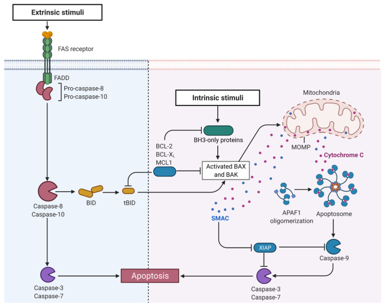
Mitochondria are involved in apoptosis, necrosis, and necroptosis [48]. Proteins of the B-cell lymphoma-2 (BCL-2) family bind voltage-dependent anion channels to accelerate the release of cytochrome c and induce apoptosis [61]. Myeloid leukemia cell differentiation protein-1 (MCL1) and BCL-xL are found in various mitochondrial subcompartments and unleash the antiapoptotic activities by competing with proapoptotic members of the BCL-2 family [48]. The BCL-2/BAX ratio is correlated with cytochrome c and apoptosis-inducing factors (AIFs), which determine the capability for mitochondria-mediated apoptosis [29]. The functional roles of BCL-2 in UC have also been studied. BCL-2 overexpression is associated with poor prognosis, early recurrence of bladder cancer [62,63,64], and resistance to gene therapy and chemotherapy [65,66]. In patients with bladder cancer receiving intravesical chemotherapy after tumor resection, early relapse can be observed in patients with a BCL-2/BAX ratio > 1 and a p53 gene mutation [62]. Patients with BCL-2-positive bladder cancer have significantly worse survival than those with BCL-2-negative tumors [63]. Recently, apoptotic protease-activating factor 1 (APAF1) in UC has been reported to be the direct target gene of miR-1270, which could induce apoptosis and enhance the cisplatin chemosensitivity of cancer cells [67]. In addition, in UC, the expression of X-linked inhibitor of apoptosis (XIAP) is higher at a later TMN stage [68]. The second mitochondria-derived activator of caspases (SMAC) competitively binds to XIAP, leading to the release of caspases and allowing the execution of apoptosis [69,70]. Figure 3 illustrates mitochondria-regulated apoptosis in UC.
Figure 3.
Mitochondria regulate apoptosis in UC. Abbreviations: APAF1, apoptotic protease-activating factor 1; BAK, Bcl-2 homologous antagonist/killer; BAX, Bcl-2-associated X protein; BCL, B-cell lymphoma; BH3, Bcl-2 homology domain 3; BID, BH3 interacting domain death agonist; FADD, Fas-associated via death domain; MCL1, myeloid leukemia cell differentiation protein-1; MOMP, mitochondrial outer membrane permeabilization; SMAC, the second mitochondria-derived activator of caspases; XIAP, X-linked inhibitor of apoptosis. The figure was created with BioRender.com.
2.7. Mitochondria Regulate Cell Proliferation in UC
A distinguished feature of cancers is their sustained cellular proliferation resulting from altered expression of constitutive telomerase that determines the maintenance of telomere length. It is known that telomerase reverse transcriptase (TERT) shuttles from the nucleus to the mitochondria upon oxidative stress, preserving mitochondrial functions and decreasing oxidative stress, thus protecting mtDNA and nuclear DNA (nDNA) from oxidative damage to avoid apoptosis [71,72]. In a recent report, mutations in the TERT promoter accounted for 84% of UC patients [73]. A meta-analysis further elucidated that bladder cancer patients carrying TERT promoter mutations have a greater risk of recurrence [74]. Using algorithmic inference from cross-sectional data, Hayashi et al. suggested that TERT promoter mutations play a role in the tumorigenesis of bladder cancer [75].
3. Therapeutic Strategies Targeting Mitochondria in UC
3.1. Targeting the TCA
Dichloroacetate (DCA) is a PDK inhibitor that can activate PDH, promote glucose oxidation, and further decrease tumor growth and angiogenesis. It has been demonstrated to decrease proliferation rates, increase pyruvate oxidation, and increase mitochondrial activity in UC [76]. Recently, metformin was shown to work synergistically with DCA to inhibit proliferation and reduce metabolic activity in a canine UC cell line [77].
Vitamin K2 has also been shown to exert anticancer effects. Recently, vitamin K2 was reported to promote glycolysis in UC cells by enhancing glucose consumption and lactate production and inhibiting the TCA cycle by reducing the amount of acetyl-CoA. This vitamin K2-induced metabolic stress triggers AMPK-dependent autophagic cell death in UC cells [78].
3.2. Restoring Mitochondria-Driven Apoptosis
Induction of apoptosis is a principal anticancer strategy used to eliminate cancer cells. Understanding apoptotic signaling pathways may assist in the discovery of novel therapeutic targets [79,80]. To date, three signaling mechanisms involving apoptosis have been discovered: the death-receptor-mediated extrinsic pathway [81], mitochondria-mediated intrinsic pathway [82], and endoplasmic reticulum (ER) stress-mediated pathway [83]. Mitochondria play an important role in apoptosis. AIF is the first caspase-independent cell death effector that interacts with DNA and induces nuclear condensation and DNA fragmentation. To explore novel and effective therapies for UC, a plethora of studies on the potential mechanisms of apoptosis have been performed.
Taking advantage of antisense oligodeoxynucleotides (AS-ODNs) to downregulate BCL-2 can partially sensitize bladder cancers to cisplatin and radiotherapy [84,85]. Studies have shown that BCL-2, BAX, and p53 contribute to drug sensitivity and apoptosis status and may help predict disease progression or recurrence [62,64]. In advanced bladder cancer, quantifying BCL-2 may help select target patients who may benefit from neoadjuvant chemotherapy [63]. For example, cisplatin is an important chemotherapeutic agent that is used to treat UC. Cisplatin induces apoptosis in a mitochondria-dependent and death-receptor-independent manner. BCL-2 overexpression inhibits cisplatin-induced BAX translocation and downstream events. Small interfering RNA (siRNA) targeting BCL-2 may help reverse cisplatin resistance in bladder cancer [66]. Bolenz et al. studied the application of AS-ODNs targeting BCL-2 and BCL-xL and revealed an effective improvement in the cytotoxicity of chemotherapeutic agents, not merely cisplatin but also gemcitabine, mitomycin C, and paclitaxel. The combined treatment resulted in notably higher death rates in nearly all cell lines [85].
Silibinin, a natural flavonoid, inhibits the growth of UC cells and induces caspase-dependent and caspase-independent apoptosis, which is associated with disruption of MMP and selective release of AIF and cytochrome c from mitochondria. In addition to inducing apoptosis via caspase activation in human UC cells, silibinin has been proven to be an intravesical chemotherapy for the inhibition of carcinogenesis and the progression of bladder cancer [86]. Additionally, the orally-fed silibinin has been reported to prevent N-butyl-N-(4-hydroxybutyl) nitrosamine (OH-BBN)-induced bladder carcinogenesis in mice. Accumulating evidence indicates that silibinin is an effective agent for chemotherapy against bladder tumor cells [86,87,88,89], as well as prostate [90,91], breast [92,93], skin [94], colon [95], lung [96], and kidney [97,98].
Baicalein is a flavone derived from the herb Huangqin, which is used in traditional Chinese medicine as an anti-inflammatory agent [99]. It induces apoptosis through a mitochondria-dependent caspase activation pathway in bladder cancer cells [100]. Wu et al. demonstrated that baicalein inhibits bladder cancer proliferation and migration in a dose-dependent manner via the reduction of phosphorylated NF-κB and MMP-2/9 expression [101].
Resveratrol is a polyphenolic compound naturally found in peanuts, mulberries, and grapes. It is an ingredient of red wine and exerts cardio- and neuroprotective effects [102,103]. In in vitro studies of UC, resveratrol has been shown to disrupt the MMP, increase ROS production, reduce ATP concentrations, provoke the release of cytochrome c from mitochondria to the cytosol, activate caspase-9 and caspase-3, and eventually induce apoptosis in cancer cells [104,105].
CXC195 also induces apoptosis by activating JNK, DP5, and PUMA, inhibiting BCL-2 and BCL-xL, and consequently inducing mitochondrial- and caspase-dependent apoptosis [79]. CXC195 is a tetramethylpyrazine (TMP) analog that displays antioxidant activity and antiapoptotic effects by inhibiting NADPH oxidase and iNOS expression and regulating the PI3K-AKT-GSK3b pathway. CXC195 is thought to be a promising anticancer drug that inhibits cell proliferation and inflammatory responses in bladder cancer [79].
Tumor necrosis factor (TNF)-related apoptosis-inducing ligand (TRAIL; Apo-2L) is a member of the TNF family and has recently gained attention because of its ability to induce apoptosis in cancers [106]. TRAIL induces apoptosis through a caspase-dependent mechanism, which can be strengthened by the release of cytochrome c and the loss of MMP [107]. TRAIL is a potent antitumor agent in preclinical studies; however, it has some limitations in potency. Combining TRAIL with other agents may improve cancer cell responsiveness. Histone deacetylase inhibitors have been shown to modulate the sensitivity of TRAIL-resistant bladder cancer cells [106].
3.3. Targeting Mitochondrial Turnover
Mitochondrial fusion, fission, and mitophagy have been examined as potential anticancer targets. Dynasore is a GTPase inhibitor of dynamin-related protein 1 (DRP1) [108]. Inhibition of mitochondrial fission by dynasore suppresses cancer cell proliferation and induces apoptosis. It inhibits migration and/or invasion in various cancer cell lines, including the bladder cancer cell line [109]. Radiation therapy may also play a role in UC treatment by causing mitochondrial damage. Shea et al. used cultured MGH-U1 (human urinary bladder carcinoma) cells and treated them with doxycycline and long-wave ultraviolet A (UVA) radiation. The cells were found to have mitochondrial damage when the UVA dose reached 1 J/cm2 and above [110].
3.4. Targeting Other Mitochondrial Modulators
Some proteins can indirectly modulate the mitochondrial function. NBR1 (a neighbor of the BRCA1 gene, an autophagy cargo receptor) is overexpressed in human UC cells. Rapamycin targeting the mammalian target of rapamycin (mTOR) kinase can regulate autophagy and has therapeutic effects in patients with cancer. In NBR1-knockdown UC cells, sensitivity to rapamycin-associated apoptosis and mitochondrial defects was enhanced. Loss of NBR1 expression changes cellular responses to rapamycin, leading to impaired ATP homeostasis and increased ROS levels. Therefore, NBR1 may be a potential therapeutic target for treating UC [86]. Table 1 summarizes mitochondria-targeted therapies for UC.
Table 1.
Mitochondria-targeted therapies for UC.
| Therapies | Strategies | Targets | References |
|---|---|---|---|
| DCA | inhibit PDK and activate PDH | mitochondrial TCA | [76] |
| vitamin K2 | promote the glycolysis | mitochondrial TCA | [78] |
| AS-ODNs | improve drug sensitivity induce apoptosis |
BCL-2, NRB1 | [84,85] |
| siRNA | improve drug sensitivity induce apoptosis |
BCL-2, NRB1 | [66] |
| metformin | induce apoptosis | mitochondria | [77] |
| silibinin | induce apoptosis | mitochondria | [86] |
| baicalein | induce apoptosis | mitochondria | [100] |
| resveratrol | induce apoptosis | mitochondria | [104,105] |
| CXC195 | induce apoptosis | TMP analog | [79] |
| TRAIL | induce apoptosis | mitochondria | [106] |
| UVA | damage mitochondria | mitochondria | [110] |
AS-ODNs, antisense oligodeoxynucleotides; siRNA, small interfering RNA; TMP, tetramethylpyrazine; TRAIL, tumor necrosis factor (TNF)-related apoptosis-inducing ligand; DCA, dichloroacetate; PDK, pyruvate dehydrogenase kinase; PDH, pyruvate dehydrogenase; UVA, ultraviolet A.
4. Conclusions and Perspectives
UC is a common but complex disease. By reviewing the available literature, we revisited the pathogenic role of mitochondria in UC. The main mechanisms by which mitochondria participate in tumorigenesis and progression of UC include mtDNA mutations, altered expression of mitochondrial proteins, metabolic reprogramming, deregulated mitochondrial ROS production and ETC activity, and mitochondria-regulated proliferation and death in cancer cells. The interplay between these different mechanisms often exists and complicates the whole process. Therapeutic strategies targeting these mitochondria-centered mechanisms are promising. They could be complementary to the current treatment modalities, including surgery, chemotherapy, and immunotherapy. Notably, the evidence summarized in this review is largely based on in vitro and animal studies. Advanced and detailed in vivo studies are required to facilitate future clinical research and clinical trials.
Author Contributions
Conceptualization, W.-C.L. and T.-W.H.; validation, C.-C.H. and H.-Y.L.; resources, W.-C.L.; writing—original draft preparation, T.-W.H., C.-C.H., and H.-Y.L.; writing—review and editing, W.-C.L.; supervision, W.-C.L.; project administration, W.-C.L. and C.-C.H.; funding acquisition, W.-C.L. and C.-C.H. All authors have read and agreed to the published version of the manuscript.
Institutional Review Board Statement
Not applicable.
Informed Consent Statement
Not applicable.
Data Availability Statement
Not applicable.
Conflicts of Interest
The authors declare that they have no conflict of interest. The funders had no role in the study design; collection, analyses, or interpretation of data; writing of the manuscript; or decision to publish the results.
Funding Statement
This review article was funded by Chang Gung Memorial Hospital (grant numbers CORPG8L0351 and CMRPG8K0011).
Footnotes
Publisher’s Note: MDPI stays neutral with regard to jurisdictional claims in published maps and institutional affiliations.
References
- 1.Green D.A., Rink M., Xylinas E., Matin S.F., Stenzl A., Roupret M., Karakiewicz P.I., Scherr D.S., Shariat S.F. Urothelial carcinoma of the bladder and the upper tract: Disparate twins. J. Urol. 2013;189:1214–1221. doi: 10.1016/j.juro.2012.05.079. [DOI] [PubMed] [Google Scholar]
- 2.Liu H.-Y., Chen Y.T., Huang S.-C., Wang H.-J., Cheng Y.-T., Kang C.H., Lee W.C., Su Y.-L., Huang C.-C., Chang Y.-L., et al. The prognostic impact of tumor architecture for upper urinary tract urothelial carcinoma: A propensity score-weighted analysis. Front. Oncol. 2021;11:613696. doi: 10.3389/fonc.2021.613696. [DOI] [PMC free article] [PubMed] [Google Scholar]
- 3.Sung H., Ferlay J., Siegel R.L., Laversanne M., Soerjomataram I., Jemal A., Bray F. Global cancer statistics 2020: Globocan estimates of incidence and mortality worldwide for 36 cancers in 185 countries. CA Cancer J. Clin. 2021;71:209–249. doi: 10.3322/caac.21660. [DOI] [PubMed] [Google Scholar]
- 4.Flaig T.W., Spiess P.E., Agarwal N., Bangs R., Boorjian S.A., Buyyounouski M.K., Chang S., Downs T.M., Efstathiou J.A., Friedlander T., et al. Bladder cancer, version 3.2020, nccn clinical practice guidelines in oncology. J. Natl. Compr. Cancer Netw. 2020;18:329–354. doi: 10.6004/jnccn.2020.0011. [DOI] [PubMed] [Google Scholar]
- 5.Chang S.S., Boorjian S.A., Chou R., Clark P.E., Daneshmand S., Konety B.R., Pruthi R., Quale D.Z., Ritch C.R., Seigne J.D., et al. Diagnosis and treatment of non-muscle invasive bladder cancer: Aua/suo guideline. J. Urol. 2016;196:1021–1029. doi: 10.1016/j.juro.2016.06.049. [DOI] [PubMed] [Google Scholar]
- 6.Aragon-Ching J.B., Werntz R.P., Zietman A.L., Steinberg G.D. Multidisciplinary management of muscle-invasive bladder cancer: Current challenges and future directions. Am. Soc. Clin. Oncol. Educ. Book. 2018;38:307–318. doi: 10.1200/EDBK_201227. [DOI] [PubMed] [Google Scholar]
- 7.Soria F., Shariat S.F., Lerner S.P., Fritsche H.M., Rink M., Kassouf W., Spiess P.E., Lotan Y., Ye D., Fernandez M.I., et al. Epidemiology, diagnosis, preoperative evaluation and prognostic assessment of upper-tract urothelial carcinoma (utuc) World J. Urol. 2017;35:379–387. doi: 10.1007/s00345-016-1928-x. [DOI] [PubMed] [Google Scholar]
- 8.Font A., Luque R., Villa J.C., Domenech M., Vazquez S., Gallardo E., Virizuela J.A., Beato C., Morales-Barrera R., Gelabert A., et al. The challenge of managing bladder cancer and upper tract urothelial carcinoma: A review with treatment recommendations from the spanish oncology genitourinary group (sogug) Target. Oncol. 2019;14:15–32. doi: 10.1007/s11523-019-00619-7. [DOI] [PubMed] [Google Scholar]
- 9.Venkatramani V., Parekh D.J. Surgery for bladder and upper tract urothelial cancer. Hematol. Oncol. Clin. N. Am. 2021;35:543–566. doi: 10.1016/j.hoc.2021.02.005. [DOI] [PubMed] [Google Scholar]
- 10.Wang Q., Zhang T., Wu J., Wen J., Tao D., Wan T., Zhu W. Prognosis and risk factors of patients with upper urinary tract urothelial carcinoma and postoperative recurrence of bladder cancer in central china. BMC Urol. 2019;19:24. doi: 10.1186/s12894-019-0457-5. [DOI] [PMC free article] [PubMed] [Google Scholar]
- 11.Joshi S.S., Quast L.L., Chang S.S., Patel S.G. Effects of tumor size and location on survival in upper tract urothelial carcinoma after nephroureterectomy. Indian J. Urol. 2018;34:68–73. doi: 10.4103/iju.IJU_216_17. [DOI] [PMC free article] [PubMed] [Google Scholar]
- 12.Eylert M.F., Hounsome L., Verne J., Bahl A., Jefferies E.R., Persad R.A. Prognosis is deteriorating for upper tract urothelial cancer: Data for england 1985-2010. BJU Int. 2013;112:E107–E113. doi: 10.1111/bju.12025. [DOI] [PubMed] [Google Scholar]
- 13.Colla Ruvolo C., Nocera L., Stolzenbach L.F., Wenzel M., Cucchiara V., Tian Z., Shariat S.F., Saad F., Longo N., Montorsi F., et al. Incidence and survival rates of contemporary patients with invasive upper tract urothelial carcinoma. Eur. Urol. Oncol. 2020;4:792–801. doi: 10.1016/j.euo.2020.11.005. [DOI] [PubMed] [Google Scholar]
- 14.Heath E.I., Rosenberg J.E. The biology and rationale of targeting nectin-4 in urothelial carcinoma. Nat. Rev. Urol. 2021;18:93–103. doi: 10.1038/s41585-020-00394-5. [DOI] [PubMed] [Google Scholar]
- 15.Sundahl N., Rottey S., De Maeseneer D., Ost P. Pembrolizumab for the treatment of bladder cancer. Expert Rev. Anticancer Ther. 2018;18:107–114. doi: 10.1080/14737140.2018.1421461. [DOI] [PubMed] [Google Scholar]
- 16.Montazeri K., Bellmunt J. Erdafitinib for the treatment of metastatic bladder cancer. Expert Rev. Clin. Pharm. 2020;13:1–6. doi: 10.1080/17512433.2020.1702025. [DOI] [PubMed] [Google Scholar]
- 17.Mehta M.M., Weinberg S.E., Chandel N.S. Mitochondrial control of immunity: Beyond atp. Nat. Rev. Immunol. 2017;17:608–620. doi: 10.1038/nri.2017.66. [DOI] [PubMed] [Google Scholar]
- 18.Galvan D.L., Green N.H., Danesh F.R. The hallmarks of mitochondrial dysfunction in chronic kidney disease. Kidney Int. 2017;92:1051–1057. doi: 10.1016/j.kint.2017.05.034. [DOI] [PMC free article] [PubMed] [Google Scholar]
- 19.Linehan W.M., Schmidt L.S., Crooks D.R., Wei D., Srinivasan R., Lang M., Ricketts C.J. The metabolic basis of kidney cancer. Cancer Discov. 2019;9:1006–1021. doi: 10.1158/2159-8290.CD-18-1354. [DOI] [PubMed] [Google Scholar]
- 20.Hervouet E., Godinot C. Mitochondrial disorders in renal tumors. Mitochondrion. 2006;6:105–117. doi: 10.1016/j.mito.2006.03.003. [DOI] [PubMed] [Google Scholar]
- 21.Meierhofer D., Mayr J.A., Foetschl U., Berger A., Fink K., Schmeller N., Hacker G.W., Hauser-Kronberger C., Kofler B., Sperl W. Decrease of mitochondrial DNA content and energy metabolism in renal cell carcinoma. Carcinogenesis. 2004;25:1005–1010. doi: 10.1093/carcin/bgh104. [DOI] [PubMed] [Google Scholar]
- 22.Nilsson H., Lindgren D., Mandahl Forsberg A., Mulder H., Axelson H., Johansson M.E. Primary clear cell renal carcinoma cells display minimal mitochondrial respiratory capacity resulting in pronounced sensitivity to glycolytic inhibition by 3-bromopyruvate. Cell Death Dis. 2015;6:e1585. doi: 10.1038/cddis.2014.545. [DOI] [PMC free article] [PubMed] [Google Scholar]
- 23.Favaretto R.L., Zequi S.C., Oliveira R.A.R., Santana T., Costa W.H., Cunha I.W., Guimaraes G.C. Tissue-based molecular markers in upper tract urothelial carcinoma and their prognostic implications. Int. Braz. J. Urol. 2018;44:22–37. doi: 10.1590/s1677-5538.ibju.2017.0204. [DOI] [PMC free article] [PubMed] [Google Scholar]
- 24.Yoshimine S., Kikuchi E., Kosaka T., Mikami S., Miyajima A., Okada Y., Oya M. Prognostic significance of bcl-xl expression and efficacy of bcl-xl targeting therapy in urothelial carcinoma. Br. J. Cancer. 2013;108:2312–2320. doi: 10.1038/bjc.2013.216. [DOI] [PMC free article] [PubMed] [Google Scholar]
- 25.DeBalsi K.L., Hoff K.E., Copeland W.C. Role of the mitochondrial DNA replication machinery in mitochondrial DNA mutagenesis, aging and age-related diseases. Ageing Res. Rev. 2017;33:89–104. doi: 10.1016/j.arr.2016.04.006. [DOI] [PMC free article] [PubMed] [Google Scholar]
- 26.Dasgupta S., Shao C., Keane T.E., Duberow D.P., Mathies R.A., Fisher P.B., Kiemeney L.A., Sidransky D. Detection of mitochondrial deoxyribonucleic acid alterations in urine from urothelial cell carcinoma patients. Int. J. Cancer. 2012;131:158–164. doi: 10.1002/ijc.26357. [DOI] [PMC free article] [PubMed] [Google Scholar]
- 27.Fliss M.S., Usadel H., Caballero O.L., Wu L., Buta M.R., Eleff S.M., Jen J., Sidransky D. Facile detection of mitochondrial DNA mutations in tumors and bodily fluids. Science. 2000;287:2017–2019. doi: 10.1126/science.287.5460.2017. [DOI] [PubMed] [Google Scholar]
- 28.Guney A.I., Ergec D.S., Tavukcu H.H., Koc G., Kirac D., Ulucan K., Javadova D., Turkeri L. Detection of mitochondrial DNA mutations in nonmuscle invasive bladder cancer. Genet. Test. Mol. Biomark. 2012;16:672–678. doi: 10.1089/gtmb.2011.0227. [DOI] [PubMed] [Google Scholar]
- 29.Dasgupta S., Hoque M.O., Upadhyay S., Sidransky D. Mitochondrial cytochrome b gene mutation promotes tumor growth in bladder cancer. Cancer Res. 2008;68:700–706. doi: 10.1158/0008-5472.CAN-07-5532. [DOI] [PubMed] [Google Scholar]
- 30.Tzen C.-Y., Mau B.-L., Wu T.-Y. Nd4 mutation in transitional cell carcinoma: Does mitochondrial mutation occur before tumorigenesis? Mitochondrion. 2007;7:273–278. doi: 10.1016/j.mito.2007.04.004. [DOI] [PubMed] [Google Scholar]
- 31.Reznik E., Miller M.L., Senbabaoglu Y., Riaz N., Sarungbam J., Tickoo S.K., Al-Ahmadie H.A., Lee W., Seshan V.E., Hakimi A.A., et al. Mitochondrial DNA copy number variation across human cancers. eLife. 2016;5:e10769. doi: 10.7554/eLife.10769. [DOI] [PMC free article] [PubMed] [Google Scholar]
- 32.Gibellini L., De Biasi S., Nasi M., Iannone A., Cossarizza A., Pinti M. Mitochondrial proteases as emerging pharmacological targets. Curr. Pharm. Des. 2016;22:2679–2688. doi: 10.2174/1381612822666160202130344. [DOI] [PubMed] [Google Scholar]
- 33.Liu Y., Lan L., Huang K., Wang R., Xu C., Shi Y., Wu X., Wu Z., Zhang J., Chen L., et al. Inhibition of lon blocks cell proliferation, enhances chemosensitivity by promoting apoptosis and decreases cellular bioenergetics of bladder cancer: Potential roles of lon as a prognostic marker and therapeutic target in baldder cancer. Oncotarget. 2014;5:11209–11224. doi: 10.18632/oncotarget.2026. [DOI] [PMC free article] [PubMed] [Google Scholar]
- 34.Mishra P., Chan D.C. Metabolic regulation of mitochondrial dynamics. J. Cell Biol. 2016;212:379–387. doi: 10.1083/jcb.201511036. [DOI] [PMC free article] [PubMed] [Google Scholar]
- 35.Lee W.C., Chiu C.H., Chen J.B., Chen C.H., Chang H.W. Mitochondrial fission increases apoptosis and decreases autophagy in renal proximal tubular epithelial cells treated with high glucose. DNA Cell Biol. 2016;35:657–665. doi: 10.1089/dna.2016.3261. [DOI] [PubMed] [Google Scholar]
- 36.Jin B., Fu G., Pan H., Cheng X., Zhou L., Lv J., Chen G., Zheng S. Anti-tumour efficacy of mitofusin-2 in urinary bladder carcinoma. Med. Oncol. 2011;28((Suppl. S1)):S373–S380. doi: 10.1007/s12032-010-9662-5. [DOI] [PubMed] [Google Scholar]
- 37.Chew K., Zhao L. Interactions of mitochondrial transcription factor a with DNA damage: Mechanistic insights and functional implications. Genes. 2021;12:1246. doi: 10.3390/genes12081246. [DOI] [PMC free article] [PubMed] [Google Scholar]
- 38.Kang D., Kim S.H., Hamasaki N. Mitochondrial transcription factor a (tfam): Roles in maintenance of mtdna and cellular functions. Mitochondrion. 2007;7:39–44. doi: 10.1016/j.mito.2006.11.017. [DOI] [PubMed] [Google Scholar]
- 39.Mo M., Peng F., Wang L., Peng L., Lan G., Yu S. Roles of mitochondrial transcription factor a and microrna-590-3p in the development of bladder cancer. Oncol. Lett. 2013;6:617–623. doi: 10.3892/ol.2013.1419. [DOI] [PMC free article] [PubMed] [Google Scholar]
- 40.Liu L., McKeehan W.L. Sequence analysis of lrpprc and its sec1 domain interaction partners suggests roles in cytoskeletal organization, vesicular trafficking, nucleocytosolic shuttling, and chromosome activity. Genomics. 2002;79:124–136. doi: 10.1006/geno.2001.6679. [DOI] [PMC free article] [PubMed] [Google Scholar]
- 41.Tsuchiya N., Fukuda H., Nakashima K., Nagao M., Sugimura T., Nakagama H. Lrp130, a single-stranded DNA/rna-binding protein, localizes at the outer nuclear and endoplasmic reticulum membrane, and interacts with mrna in vivo. Biochem. Biophys. Res. Commun. 2004;317:736–743. doi: 10.1016/j.bbrc.2004.03.103. [DOI] [PubMed] [Google Scholar]
- 42.Wei W.S., Wang N., Deng M.H., Dong P., Liu J.Y., Xiang Z., Li X.D., Li Z.Y., Liu Z.H., Peng Y.L., et al. Lrpprc regulates redox homeostasis via the circankhd1/foxm1 axis to enhance bladder urothelial carcinoma tumorigenesis. Redox Biol. 2021;48:102201. doi: 10.1016/j.redox.2021.102201. [DOI] [PMC free article] [PubMed] [Google Scholar]
- 43.Luan T., Fu S., Huang L., Zuo Y., Ding M., Li N., Chen J., Wang H., Wang J. Microrna-98 promotes drug resistance and regulates mitochondrial dynamics by targeting lass2 in bladder cancer cells. Exp. Cell Res. 2018;373:188–197. doi: 10.1016/j.yexcr.2018.10.013. [DOI] [PubMed] [Google Scholar]
- 44.Zhao Q., Wang H., Yang M., Yang D., Zuo Y., Wang J. Expression of a tumor-associated gene, lass2, in the human bladder carcinoma cell lines biu-87, t24, ej and ej-m3. Exp. Ther. Med. 2013;5:942–946. doi: 10.3892/etm.2013.892. [DOI] [PMC free article] [PubMed] [Google Scholar]
- 45.Wang H., Wang J., Zuo Y., Ding M., Yan R., Yang D., Ke C. Expression and prognostic significance of a new tumor metastasis suppressor gene lass2 in human bladder carcinoma. Med. Oncol. 2012;29:1921–1927. doi: 10.1007/s12032-011-0026-6. [DOI] [PubMed] [Google Scholar]
- 46.Warburg O., Wind F., Negelein E. über den stoffwechsel von tumoren im körper. Klin. Wochenschr. 1926;5:829–832. doi: 10.1007/BF01726240. [DOI] [Google Scholar]
- 47.Kuo Y.H., Chan T.C., Lai H.Y., Chen T.J., Wu L.C., Hsing C.H., Li C.F. Overexpression of pyruvate dehydrogenase kinase-3 predicts poor prognosis in urothelial carcinoma. Front. Oncol. 2021;11:749142. doi: 10.3389/fonc.2021.749142. [DOI] [PMC free article] [PubMed] [Google Scholar]
- 48.Grasso D., Zampieri L.X., Capeloa T., Van de Velde J.A., Sonveaux P. Mitochondria in cancer. Cell Stress. 2020;4:114–146. doi: 10.15698/cst2020.06.221. [DOI] [PMC free article] [PubMed] [Google Scholar]
- 49.Draoui N., Schicke O., Seront E., Bouzin C., Sonveaux P., Riant O., Feron O. Antitumor activity of 7-aminocarboxycoumarin derivatives, a new class of potent inhibitors of lactate influx but not efflux. Mol. Cancer Ther. 2014;13:1410–1418. doi: 10.1158/1535-7163.MCT-13-0653. [DOI] [PubMed] [Google Scholar]
- 50.Ward P.S., Thompson C.B. Metabolic reprogramming: A cancer hallmark even warburg did not anticipate. Cancer Cell. 2012;21:297–308. doi: 10.1016/j.ccr.2012.02.014. [DOI] [PMC free article] [PubMed] [Google Scholar]
- 51.Daye D., Wellen K.E. Metabolic reprogramming in cancer: Unraveling the role of glutamine in tumorigenesis. Semin. Cell Dev. Biol. 2012;23:362–369. doi: 10.1016/j.semcdb.2012.02.002. [DOI] [PubMed] [Google Scholar]
- 52.Sun N., Liang Y., Chen Y., Wang L., Li D., Liang Z., Sun L., Wang Y., Niu H. Glutamine affects t24 bladder cancer cell proliferation by activating stat3 through ros and glutaminolysis. Int. J. Mol. Med. 2019;44:2189–2200. doi: 10.3892/ijmm.2019.4385. [DOI] [PMC free article] [PubMed] [Google Scholar]
- 53.Zhao H., Wu W., Li X., Chen W. Long noncoding rna uca1 promotes glutamine-driven anaplerosis of bladder cancer by interacting with hnrnp i/l to upregulate gpt2 expression. Transl. Oncol. 2022;17:101340. doi: 10.1016/j.tranon.2022.101340. [DOI] [PMC free article] [PubMed] [Google Scholar]
- 54.Kumari S., Badana A.K., Murali Mohan G., Shailender G., Malla R. Reactive oxygen species: A key constituent in cancer survival. Biomark. Insights. 2018;13:1177271918755391. doi: 10.1177/1177271918755391. [DOI] [PMC free article] [PubMed] [Google Scholar]
- 55.Murphy M.P. How mitochondria produce reactive oxygen species. Biochem. J. 2009;417:1–13. doi: 10.1042/BJ20081386. [DOI] [PMC free article] [PubMed] [Google Scholar]
- 56.Sena L.A., Chandel N.S. Physiological roles of mitochondrial reactive oxygen species. Mol. Cell. 2012;48:158–167. doi: 10.1016/j.molcel.2012.09.025. [DOI] [PMC free article] [PubMed] [Google Scholar]
- 57.Huang C.N., Tsai J.L., Chen M.T., Wu W.J., Kuo K.W., Huang C.H. Changes in the activities of mitochondrial enzymes in the progress of tumorigenesis of bladder cancer. Biochem. Mol. Biol. Int. 1998;46:375–383. doi: 10.1080/15216549800203882. [DOI] [PubMed] [Google Scholar]
- 58.Seo J.M., Cho K.J., Kim E.Y., Choi M.H., Chung B.C., Kim J.H. Up-regulation of blt2 is critical for the survival of bladder cancer cells. Exp. Mol. Med. 2011;43:129–137. doi: 10.3858/emm.2011.43.3.014. [DOI] [PMC free article] [PubMed] [Google Scholar]
- 59.Kim E.Y., Seo J.M., Kim C., Lee J.E., Lee K.M., Kim J.H. Blt2 promotes the invasion and metastasis of aggressive bladder cancer cells through a reactive oxygen species-linked pathway. Free Radic. Biol. Med. 2010;49:1072–1081. doi: 10.1016/j.freeradbiomed.2010.06.023. [DOI] [PubMed] [Google Scholar]
- 60.Shimada K., Nakamura M., Anai S., De Velasco M., Tanaka M., Tsujikawa K., Ouji Y., Konishi N. A novel human alkb homologue, alkbh8, contributes to human bladder cancer progression. Cancer Res. 2009;69:3157–3164. doi: 10.1158/0008-5472.CAN-08-3530. [DOI] [PubMed] [Google Scholar]
- 61.Shimizu S., Narita M., Tsujimoto Y. Bcl-2 family proteins regulate the release of apoptogenic cytochrome c by the mitochondrial channel vdac. Nature. 1999;399:483–487. doi: 10.1038/20959. [DOI] [PubMed] [Google Scholar]
- 62.Ye D., Li H., Qian S., Sun Y., Zheng J., Ma Y. Bcl-2/bax expression and p53 gene status in human bladder cancer: Relationship to early recurrence with intravesical chemotherapy after resection. J. Urol. 1998;160:2025–2028; discussion 2029. doi: 10.1016/S0022-5347(01)62233-X. [DOI] [PubMed] [Google Scholar]
- 63.Cooke P.W., James N.D., Ganesan R., Burton A., Young L.S., Wallace D.M. Bcl-2 expression identifies patients with advanced bladder cancer treated by radiotherapy who benefit from neoadjuvant chemotherapy. BJU Int. 2000;85:829–835. doi: 10.1046/j.1464-410x.2000.00612.x. [DOI] [PubMed] [Google Scholar]
- 64.Karam J.A., Lotan Y., Karakiewicz P.I., Ashfaq R., Sagalowsky A.I., Roehrborn C.G., Shariat S.F. Use of combined apoptosis biomarkers for prediction of bladder cancer recurrence and mortality after radical cystectomy. Lancet Oncol. 2007;8:128–136. doi: 10.1016/S1470-2045(07)70002-5. [DOI] [PubMed] [Google Scholar]
- 65.Miyake H., Hanada N., Nakamura H., Kagawa S., Fujiwara T., Hara I., Eto H., Gohji K., Arakawa S., Kamidono S., et al. Overexpression of bcl-2 in bladder cancer cells inhibits apoptosis induced by cisplatin and adenoviral-mediated p53 gene transfer. Oncogene. 1998;16:933–943. doi: 10.1038/sj.onc.1201602. [DOI] [PubMed] [Google Scholar]
- 66.Cho H.J., Kim J.K., Kim K.D., Yoon H.K., Cho M.Y., Park Y.P., Jeon J.H., Lee E.S., Byun S.S., Lim H.M., et al. Upregulation of bcl-2 is associated with cisplatin-resistance via inhibition of bax translocation in human bladder cancer cells. Cancer Lett. 2006;237:56–66. doi: 10.1016/j.canlet.2005.05.039. [DOI] [PubMed] [Google Scholar]
- 67.Yuan W., Zhou R., Wang J., Han J., Yang X., Yu H., Lu H., Zhang X., Li P., Tao J., et al. Circular rna cdr1as sensitizes bladder cancer to cisplatin by upregulating apaf1 expression through mir-1270 inhibition. Mol. Oncol. 2019;13:1559–1576. doi: 10.1002/1878-0261.12523. [DOI] [PMC free article] [PubMed] [Google Scholar]
- 68.Li M., Song T., Yin Z.F., Na Y.Q. Xiap as a prognostic marker of early recurrence of nonmuscular invasive bladder cancer. Chin. Med. J. 2007;120:469–473. doi: 10.1097/00029330-200703020-00007. [DOI] [PubMed] [Google Scholar]
- 69.Abbas R., Larisch S. Targeting xiap for promoting cancer cell death-the story of arts and smac. Cells. 2020;9:663. doi: 10.3390/cells9030663. [DOI] [PMC free article] [PubMed] [Google Scholar]
- 70.Flanagan L., Sebastia J., Tuffy L.P., Spring A., Lichawska A., Devocelle M., Prehn J.H., Rehm M. Xiap impairs smac release from the mitochondria during apoptosis. Cell Death Dis. 2010;1:e49. doi: 10.1038/cddis.2010.26. [DOI] [PMC free article] [PubMed] [Google Scholar]
- 71.Dong C.K., Masutomi K., Hahn W.C. Telomerase: Regulation, function and transformation. Crit. Rev. Oncol. Hematol. 2005;54:85–93. doi: 10.1016/j.critrevonc.2004.12.005. [DOI] [PubMed] [Google Scholar]
- 72.Zheng Q., Huang J., Wang G. Mitochondria, telomeres and telomerase subunits. Front. Cell Dev. Biol. 2019;7:274. doi: 10.3389/fcell.2019.00274. [DOI] [PMC free article] [PubMed] [Google Scholar]
- 73.Roggisch J., Ecke T., Koch S. Molecular identification of telomerase reverse transcriptase (tert) promotor mutations in primary and recurrent tumors of invasive and noninvasive urothelial bladder cancer. Urol. Oncol. 2020;38:77.e17–77.e25. doi: 10.1016/j.urolonc.2019.08.007. [DOI] [PubMed] [Google Scholar]
- 74.Wan S., Liu X., Hua W., Xi M., Zhou Y., Wan Y. The role of telomerase reverse transcriptase (tert) promoter mutations in prognosis in bladder cancer. Bioengineered. 2021;12:1495–1504. doi: 10.1080/21655979.2021.1915725. [DOI] [PMC free article] [PubMed] [Google Scholar]
- 75.Hayashi Y., Fujita K., Banno E., Eich M.L., Netto G.J., Nonomura N. Telomerase reverse transcriptase promoter mutation in tumorigenesis of bladder cancer: Evolutionary trajectory by algorithmic inference from cross-sectional data. Int. J. Urol. 2021;28:774–776. doi: 10.1111/iju.14574. [DOI] [PubMed] [Google Scholar]
- 76.Harting T., Stubbendorff M., Willenbrock S., Wagner S., Schadzek P., Ngezahayo A., Murua Escobar H.M., Nolte I. The effect of dichloroacetate in canine prostate adenocarcinomas and transitional cell carcinomas in vitro. Int. J. Oncol. 2016;49:2341–2350. doi: 10.3892/ijo.2016.3720. [DOI] [PubMed] [Google Scholar]
- 77.Klose K., Packeiser E.M., Muller P., Granados-Soler J.L., Schille J.T., Goericke-Pesch S., Kietzmann M., Murua Escobar H., Nolte I. Metformin and sodium dichloroacetate effects on proliferation, apoptosis, and metabolic activity tested alone and in combination in a canine prostate and a bladder cancer cell line. PLoS ONE. 2021;16:e0257403. doi: 10.1371/journal.pone.0257403. [DOI] [PMC free article] [PubMed] [Google Scholar]
- 78.Duan F., Mei C., Yang L., Zheng J., Lu H., Xia Y., Hsu S., Liang H., Hong L. Vitamin k2 promotes pi3k/akt/hif-1alpha-mediated glycolysis that leads to ampk-dependent autophagic cell death in bladder cancer cells. Sci. Rep. 2020;10:7714. doi: 10.1038/s41598-020-64880-x. [DOI] [PMC free article] [PubMed] [Google Scholar]
- 79.Zeng T., Peng L., Chao H., Xi H., Fu B., Wang Y., Zhu Z., Wang G. Ire1α-traf2-ask1 complex-mediated endoplasmic reticulum stress and mitochondrial dysfunction contribute to cxc195-induced apoptosis in human bladder carcinoma t24 cells. Biochem. Biophys. Res. Commun. 2015;460:530–536. doi: 10.1016/j.bbrc.2015.03.064. [DOI] [PubMed] [Google Scholar]
- 80.Ghobrial I.M., Witzig T.E., Adjei A.A. Targeting apoptosis pathways in cancer therapy. CA A Cancer J. Clin. 2005;55:178–194. doi: 10.3322/canjclin.55.3.178. [DOI] [PubMed] [Google Scholar]
- 81.Wang J., Hansen K., Edwards R., Van Houten B., Qian W. Mitochondrial division inhibitor 1 (mdivi-1) enhances death receptor-mediated apoptosis in human ovarian cancer cells. Biochem. Biophys. Res. Commun. 2015;456:7–12. doi: 10.1016/j.bbrc.2014.11.010. [DOI] [PMC free article] [PubMed] [Google Scholar]
- 82.Youle R.J., Strasser A. The bcl-2 protein family: Opposing activities that mediate cell death. Nat. Rev. Mol. Cell Biol. 2008;9:47–59. doi: 10.1038/nrm2308. [DOI] [PubMed] [Google Scholar]
- 83.Urano F., Wang X., Bertolotti A., Zhang Y., Chung P., Harding H.P., Ron D. Coupling of stress in the er to activation of jnk protein kinases by transmembrane protein kinase ire1. Science. 2000;287:664–666. doi: 10.1126/science.287.5453.664. [DOI] [PubMed] [Google Scholar]
- 84.Duggan B.J., Maxwell P., Kelly J.D., Canning P., Anderson N.H., Keane P.F., Johnston S.R., Williamson K.E. The effect of antisense bcl-2 oligonucleotides on bcl-2 protein expression and apoptosis in human bladder transitional cell carcinoma. J. Urol. 2001;166:1098–1105. doi: 10.1016/S0022-5347(05)65929-0. [DOI] [PubMed] [Google Scholar]
- 85.Bolenz C., Becker A., Trojan L., Schaaf A., Cao Y., Weiss C., Alken P., Michel M.S. Optimizing chemotherapy for transitional cell carcinoma by application of bcl-2 and bcl-xl antisense oligodeoxynucleotides. Urol. Oncol. Semin. Orig. Investig. 2007;25:476–482. doi: 10.1016/j.urolonc.2007.01.017. [DOI] [PubMed] [Google Scholar]
- 86.Zeng J., Sun Y., Wu K., Li L., Zhang G., Yang Z., Wang Z., Zhang D., Xue Y., Chen Y., et al. Chemopreventive and chemotherapeutic effects of intravesical silibinin against bladder cancer by acting on mitochondria. Mol. Cancer Ther. 2011;10:104–116. doi: 10.1158/1535-7163.MCT-10-0577. [DOI] [PubMed] [Google Scholar]
- 87.Tyagi A., Agarwal C., Harrison G., Glode L.M., Agarwal R. Silibinin causes cell cycle arrest and apoptosis in human bladder transitional cell carcinoma cells by regulating cdki-cdk-cyclin cascade, and caspase 3 and parp cleavages. Carcinogenesis. 2004;25:1711–1720. doi: 10.1093/carcin/bgh180. [DOI] [PubMed] [Google Scholar]
- 88.Tyagi A., Singh R.P., Agarwal C., Agarwal R. Silibinin activates p53-caspase 2 pathway and causes caspase-mediated cleavage of cip1/p21 in apoptosis induction in bladder transitional-cell papilloma rt4 cells: Evidence for a regulatory loop between p53 and caspase 2. Carcinogenesis. 2006;27:2269–2280. doi: 10.1093/carcin/bgl098. [DOI] [PubMed] [Google Scholar]
- 89.Singh R.P., Tyagi A., Sharma G., Mohan S., Agarwal R. Oral silibinin inhibits in vivo human bladder tumor xenograft growth involving down-regulation of survivin. Clin. Cancer Res. 2008;14:300–308. doi: 10.1158/1078-0432.CCR-07-1565. [DOI] [PubMed] [Google Scholar]
- 90.Wu K.J., Zeng J., Zhu G.D., Zhang L.L., Zhang D., Li L., Fan J.H., Wang X.Y., He D.L. Silibinin inhibits prostate cancer invasion, motility and migration by suppressing vimentin and mmp-2 expression. Acta Pharm. Sin. 2009;30:1162–1168. doi: 10.1038/aps.2009.94. [DOI] [PMC free article] [PubMed] [Google Scholar]
- 91.Roy S., Kaur M., Agarwal C., Tecklenburg M., Sclafani R.A., Agarwal R. P21 and p27 induction by silibinin is essential for its cell cycle arrest effect in prostate carcinoma cells. Mol. Cancer Ther. 2007;6:2696–2707. doi: 10.1158/1535-7163.MCT-07-0104. [DOI] [PubMed] [Google Scholar]
- 92.Tyagi A.K., Agarwal C., Chan D.C., Agarwal R. Synergistic anti-cancer effects of silibinin with conventional cytotoxic agents doxorubicin, cisplatin and carboplatin against human breast carcinoma mcf-7 and mda-mb468 cells. Oncol. Rep. 2004;11:493–499. doi: 10.3892/or.11.2.493. [DOI] [PubMed] [Google Scholar]
- 93.Lee S.O., Jeong Y.J., Im H.G., Kim C.H., Chang Y.C., Lee I.S. Silibinin suppresses pma-induced mmp-9 expression by blocking the ap-1 activation via mapk signaling pathways in mcf-7 human breast carcinoma cells. Biochem. Biophys. Res. Commun. 2007;354:165–171. doi: 10.1016/j.bbrc.2006.12.181. [DOI] [PubMed] [Google Scholar]
- 94.Gu M., Singh R.P., Dhanalakshmi S., Agarwal C., Agarwal R. Silibinin inhibits inflammatory and angiogenic attributes in photocarcinogenesis in skh-1 hairless mice. Cancer Res. 2007;67:3483–3491. doi: 10.1158/0008-5472.CAN-06-3955. [DOI] [PubMed] [Google Scholar]
- 95.Singh R.P., Gu M., Agarwal R. Silibinin inhibits colorectal cancer growth by inhibiting tumor cell proliferation and angiogenesis. Cancer Res. 2008;68:2043–2050. doi: 10.1158/0008-5472.CAN-07-6247. [DOI] [PubMed] [Google Scholar]
- 96.Singh R.P., Deep G., Chittezhath M., Kaur M., Dwyer-Nield L.D., Malkinson A.M., Agarwal R. Effect of silibinin on the growth and progression of primary lung tumors in mice. J. Natl. Cancer Inst. 2006;98:846–855. doi: 10.1093/jnci/djj231. [DOI] [PubMed] [Google Scholar]
- 97.Cheung C.W., Taylor P.J., Kirkpatrick C.M., Vesey D.A., Gobe G.C., Winterford C., Nicol D.L., Johnson D.W. Therapeutic value of orally administered silibinin in renal cell carcinoma: Manipulation of insulin-like growth factor binding protein-3 levels. BJU Int. 2007;100:438–444. doi: 10.1111/j.1464-410X.2007.07012.x. [DOI] [PubMed] [Google Scholar]
- 98.Li L., Gao Y., Zhang L., Zeng J., He D., Sun Y. Silibinin inhibits cell growth and induces apoptosis by caspase activation, down-regulating survivin and blocking egfr-erk activation in renal cell carcinoma. Cancer Lett. 2008;272:61–69. doi: 10.1016/j.canlet.2008.06.033. [DOI] [PubMed] [Google Scholar]
- 99.Liu H., Dong Y., Gao Y., Du Z., Wang Y., Cheng P., Chen A., Huang H. The fascinating effects of baicalein on cancer: A review. Int. J. Mol. Sci. 2016;17:1681. doi: 10.3390/ijms17101681. [DOI] [PMC free article] [PubMed] [Google Scholar]
- 100.Li H.L., Zhang S., Wang Y., Liang R.R., Li J., An P., Wang Z.M., Yang J., Li Z.F. Baicalein induces apoptosis via a mitochondrial-dependent caspase activation pathway in t24 bladder cancer cells. Mol. Med. Rep. 2013;7:266–270. doi: 10.3892/mmr.2012.1123. [DOI] [PubMed] [Google Scholar]
- 101.Wu J.Y., Tsai K.W., Li Y.Z., Chang Y.S., Lai Y.C., Laio Y.H., Wu J.D., Liu Y.W. Anti-bladder-tumor effect of baicalein from scutellaria baicalensis georgi and its application in vivo. Evid.-Based Complement. Altern. Med. 2013;2013:579751. doi: 10.1155/2013/579751. [DOI] [PMC free article] [PubMed] [Google Scholar]
- 102.Burns J., Yokota T., Ashihara H., Lean M.E., Crozier A. Plant foods and herbal sources of resveratrol. J. Agric. Food Chem. 2002;50:3337–3340. doi: 10.1021/jf0112973. [DOI] [PubMed] [Google Scholar]
- 103.Lin K.L., Lin K.J., Wang P.W., Chuang J.H., Lin H.Y., Chen S.D., Chuang Y.C., Huang S.T., Tiao M.M., Chen J.B., et al. Resveratrol provides neuroprotective effects through modulation of mitochondrial dynamics and erk1/2 regulated autophagy. Free Radic. Res. 2018;52:1371–1386. doi: 10.1080/10715762.2018.1489128. [DOI] [PubMed] [Google Scholar]
- 104.Lin X., Wu G., Huo W.Q., Zhang Y., Jin F.S. Resveratrol induces apoptosis associated with mitochondrial dysfunction in bladder carcinoma cells. Int. J. Urol. 2012;19:757–764. doi: 10.1111/j.1442-2042.2012.03024.x. [DOI] [PubMed] [Google Scholar]
- 105.Stocco B., Toledo K., Salvador M., Paulo M., Koyama N., Torqueti Toloi M.R. Dose-dependent effect of resveratrol on bladder cancer cells: Chemoprevention and oxidative stress. Maturitas. 2012;72:72–78. doi: 10.1016/j.maturitas.2012.02.004. [DOI] [PubMed] [Google Scholar]
- 106.Earel J.K., Jr., VanOosten R.L., Griffith T.S. Histone deacetylase inhibitors modulate the sensitivity of tumor necrosis factor-related apoptosis-inducing ligand-resistant bladder tumor cells. Cancer Res. 2006;66:499–507. doi: 10.1158/0008-5472.CAN-05-3017. [DOI] [PubMed] [Google Scholar]
- 107.Zhang L., Fang B. Mechanisms of resistance to trail-induced apoptosis in cancer. Cancer Gene Ther. 2005;12:228–237. doi: 10.1038/sj.cgt.7700792. [DOI] [PubMed] [Google Scholar]
- 108.Macia E., Ehrlich M., Massol R., Boucrot E., Brunner C., Kirchhausen T. Dynasore, a cell-permeable inhibitor of dynamin. Dev. Cell. 2006;10:839–850. doi: 10.1016/j.devcel.2006.04.002. [DOI] [PubMed] [Google Scholar]
- 109.Zhang Y., Nolan M., Yamada H., Watanabe M., Nasu Y., Takei K., Takeda T. Dynamin2 gtpase contributes to invadopodia formation in invasive bladder cancer cells. Biochem. Biophys. Res. Commun. 2016;480:409–414. doi: 10.1016/j.bbrc.2016.10.063. [DOI] [PubMed] [Google Scholar]
- 110.Shea C.R., Wimberly J., Hasan T. Mitochondrial phototoxicity sensitized by doxycycline in cultured human carcinoma cells. J. Investig. Derm. 1986;87:338–342. doi: 10.1111/1523-1747.ep12524430. [DOI] [PubMed] [Google Scholar]
Associated Data
This section collects any data citations, data availability statements, or supplementary materials included in this article.
Data Availability Statement
Not applicable.


