FIG 1.
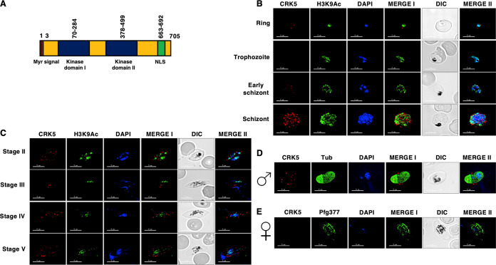
Expression and localization of PfCRK5 in asexual and sexual stages. (A) Schematic for various motifs and domains of PfCRK5 showing an N-terminal myristoylation signal (red) followed by 2 bipartite domains (blue) and nuclear localization signal (NLS) (in green). (B) Immunofluorescence assays were performed on WT NF54 asexual blood stages (ring, trophozoite and schizont) to colocalize PfCRK5 (red) in combination with Histone marker H3K9Ac (green). The parasite nucleus was localized with 4′,6-diamidino-2-phenylindole (DAPI) (in blue). Scale bar = 5 μm. (C) Immunofluorescence assays were performed on WT NF54 sexual (stage II-V gametocytes) using thin culture smears and anti-PfCRK5 antisera (in red) in combination with H3K9Ac (green). (D) and (E) Immunofluorescence assays were performed on stage V gametocytes using thin smears and anti-PfCRK5 antisera (in red) either in combination with α-Tubulin (marker for male gametocytes, in green) or anti- Pfg377 (marker for female gametocytes, in green). Parasite nucleus was visualized with DAPI (blue). Scale bar = 5 μm.

