ABSTRACT
Staphylococcus aureus represents a major human pathogen that is frequently involved in polymicrobial infections. However, the prevalence and role of co-infectious microbes on the pathogenesis and fitness essentiality of S. aureus in vivo remain largely unknown. In this study, we firstly performed a retrospective surveillance of 760 clinical samples and revealed a notable predominance of co-infection with S. aureus and Acinetobacter baumannii. The high-density S. aureus transposon mutant library coupled to transposon insertion sequencing (Tn-Seq) further identified a core set of genes enriched in metabolism of inorganic ions, amino acids, and carbohydrates, which are essential for infection and tissue colonization of S. aureus in the murine systemic infection model. Notably, we revealed a differential requirement of fitness factors for S. aureus in tissue-specific (liver and kidney) and infection-type-specific manner (mono- and co-infection). Co-infection with A. baumannii dramatically altered the fitness requirements of S. aureus in vivo; 49% of the mono-infection fitness genes in S. aureus strain Newman were converted to non-essential, and the functionality of ATP-binding cassette (ABC) transporters was significantly elicited during co-infection. Furthermore, the number of genes essential during co-infection (503) outnumbers the genes essential during mono-infection (362). In addition, the roles of 3 infection-type-specific genes in S. aureus during mono-infection or co-infection with A. baumannii were validated with competitive experiments in vivo. Our data indicated a high incidence and clinical relevance of S. aureus and A. baumannii co-infection, and provided novel insights into establishing antimicrobial regimens to control co-infections.
IMPORTANCE Polymicrobial infections are widespread in clinical settings, which potentially correlate with increased infection severity and poor clinical outcomes. Staphylococcus aureus is a formidable human pathogen that causes a variety of diseases in polymicrobial nature. Co-infection and interaction of S. aureus have been described with limited pathogens, mainly including Pseudomonas aeruginosa, Candida albicans, and influenza A virus. Thus far, the prevalence and role of co-infectious microbes on the pathogenesis and fitness essentiality of S. aureus in vivo remain largely unknown. Understanding the polymicrobial composition and interaction, from a community and genome-wide perspective, is thus crucial to shed light on S. aureus pathogenesis strategy. Here, our findings demonstrated, for the first time, that a high incidence rate and clinical relevance of co-infection was caused by S. aureus and Acinetobacter baumannii, illustrating the importance of polymicrobial nature in investigating S. aureus pathogenesis. The infection-type-specific genes likely serve as potential therapeutic targets to control S. aureus infections, either in mono- or co-infection situation, providing novel insights into the development of antimicrobial regimens to control co-infections.
KEYWORDS: Staphylococcus aureus, Acinetobacter baumannii, co-infection, Tn-seq, essential fitness
INTRODUCTION
Infections caused by multiple species of microorganisms, also known as polymicrobial infections, are prevalent in the clinical setting and account for approximately 25% of clinical infections (1). Among polymicrobial communities, co-infectious microbes may develop diverse interactions, either mutualistic or competitive, in response to physicochemical microenvironments and nutrient availability, which ultimately shapes the spatial organization, pathogenic potential, and disease capability of the community (2, 3). For instance, the “food for detoxification” relationship was established for the oral opportunistic pathogen Aggregatibacter actinomycetemcomitans and the commensal Streptococcus gordonii, where A. actinomycetemcomitans spatially colocalizes around, but maintains an optimal distance (> 4 μm) from S. gordonii, which allows for the metabolic cross-feeding of L-lactate and the simultaneous reduction of peroxide—both of which are produced by S. gordonii (2, 4). This mutualistic synergy between the 2 species results in increased bacterial burden and augmented virulence during abscess formation as compared with infection by each single-species alone (2, 4, 5). Similar fine-scale polymicrobial interactions have been depicted in various microbes (6–10), which have not only broadened our understanding of bacterial pathogenesis strategies, but also revealed novel potential interventions contributing to the control and elimination of polymicrobial infections.
Staphylococcus aureus is a formidable human pathogen that causes a variety of diseases of polymicrobial nature, such as polymicrobial pneumonia, diabetic foot ulcers, and prosthetic joint infections (11, 12). The co-infectious microbes could serve as specific stress factors, exerting pleiotropic effects on the behavior and fitness of S. aureus, which results in altered repertoires related to multispecies competition (13), antibiotic resistance (14, 15), virulence (16, 17), and/or host immune evasion (18). One well-studied model is exemplified by co-infection of S. aureus and Pseudomonas aeruginosa in the lungs of cystic fibrosis (CF) individuals (19). Adaptation to the CF environment modulates the interaction patterns and elicits either a coexisting or competitive status between S. aureus and P. aeruginosa. In addition, co-infections and interactions of S. aureus with other microbes have also been described, either phenotypically or mechanistically, such as Candida albicans (14, 20, 21), influenza A virus (22–25), and even severe acute respiratory syndrome coronavirus 2 (SARS-CoV2) (26–28). Notably, S. aureus co-infections have always demonstrated increased infectious severity and poor clinical outcomes. Nonetheless, considering the important clinical relevance and diverse microbes potentially cohabitating with S. aureus, the prevalence and effect of co-infection microbes on the pathogenesis and in vivo fitness essentiality of S. aureus remain poorly understood. Thus, a comprehensive understanding of polymicrobial infections is needed in order to thoroughly investigate S. aureus pathogenesis.
In this study, we firstly performed a retrospective surveillance of 760 infection samples recovered from 208 burn patients hospitalized in the intensive care unit, and the infection types as well as microbial compositions were analyzed. Notably, co-infection caused by S. aureus and Acinetobacter baumannii (a non-fermenting Gram-negative pathogen) predominated in collected samples. Then we utilized transposon insertion mutagenesis coupled with high-throughput sequencing (Tn-seq) to determine the in vivo interactions between S. aureus and A. baumannii. The gene essentiality for S. aureus during co-infection with A. baumannii was probed at the genome-wide scale using a murine systemic infection model. The results showed that the fitness requirements of S. aureus were dramatically altered during co-infection with A. baumannii, with 49% of the essential genes needed during mono-infection converted to non-essential during co-infection. Our work illustrates the high incidence rate and clinical relevance of studying S. aureus in polymicrobial infections and provides novel insights into S. aureus virulence strategies in vivo.
RESULTS
Co-infection of S. aureus and A. baumannii predominated in collected clinical samples.
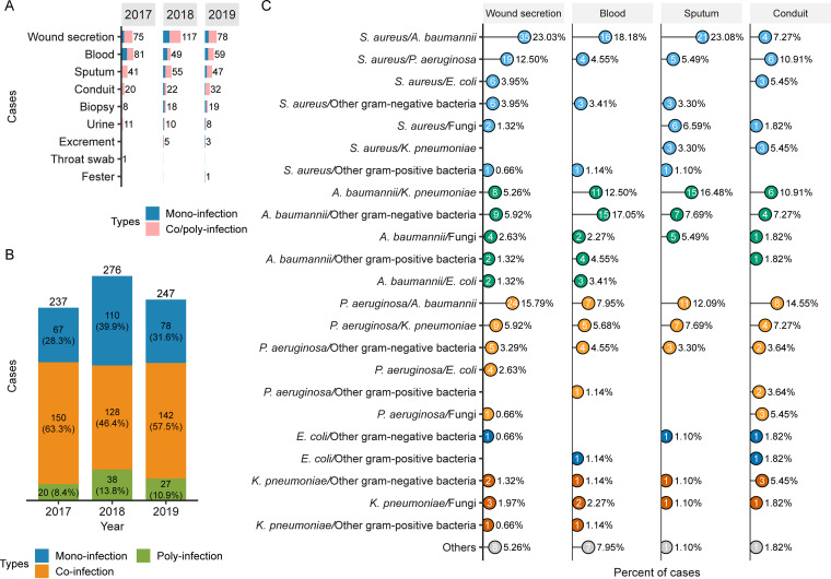
S. aureus represents a common cause of polymicrobial infections (2, 3); however, it is mostly studied in single-species infections. To further survey the infection type and microbial composition of S. aureus-related multispecies infections, we first carried out a retrospective study, where 760 clinical samples were collected from various tissues of 208 burn patients who were hospitalized in the intensive care unit during 2017 to 2019. The specimens of various origins, such as wound secretion, blood, sputum, conduit, biopsy, and urine, exhibited preferred multispecies infections versus single-species infections (Fig. 1A). In addition, the samples were further grouped into 3 major categories: mono-infection (caused by single-species), co-infection (caused by 2 different species), and poly-infection (caused by 3 or more different species). The incidence of mono-infection in 2017, 2018, and 2019 was 28.3%, 39.9%, and 31.6%, respectively; the incidence of poly-infection was 8.4%, 13.8%, and 10.9%, respectively (Fig. 1B). In comparison, co-infections predominated in collected samples with an incidence rate of 63.3%, 46.4%, and 57.5% in 2017, 2018, and 2019, respectively (Fig. 1B). These results indicated that multispecies infections constitute a prevalent situation in clinical settings, which deserves more attention and further investigation.
FIG 1.
Statistics of the collected clinical samples and bacterial diversity. (A) A total of 760 clinical samples were recovered from 208 burn patients that were hospitalized in the intensive care unit during 2017 to 2019. The collected samples had diverse origins for both mono-infections and co/poly-infections. (B) The 760 clinical samples were classified into three major types, namely, mono-infection, co-infection, and poly-infection, according to the bacterial species isolated from each sample. Multi-species infections are prevalent in clinical settings, where co-infections dominate the clinical samples. (C) Statistics of the bacterial composition for co-infections according to the sample origins. The co-infecting microbes were assigned to various distinct species, including the antibiotic-resistant ESKAPE pathogens (S. aureus, P. aeruginosa, A. baumannii, K. pneumoniae). Co-infection of S. aureus and A. baumannii showed the highest incidence rates and accounted for 23.03%, 18.18%, and 23.08% of the samples collected from wound secretion, blood, and sputum, respectively.
Out of 760 clinical samples, 420 (55%) were classified into co-infection type. To elucidate the diversity of co-infections, the co-infecting microbial pairs were further analyzed in detail according to the sample origins, including wound secretion, blood, sputum, and conduit (Fig. 1C). Note that the numbers of co-infection samples collected from other origins (biopsy, urine, excrement, throat swab, and fester) were all less than 10, so we did not analyze these samples further. The results showed that the co-infecting microbes were assigned to distinct species, including the antibiotic-resistant Enterococcus faecium, S. aureus, Klebsiella pneumoniae, A. baumannii, P. aeruginosa, Enterobacter spp. (ESKAPE) pathogens and/or fungus; whereby the diversified combinations of co-infection were identified (Fig. 1C). Notably, the combination of co-infection with S. aureus and A. baumannii was mostly recovered and accounted for 23.03%, 18.18%, and 23.08% of the samples collected from wound secretion, blood, and sputum, respectively (Fig. 1C). These data suggested that co-infections predominate in collected samples with potential clinical relevance. Given the highest incidence of co-infection with S. aureus and A. baumannii identified in this study and the limited understanding of the interaction between the two species, we mainly focused our study on the infections caused by S. aureus and A. baumannii, while other co-infection combinations also deserve further consideration.
Construction and characterization of a high-density transposon insertion mutant library in S. aureus strain Newman.
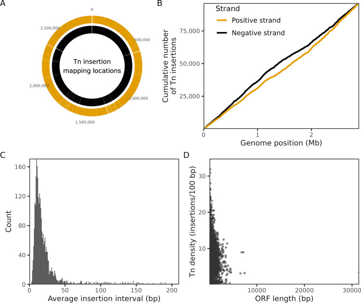
Transposon mutagenesis represents a robust tool that enables unbiased genome-wide identification of gene essentiality under specific in vitro and/or in vivo conditions (29, 30). Here, we firstly constructed a high-density transposon insertion mutant library in S. aureus reference strain Newman using the transposon mariner, which enable to insert efficiently into the TA dinucleotides and generates thousands of mutants across a genome (31). Combined with next-generation high-throughput sequencing, the library obtained 171,620 specific insertions that were specifically mapped to the genome of strain Newman, with an average distance of 31-bp between the 2 successive transposon insertions (Fig. 2 and Table S1). S. aureus strain Newman has 2,696 putative genes in total, including 2,624 genes coding for proteins and 72 for RNAs. By comparing the transposon insertion frequency across the Newman genome, 174 genes encoding proteins were identified as essential for S. aureus survival under in vitro conditions based on the absence or underrepresentation of insertions in the transposon library (Table S2). Notably, these genes were functionally enriched for central cellular processes, such as ribosome activity, DNA replication, cell division, and central metabolism (Table S2).
FIG 2.
Quality assessment of the constructed Tn insertion mutant library. (A) Distribution of the genome-wide Tn insertion locations mapped to the genome of S. aureus strain Newman. The outer ring (red) represents the positive strand, and the inner ring (blue) represents the negative strand. (B) Cumulative number of Tn insertions in positive strand (red) and negative strand (blue). (C) Statistics of the Tn insertion number per interval (bp). The peak indicates an average distance of 31-bp between the two successive transposon insertions. (D) Statistics of the Tn density (number of insertions per 100-bp).
Sequencing information for all Tn-seq replicates. Statistics of the Tn-seq data recovered from input, mono-infection of S. aureus, and co-infection of S. aureus and A. baumannii. Download Table S1, XLSX file, 0.01 MB (14.4KB, xlsx) .
Copyright © 2022 Li et al.
This content is distributed under the terms of the Creative Commons Attribution 4.0 International license.
Statistics of S. aureus essential genes for mono-infection and co-infection with A. baumannii. The essential genes were categorized into the groups of In vitro essential genes, Mono-infection essential genes, Co-infection essential genes, Essential for both Mono- and Co-infections, Unique to Mono-infection, and Unique to Co-infection. Download Table S2, XLS file, 0.2 MB (216KB, xls) .
Copyright © 2022 Li et al.
This content is distributed under the terms of the Creative Commons Attribution 4.0 International license.
Analysis of fitness determinants required for S. aureus mono-infection.
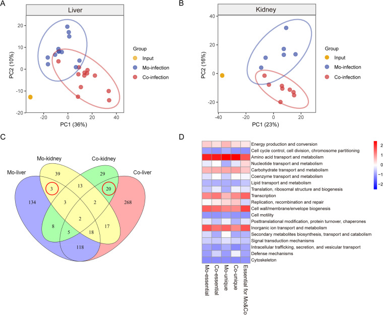
Infectious studies were conducted to screen for fitness determinants required for S. aureus mono-infection using a murine model. BALB/c mice were inoculated intravenously via the tail vein with the Newman transposon library (1 × 10⁷ CFU per mouse). The livers and kidneys of the infected mice at 5 days post-injection were recovered, homogenized, and prepared for Tn-seq. K-means clustering of the principal-component analysis (PCA) highlighted that the Tn-seq data generated from input and mono-infection clustered independently from each other, both in the tissue colonization of livers and kidneys (Fig. 3A and B). To specifically focus on the in vivo essential factors, the in vitro essential genes (174) were removed from the transposon input pool, and excluded from all subsequent analyses.
FIG 3.
Clustering and functional annotation of Tn-seq data. (A) Principal-component analysis of the normalized Tn-seq counts recovered from the liver in three conditions: input (orange, n = 4, the points are overlapped), mono-infection (blue, n = 12), and co-infection (red, n = 12). (B) Principal-component analysis of the normalized Tn-seq counts recovered from the kidney in three conditions: input (orange, n = 4, the points are overlapped), mono-infection (blue, n = 6), and co-infection (red, n = 8). (C) Venn diagram of the essential genes of S. aureus required for in vivo colonization of the liver and kidney both in mono-infection and co-infection with A. baumannii. The red circle depicts the genes essential for colonization of both the liver and kidney while also being divergently required for mono-infection or co-infection. (D) Heatmap of COG functional categories of the essential in vivo genes of S. aureus. Conserved COG functionality was assigned to both infection conditions, while differentiated requirements were observed for genes unique to mono-infection and co-infection conditions.
Among the 2,522 genes that were non-essential for S. aureus viability in vitro, a total of 362 genes were identified essential for S. aureus mono-infection (Fig. 3C and Table S2). Specifically, 26 (7%) genes encode fitness factors required for the colonization of both the liver and kidney, representing a core set of genes essential for S. aureus in vivo mono-infection, 265 (73%) genes encoded liver-specific fitness factors, and 71 (20%) were kidney-specific (Fig. 3C and Table S2). Furthermore, 326 mono-infection essential genes could be annotated according to the classification of the gene category within the Clusters of Orthologous Groups (COG) database. In addition to the COG category of function unknown, these genes were mostly enriched in the COG categories for amino acid, inorganic ion, and carbohydrate metabolism, as well as cell envelope biogenesis (Fig. 3D).
Analysis of fitness determinants required for S. aureus co-infection with A. baumannii.
Given that S. aureus and A. baumannii were the most commonly co-isolated microbes in various infection samples in this study, we next assessed the fitness factors crucial for S. aureus during the co-infection with A. baumannii. A mixture of Newman transposon insertion library and 10-fold less A. baumannii, which attempts to avoid the unexpected effect caused by increased inoculum, was inoculated into mice. Liver and kidney samples were collected and subjected to Tn-seq analysis. The data of co-infection exhibited distinct PCA clustering from that of both the input and mono-infection (Fig. 3A and B), indicating that the presence of A. baumannii alters the gene essentiality for S. aureus.
Tn-seq identified 503 genes in total that were essential for S. aureus co-infection with A. baumannii in vivo (Fig. 3C and Table S2), which outnumber the genes essential during mono-infection (362). Further examination revealed that 421 (84%) genes were essential for S. aureus colonization of the liver, 53 (10%) genes specific for kidney, and only 29 (6%) genes were required for colonization of both tissues (Fig. 3C and Table S2), indicating that distinct subsets of S. aureus genes were required for different tissue colonization. Similarly, the genes essential for co-infection were mostly enriched in the COG functional categories of metabolism for amino acid, inorganic ion, carbohydrate, as well as the biological process for transcription and cell envelope biogenesis (Fig. 3D).
Presence of A. baumannii dramatically alters the fitness requirements for S. aureus in vivo.
Given the limited information regarding the in vivo interplay between S. aureus and A. baumannii, we further assessed how the presence of A. baumannii impacts the fitness requirements of S. aureus in a murine systemic infection model. Notably, integrated analysis of the data recovered from both mono-infection and co-infection conditions revealed that 176 (26%) genes were required specifically for S. aureus mono-infection (designated hereinafter as genes unique to mono-infection), 317 (47%) genes were required exclusively for co-infection condition (designated hereinafter as genes unique to co-infection), and 186 (27%) genes were essential for both infection types (Fig. 3C and Table S2). Three genes unique to mono-infection were required for colonization of both the liver and kidney (Fig. 3C), including the gene encoding ornithine cyclodeaminase SbnB (NWMN_0061), competence protein ComGB (NWMN_1447), and hypothetical protein (NWMN_0560). Twenty genes unique to co-infection, such as phosphotransferase system protein TreP (NWMN_0438), oxidoreductase (NWMN_2478), deoxynucleoside kinase (NWMN_0518), Na+/H+ antiporter MnhG (NWMN_0599), and oligopeptide transport permease (NWMN_0856), were required for the colonization of both tissues (Fig. 3C).
Interestingly, genes encoding known virulence-associated factors, including exotoxins, cofactors and enzymes, adhesins, biofilm, and global regulators, exhibited distinct essentiality during mono-infection and co-infection conditions. Several factors were required for both infection types, such as the staphylococcal accessory gene regulator AgrC, fibrinogen-binding protein ClfA, biofilm-associated N-glucosaminyltransferase IcaA, sortase SrtA, and iron-regulated heme-iron binding protein IsdA (Table S2). In contrast, co-infection with A. baumannii elicited additional requirement of several virulence factors, particularly for the two-component system members (SaeR, ArlR, HssS, and KdpE), cysteine protease SspB, and hyaluronate lyase HysA (Table S2). These results indicated that the essential fitness factors of S. aureus were dramatically changed during co-infection with A. baumannii, and about 49% of the essential genes (176) for S. aureus mono-infection were converted to non-essential in the presence of A. baumannii. Meanwhile, a total of 317 non-essential genes during mono-infection were additionally required during co-infection. The transition of essential genes between mono-infection and co-infection conditions depicts an interesting reciprocal relationship.
Furthermore, 186 genes essential for S. aureus across both infection types represented a core set of genes and were mostly enriched in the COG functional categories of transport and metabolism of inorganic ions, amino acids, carbohydrates, and nucleotides (Fig. 3C and D), elucidating a critical role of metabolism for S. aureus during in vivo infection. In addition to shared metabolism activities, the COG annotation of infection-type-specific fitness genes resulted in divergent functional enrichments for co-infection compared to mono-infection conditions, mainly exhibiting the overrepresentation of the COG category of nucleotide and coenzyme metabolism, translation, and defense mechanisms, together with the alleviation of secondary metabolites catabolism (Fig. 3D).
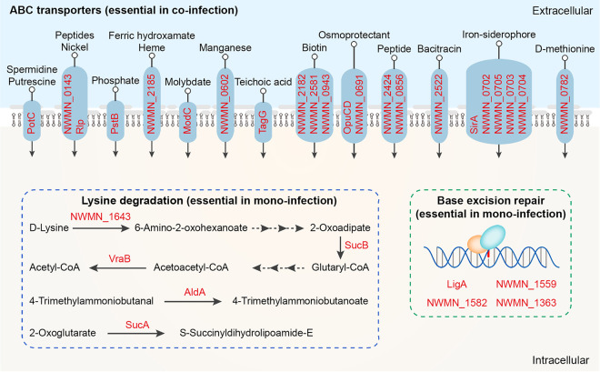
Kyoto Encyclopedia of Genes and Genomes (KEGG) pathways analysis revealed further insights into the distinct functions of in vivo essential genes either unique to mono-infection or to co-infection. Notably, KEGG pathways of lysine degradation and base excision repair predominated genes specific for mono-infection (Fig. 4), suggesting these functions might be compensated by A. baumannii during co-infection. Of note, 5 genes involved in lysine degradation were identified, which include genes encoding aldehyde dehydrogenase AldA (NWMN_0113), dihydrolipoamide succinyltransferase SucB (NWMN_1325), 2-oxoglutarate dehydrogenase SucA (NWMN_1326), d-alanine aminotransferase (NWMN_1643), and acetyl-CoA C-acetyltransferase VraB (NWMN_0539) (Fig. 4). Four genes were identified to be related to base excision repair, including endonuclease III (NWMN_1363), DNA-3-methyladenine glycosidase (NWMN_1559), formamidopyrimidine-DNA glycosylase (NWMN_1582), and DNA ligase (NWMN_1842) (Fig. 4). Additionally, fitness factors unique to co-infection were significantly enriched in the KEGG pathway of ATP-binding cassette (ABC) transporters (P = 0.02) (Fig. 4). Consistently, 27 genes encoding ABC transporters, which potentially participate in the transportation of diverse substrates, such as peptides, phosphate, biotin, siderophore, heme, and bacitracin, were screened out of the 317 genes unique to co-infection (Fig. 4). The overrepresentation of ABC transporters for genes specific to co-infection suggested that the presence of A. baumannii elicited additional functions, i.e., nutrients competition and/or resilience toward harmful antibacterial agents, required for S. aureus to successfully establish in vivo infection and tissue colonization.
FIG 4.
Functionality analysis of essential S. aureus in vivo genes unique to mono-infection and co-infection. Schematic diagram of the functionality of enriched genes (labeled as red color). The genes required exclusively for mono-infection were mostly enriched for KEGG pathways of lysine degradation (P-adj value of 0.06) and base excision repair (P-adj value of 0.20). Co-infection-unique genes were significantly enriched in KEGG pathway of ABC transporters (P-adj value of 0.02). The genes encoding ABC transporters participate in the transportation of diverse substrates. Note that 5 genes with unknown substrate specificity, including NWMN_1203, NWMN_1758, NWMN_0251, NWMN_0654, NWMN_2340, were not drawn in the diagram.
Validation of candidate fitness factors required in type-specific infection.
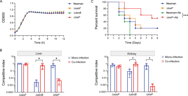
To verify the accuracy of the genome-scale Tn-seq analyses, 3 individual genes, including the mono-infection-unique essential gene sbnB (NWMN_0061) encoding ornithine cyclodeaminase, the co-infection-unique essential gene treP (NWMN_0438) encoding trehalose-specific transporter, and the in vivo non-essential gene sasF (NWMN_2545) encoding cell-wall-anchored protein SasF, were selected based on the fold change of Tn-seq reads of differentially screened genes. Note that gene sbnB and treP was required for distinct infection types but essential for colonization of both the liver and kidney, allowing to assess in detail the gene essentiality in specific infection-type manner. Then, the corresponding mutants (ΔsbnB, ΔtreP, and ΔsasF) were generated in S. aureus Newman background. Bacterial growth curve determination revealed that all 3 mutants exhibited similar growth patterns compared to the wild-type (WT) strain Newman (Fig. 5A). We then co-infected each of the mutants against strain Newman with or without A. baumannii in the murine systemic model. The livers and kidneys were collected 5 days postinfection and processed for viable CFU counts of S. aureus. As expected, mutation of gene sasF resulted in no significant difference in the CFU recovered from both tissues between ΔsasF and WT, regardless of the presence of A. baumannii (Fig. 5B). Decreased colonization in both the liver and kidney was observed when mutant ΔsbnB competed against WT, exclusively in mono-infection but not in co-infection with A. baumannii (Fig. 5B). By contrast, gene treP was crucial for efficient colonization of S. aureus in the liver and kidney during co-infection with A. baumannii, but not required for mono-infection condition (Fig. 5B).
FIG 5.
Validation of selected fitness factors for S. aureus in vivo pathogenesis. (A) In vitro growth monitoring of S. aureus strain Newman and three gene mutants. Note that gene sasF is non-essential for S. aureus in vivo infection, gene sbnB is required for mono-infection, and treP is required for co-infection. No obvious difference was observed in the growth of the mutants compared to the WT strain Newman. (B) In vivo competition assay. The 3 infection-specific gene mutants were used in competition assays with the WT strain Newman in mono- and co-infection with A. baumannii in a murine systemic infection model. Bacterial CFU were recovered from both the livers and kidneys 5 days postinfection, and the competitive index for each gene mutant was calculated using WT as the control. The results are presented as the mean ± SD (standard deviation) from 3 biological replicates. Statistical analysis was performed using a Student's t test, *, P < 0.05. (C) Mice survival curves. Mice (n = 10) were inoculated via tail vein injection with a bacterial suspension at 2 × 10⁷ CFU of either WT or mutant, or 2 × 10⁷ S. aureus and 2 × 10⁶ A. baumannii for co-infection. Survival of the mice was monitored for 9 days post-injection. Ab, A. baumannii. Statistical analysis was performed by log-rank (Mantel-Cox) test, ***, P < 0.001.
To gain further insights into the differential requirement of the gene treP for S. aureus infection in vivo, mice were inoculated with WT or mutant ΔtreP, either as a single-species injection or co-injected with A. baumannii, and the survival rates of mice were monitored. No obvious difference regarding the survival of the mice was found between the WT and ΔtreP (Fig. 5C). In comparison, the presence of A. baumannii augmented the virulence of the WT strain, while it significantly alleviated the virulence of the mutant ΔtreP, which led to increased and prolonged mouse survival under tested conditions (Fig. 5C). Together, these results showing direct in vivo co-infection with the fitness gene mutant and WT strain Newman confirmed the infection-type-specific phenotypes, which were consistent with the primary Tn-seq results in this study.
DISCUSSION
Polymicrobial infections have been historically recognized (2, 9, 32); however, the detailed interactions and pathogenesis strategies between different species remain largely unknown. S. aureus is a major human pathogen that is frequently involved in polymicrobial infections, and the presence of co-infectious microbes complicates the in vivo behaviors of S. aureus compared to mono-infection conditions. To date, the majority of studies regarding co-infections and interactions between S. aureus and other microbial community members have focused on S. aureus and P. aeruginosa (19, 33), as well as the pathogenic fungus C. albicans (14, 20), highlighting the importance of polymicrobial infections in investigating S. aureus pathogenesis. In this study, we discovered a widespread incidence of co-infections caused by S. aureus and A. baumannii through a retrospective surveillance of clinical samples recovered from burn patients. The results of this study broaden our knowledge of the diverse species that cause co-infections with S. aureus.
Despite the high prevalence of both S. aureus and A. baumannii in the same infection niches, the interplay between the two species has been poorly explored. A previous study cocultured clinical strains of A. baumannii and S. aureus that were recovered from the same soft tissue of a diabetic patient and found that these strains exhibit a state of commensalism in vitro, without effects on each other either beneficially or detrimentally (34). Another study demonstrated that the presence of A. baumannii alters the pharmacodynamics and modulates the killing of S. aureus by meropenem in vitro, pointing out that dose-optimized beta-lactams represent a therapeutic option for control of co-infections involving S. aureus and A. baumannii (35). Here, we firstly investigated the fitness requirement for S. aureus to establish infection and colonize tissue in vivo. Co-infection with A. baumannii dramatically altered the fitness essentiality of S. aureus by increasing the co-infection-unique genes (317) and alleviation of the mono-infection-unique genes (176). Specifically, approximately 49% of the mono-infection essential genes converted to non-essential during co-infection. Similarly, a previous study showed via a murine model of chronic surgical wound infection that co-infection with P. aeruginosa results in conversion of ~25% of the monococulture essential genes in S. aureus to non-essential (36), revealing the complicated interactions between S. aureus and P. aeruginosa in vivo. In addition, while the transition of essential genes between mono-infection and co-infection was observed in the presence of A. baumannii, the cues and underlying mechanism eliciting this transition remain to be fully explored. We speculate that the differential requirement of essential genes might derive from the direct interaction between the two species, but also raise the possibility that host factors function in this transition, such as limited nutrients might indirectly shape the interaction. Further integrated analysis of the altered essential genes in vivo and the direct interaction of S. aureus and A. baumannii in vitro may contribute to the comprehensive understanding of this crucial process.
In this study, 186 genes were identified as in vivo fitness determinants for S. aureus during both single-species infection and co-infection with A. baumannii, suggesting that these genes likely encode functions crucial for S. aureus pathogenesis and tissue colonization. Among them, the core set of fitness factors was mostly assigned to the COG functionality of metabolism of inorganic ions, amino acids, carbohydrates, and nucleotides, coinciding with findings of other studies where central metabolism plays a critical role for S. aureus infection in vivo (36, 37). In addition, 176 genes identified as unique to S. aureus mono-infection were converted to non-essential during co-infection with A. baumannii, and these genes were mostly enriched in KEGG pathways of lysine degradation and base excision repair. Notably, metabolism of lysine has been shown to have diverse effects on S. aureus physiology, resistance, and pathogenesis. For example, modification of membrane lipids with l-lysine confers S. aureus with resistance to human defensins and evasion of neutrophil killing (38), and increased lysine amounts have been shown to benefit adaptation of S. aureus after internalization by human lung epithelial cells (S9 and A549) and human embryonic kidney cells (HEK293) (39). Furthermore, a widespread bacterial lysine degradation pathway that converts glutarate to succinate provides an important link in central carbon and energy metabolism (40). Thus, an interesting question is how the presence of A. baumannii contributes to the lysine degradation of S. aureus during co-infection. The specific impact on S. aureus virulence deserves further investigation.
The ability of S. aureus to withstand damage caused by the host immune defense is crucial for the successful establishment of an infection, in which DNA is a common target of host-producing antimicrobials. A previous study demonstrated that neutrophils cause DNA double-strand breaks in the genome of S. aureus through reactive oxygen species produced by the oxidative burst, and such DNA damage can be repaired by an AddAB-family of helicase/nuclease complexes (RexAB) (41). The repair of DNA damage enables the survival of S. aureus in host tissues during infection, indicating that DNA damage repair represents an important mechanism by which S. aureus withstands host defense (41). In this study, 4 genes involved in the functionality of base excision repair were identified unique to S. aureus mono-infection, but were not required for co-infection with A. baumannii, suggesting that A. baumannii may provide functional compensation for S. aureus to avoid and/or cope with the DNA damage encountered within the same infection microenvironment. However, the mechanisms underlying this relationship are unclear and need further investigation.
Another interesting finding is that among the 317 genes essential exclusively during co-infection, a total of 27 genes encoding transporters were significantly enriched in the KEGG pathway of ABC transporters (P = 0.02). Moreover, these ABC transporters potentially participate in the transportation of a broad spectrum of substrates, including peptides, phosphate, biotin, siderophore, heme, and bacitracin. Similar results were obtained in a previous study showing that co-infection with S. gordonii imposes unique metabolic stresses on A. actinomycetemcomitans as multiple nutrient transporters were solely required during co-infection (36). Membrane transport systems are abundant membrane proteins in S. aureus, with niche- and environment-specific roles in sustaining cell integrity and metabolic homeostasis, contributing to bacterial growth, antibiotic resistance, and virulence (42). The additional requirement of ABC transporters for S. aureus during co-infection with A. baumannii may be explained by 2 aspects, namely, competition for limited nutrients in a specific niche and/or efflux of harmful factors generated by A. baumannii.
Furthermore, this study revealed differential requirements of fitness factors for S. aureus colonization in the liver and kidney. Among the 679 genes essential for S. aureus infection in vivo, 520 (77%) genes were unique to colonization of the liver, 81 (12%) were unique to kidneys, and only 2 genes (NWMN_0574 and NWMN_0639) were required for colonization in both tissues and mono/co-infection types. A Tn-seq screen using a murine model of catheter-associated urinary tract infection revealed distinct fitness requirements for the opportunistic pathogen Providencia stuartii colonization in either the bladder or kidneys during single-species infection or co-infection with a common co-colonizer, Proteus mirabilis (43). A previous study identified a LysR-type transcriptional regulator (LTTR) required for efficient colonization of S. aureus in the kidney, but not in the liver, in a murine model of metastatic bloodstream infection (44). The requirement of different subsets of genes as depicted in this study and previous reports highlights the remarkable plasticity of S. aureus to coordinate its expression of fitness factors, which in turn contribute to adaptation to the niche-specific microenvironment and nutrient availability during infection.
In addition, since the first report of Tn mutagenesis in S. aureus using the Enterococcus faecalis transposon Tn917 in 1997 (45), Tn-seq has emerged as a powerful high-throughput technique that is widely applied to identify infection-specific fitness determinants of S. aureus (29). Given the nature of the transposons mostly used, several common limitations remain to be addressed, including inability to accurately assess the fitness contribution of secreted factors, mainly due to cross-complementation, which demonstrated, in our study and others, that genes encoding exoproducts were poorly identified in Tn-seq screens (29, 36). In general, with the introduction of improved Tn methodologies, including a bacteriophage-based transposition system (46) and CRISPR-Cas12k-based, RNA-guided mutagenesis (47), Tn-seq may contribute to the future understanding of the physiology, adaptation, and evolution of S. aureus, such as the key factors responsible for S. aureus virulence in dynamic environments (e.g., as a time series, or with specific niche relevance), transmission from livestock to humans, and genetic mutations or phenotypic variations critical for vancomycin-intermediate resistance.
Overall, this study revealed a high incidence rate and important clinical relevance of co-infections caused by S. aureus and A. baumannii. The presence of A. baumannii dramatically alters the fitness determinants essential for S. aureus infection in vivo, particularly the functional compensation of lysine degradation and DNA repair, as well as the additional requirement of factors encoding ABC transporters. The exact mechanism by which the presence of co-infecting A. baumannii leads to altered fitness requirements will be an interesting subject of future studies. Moreover, the infection-type-specific genes contributing to S. aureus in vivo pathogenesis likely represent promising targets for the development of potential therapeutic interventions aimed at controlling both single-species and polymicrobial infections.
MATERIALS AND METHODS
Bacterial strains and culture conditions.
Bacterial strains and plasmids used in this study are listed in Table S3. Clinical samples were procured with approval from the Ethics Committee of the First Affiliated Hospital of Army Medical University, PLA, under the ethics statement number KY201991. Clinical samples were collected from various tissues of 208 burn patients hospitalized in the intensive care unit of the Institute of Burn Research of Southwest Hospital of Army Medical University from years 2017 to 2019. The recovered bacteria were further identified using matrix-assisted laser desorption/ionization time-of-flight mass spectrometry (MALDI-TOF MS). Repetitive samples with the same microbes from the same tissue of the same patient were excluded, and samples with the same microbes from different sites of the same patient were included. Unless otherwise specified, S. aureus strain Newman and A. baumannii strain ATCC19606 used in this study was cultured in brain heart infusion broth (BHI) and LB broth at 37°C with shaking at 200 rpm, respectively.
Strains and plasmids used in this study. Download Table S3, DOCX file, 0.02 MB (17.7KB, docx) .
Copyright © 2022 Li et al.
This content is distributed under the terms of the Creative Commons Attribution 4.0 International license.
Construction of the transposon insertion library in S. aureus strain Newman.
The plasmid MA15 possesses a temperature-sensitive replicon and a mariner transposon, with a kanamycin resistance gene and an MmeI restriction site within each inverted repeat, and was used to construct the transposon insertion mutant library in the S. aureus strain Newman as described previously (48). Approximately 60,000 transposon insertion colonies were collected and stored in 20% glycerol at −80°C. Total DNA was extracted from the library freezer stock using the TIANamp Bacteria DNA Kit (Tiangen) according to the manufacturer’s recommendations, and subjected to Tn-Seq libraries preparation as the input.
Murine systemic infection.
Murine systemic infections were performed with 6–8 week old female BALB/c mice via tail vein injection. The mice were purchased from HUNAN SJA laboratory animal Co., LTD (Hunan, China) and housed in two-way housing on a 12-h light/dark cycle. All animal experiments were performed in strict accordance with protocols approved by the Laboratory Animal Welfare and Ethics Committee of the Third Military Medical University and complied with Institutional Animal Welfare and Ethical guidelines (AMUWEC2019421).
The preparation of the mutant library for injection was performed as previously described (49). Briefly, a 100 μL aliquot of the mutant library freezer stock was inoculated into 100 mL of BHI without antibiotic selection and cultured overnight at 37°C. To isolate cells in exponential growth phase, the overnight culture was inoculated 1:1000 into fresh BHI without antibiotics, and bacterial growth was monitored until an optical density of 600 nm (OD600) of 0.8 was achieved. For Tn-seq experiments, 1 × 10⁷ CFU of the S. aureus Newman transposon mutant library was used for mono-infection (n = 12 mice), and a mixture of 5 × 10⁶ CFU of the S. aureus library and 5 × 10⁵ CFU of the A. baumannii strain ATCC19606 were used for co-infection (n = 12 mice). Here, 10-fold less A. baumannii than S. aureus was used in the inoculum to ensure that the effects observed were not due to increased bacterial inoculum as previously described (36).
Five days postinfection, the livers and kidneys were collected from the infected mice and snap-frozen in liquid nitrogen. The liver and kidney tissues were then ground carefully into a powder in liquid nitrogen, resuspended in TE buffer in Sarstedt 2 mL microtubes prefilled with 1 g of 0.1 mm zirconia beads (BioSpec), and homogenized with a Mini-Beadbeater (Biospec). The tissue lysates were then subjected to genomic DNA extraction using the TIANamp Bacteria DNA Kit (Tiangen).
Preparation of Tn-seq libraries for sequencing.
Genomic DNA extracted from the livers and kidneys of mice subjected to the mono- or co-infection murine systemic infection models were prepared for Tn-seq analysis as described previously (30). Briefly, genomic DNA was digested with MmeI (Thermo Scientific), treated with calf intestinal alkaline phosphatase (NEB), and purified by the Wizard SV Gel and PCR Clean-Up System (Promega). Then, a double-stranded adapter, generated by annealing the primers of 5′-phosphorylated oligonucleotide A and oligonucleotide B (Table S4), was ligated to the purified DNA fragments. The DNA fragments were then subsequently amplified using the primer pair P1-transposon-MmeI and P2-tn-seq-PCR (Table S4), in which the barcodes for multiplexing were added simultaneously, and a 103-bp sequence with 20-bp of bacterial-derived DNA was obtained. The PCR products were separated on a 2% agarose gel, extracted with the gel purification column (Promega), and then processed via high-throughput sequencing using an Illumina Hi-Seq platform (Novogene). After sequencing, different samples were distinguished based on barcode sequence, and the 20-bp bacterial-specific sequences were mapped to the genome of strain Newman. The distribution of Tn insertions across the entire genome was calculated.
Primers used in this study. Download Table S4, DOCX file, 0.02 MB (19.1KB, docx) .
Copyright © 2022 Li et al.
This content is distributed under the terms of the Creative Commons Attribution 4.0 International license.
Tn-seq bioinformatics analysis.
Raw Tn-seq reads were cleaned with fastp v0.20.1 (50) and demultiplexed with SeqKit v0.13.0 (51). The 20-bp genomic sequences were extracted using SeqKit and mapped to the genome of S. aureus strain Newman using SeqKit. In-house scripts were used to filter unique mapped sequence with at most one mismatch. Gene mutations were summarized with Bedtools v2.29.2 (52). Essential genes were determined with DESeq2 v1.30.1 (53) with an adjusted P value < 0.05 and log2 (fold change) < −1. KEGG pathway enrichment was analyzed by clusterProfiler v3.14.3 (54) with an adjusted P value < 0.05.
Construction of S. aureus gene mutants.
For mutation of genes sasF, treP, and sbnB in the strain Newman, approximately 1000-bp regions upstream and downstream of the specific genes were amplified from Newman genomic DNA. The KanR cassette-encoding gene was amplified using the plasmid MA15 as the template (primer sequence in Table S4). The 3 DNA fragments in the upstream-KanR cassette-downstream orientation were then cloned into the temperature-sensitive plasmid pBT2 via Gibson assembly (NEB). Knock-in mutations of the target genes replaced by the KanR cassette were generated via homologous recombination as previously described (55), which were then ultimately verified by sequencing.
Validation of mutant fitness using the in vivo systemic infection model.
Log-phase cultures of S. aureus strain Newman or the KanR cassette knock-in mutants were washed, normalized to an equal OD600 reading in PBS, and mixed at a ratio of 1:1 of mutant to WT. Mice (n = 5 per group) were inoculated via tail vein injection with a mixture of 1 × 10⁷ CFU for mono-infection or 5 × 10⁶ CFU S. aureus and 5 × 10⁵ CFU A. baumannii for co-infections. Both the livers and kidneys were recovered 5 days postinfection and homogenized with a tissue grinder (SCIENTZ-48L, SCIENTZ). The homogenate was 10-fold serially diluted in PBS and plated on BHI agar (to quantify all S. aureus) and BHI agar supplemented with 100 μg/mL kanamycin (to quantify S. aureus mutants). For co-infection conditions, both S. aureus and A. baumannii formed colonies on BHI agar but could be easily differentiated by colony color due to the yellow pigment staphyloxanthin secreted by S. aureus. Competitive indices for each mutant in each infection type were calculated as ([CFU of mutant S. aureus/tissue]/[CFU of WT S. aureus/tissue]).
For mouse survival monitoring, log-phase cultures of S. aureus strain Newman and the individual fitness gene mutant were washed and normalized to an equal OD600 reading in PBS, respectively. Mice (n = 10 per group) were inoculated via tail vein injection with 2 × 10⁷ CFU of either WT or mutant or 2 × 10⁷ CFU S. aureus and 2 × 10⁶ CFU A. baumannii for co-infection. Growth and survival of mice were monitored for 9 days post-injection.
Statistics.
Unless noted otherwise, data were analyzed by GraphPad Prism v8.0 (GraphPad Software Inc.). Significant differences were determined by a Student's t test for comparison of two independent data sets or with one-way analysis of variance (ANOVA) followed by Tukey’s multiple comparison test for multiple comparisons. A P value of less than 0.05 was considered significant.
Data availability.
The raw Tn-seq data have been deposited into the NCBI Sequence Read Archive under accession number PRJNA787005. The genome sequence and annotation file of S. aureus strain Newman is available under GenBank accession number AP009351.
Supplementary Material
ACKNOWLEDGMENTS
This work was funded by the National Natural Science Foundation of China (grant 31900145 to G.L.), the Chongqing Municipal Natural Science Foundation of China (grant cstc2019jcyj-msxmX0142 to Y.Z.), and the Science Foundation of the Army Medical University (grant 2019JCLC02 to M.L.).
We thank LetPub (www.letpub.com) for linguistic assistance and pre-submission expert review. No potential conflict of interest was reported by the authors.
Contributor Information
Yan Zhao, Email: hnyanyanxp@aliyun.com.
Gilles P. van Wezel, Leiden University
REFERENCES
- 1.Magill SS, Edwards JR, Bamberg W, Beldavs ZG, Dumyati G, Kainer MA, Lynfield R, Maloney M, McAllister-Hollod L, Nadle J, Ray SM, Thompson DL, Wilson LE, Fridkin SK, Emerging Infections Program Healthcare-Associated Infections and Antimicrobial Use Prevalence Survey Team . 2014. Multistate point-prevalence survey of health care-associated infections. N Engl J Med 370:1198–1208. doi: 10.1056/NEJMoa1306801. [DOI] [PMC free article] [PubMed] [Google Scholar]
- 2.Stacy A, McNally L, Darch SE, Brown SP, Whiteley M. 2016. The biogeography of polymicrobial infection. Nat Rev Microbiol 14:93–105. doi: 10.1038/nrmicro.2015.8. [DOI] [PMC free article] [PubMed] [Google Scholar]
- 3.Gabrilska RA, Rumbaugh KP. 2015. Biofilm models of polymicrobial infection. Future Microbiol 10:1997–2015. doi: 10.2217/fmb.15.109. [DOI] [PMC free article] [PubMed] [Google Scholar]
- 4.Stacy A, Everett J, Jorth P, Trivedi U, Rumbaugh KP, Whiteley M. 2014. Bacterial fight-and-flight responses enhance virulence in a polymicrobial infection. Proc Natl Acad Sci USA 111:7819–7824. doi: 10.1073/pnas.1400586111. [DOI] [PMC free article] [PubMed] [Google Scholar]
- 5.Ramsey MM, Rumbaugh KP, Whiteley M. 2011. Metabolite cross-feeding enhances virulence in a model polymicrobial infection. PLoS Pathog 7:e1002012. doi: 10.1371/journal.ppat.1002012. [DOI] [PMC free article] [PubMed] [Google Scholar]
- 6.Kuboniwa M, Houser JR, Hendrickson EL, Wang Q, Alghamdi SA, Sakanaka A, Miller DP, Hutcherson JA, Wang T, Beck DAC, Whiteley M, Amano A, Wang H, Marcotte EM, Hackett M, Lamont RJ. 2017. Metabolic crosstalk regulates Porphyromonas gingivalis colonization and virulence during oral polymicrobial infection. Nat Microbiol 2:1493–1499. doi: 10.1038/s41564-017-0021-6. [DOI] [PMC free article] [PubMed] [Google Scholar]
- 7.Keogh D, Tay WH, Ho YY, Dale JL, Chen S, Umashankar S, Williams RBH, Chen SL, Dunny GM, Kline KA. 2016. Enterococcal metabolite cues facilitate interspecies niche modulation and polymicrobial infection. Cell Host Microbe 20:493–503. doi: 10.1016/j.chom.2016.09.004. [DOI] [PMC free article] [PubMed] [Google Scholar]
- 8.Alteri CJ, Himpsl SD, Mobley HL. 2015. Preferential use of central metabolism in vivo reveals a nutritional basis for polymicrobial infection. PLoS Pathog 11:e1004601. doi: 10.1371/journal.ppat.1004601. [DOI] [PMC free article] [PubMed] [Google Scholar]
- 9.Doolittle JM, Webster-Cyriaque J. 2014. Polymicrobial infection and bacterium-mediated epigenetic modification of DNA tumor viruses contribute to pathogenesis. mBio 5:e01015-14. doi: 10.1128/mBio.01015-14. [DOI] [PMC free article] [PubMed] [Google Scholar]
- 10.Liao YT, Kuo SC, Lee YT, Chen CP, Lin SW, Shen LJ, Fung CP, Cho WL, Chen TL. 2014. Sheltering effect and indirect pathogenesis of carbapenem-resistant Acinetobacter baumannii in polymicrobial infection. Antimicrob Agents Chemother 58:3983–3990. doi: 10.1128/AAC.02636-13. [DOI] [PMC free article] [PubMed] [Google Scholar]
- 11.Turner NA, Sharma-Kuinkel BK, Maskarinec SA, Eichenberger EM, Shah PP, Carugati M, Holland TL, Fowler VG, Jr. 2019. Methicillin-resistant Staphylococcus aureus: an overview of basic and clinical research. Nat Rev Microbiol 17:203–218. doi: 10.1038/s41579-018-0147-4. [DOI] [PMC free article] [PubMed] [Google Scholar]
- 12.Balasubramanian D, Harper L, Shopsin B, Torres VJ. 2017. Staphylococcus aureus pathogenesis in diverse host environments. Pathog Dis 75:ftx005. doi: 10.1093/femspd/ftx005. [DOI] [PMC free article] [PubMed] [Google Scholar]
- 13.Korgaonkar A, Trivedi U, Rumbaugh KP, Whiteley M. 2013. Community surveillance enhances Pseudomonas aeruginosa virulence during polymicrobial infection. Proc Natl Acad Sci USA 110:1059–1064. doi: 10.1073/pnas.1214550110. [DOI] [PMC free article] [PubMed] [Google Scholar]
- 14.Vila T, Kong EF, Montelongo-Jauregui D, Van Dijck P, Shetty AC, McCracken C, Bruno VM, Jabra-Rizk MA. 2021. Therapeutic implications of C. albicans-S. aureus mixed biofilm in a murine subcutaneous catheter model of polymicrobial infection. Virulence 12:835–851. doi: 10.1080/21505594.2021.1894834. [DOI] [PMC free article] [PubMed] [Google Scholar]
- 15.Michelsen CF, Christensen AM, Bojer MS, Hoiby N, Ingmer H, Jelsbak L. 2014. Staphylococcus aureus alters growth activity, autolysis, and antibiotic tolerance in a human host-adapted Pseudomonas aeruginosa lineage. J Bacteriol 196:3903–3911. doi: 10.1128/JB.02006-14. [DOI] [PMC free article] [PubMed] [Google Scholar]
- 16.Sheehan G, Tully L, Kavanagh KA. 2020. Candida albicans increases the pathogenicity of Staphylococcus aureus during polymicrobial infection of Galleria mellonella larvae. Microbiology 166:375–385. doi: 10.1099/mic.0.000892. [DOI] [PMC free article] [PubMed] [Google Scholar]
- 17.DeLeon S, Clinton A, Fowler H, Everett J, Horswill AR, Rumbaugh KP. 2014. Synergistic interactions of Pseudomonas aeruginosa and Staphylococcus aureus in an in vitro wound model. Infect Immun 82:4718–4728. doi: 10.1128/IAI.02198-14. [DOI] [PMC free article] [PubMed] [Google Scholar]
- 18.Lee J, Zilm PS, Kidd SP. 2020. Novel research models for Staphylococcus aureus small colony variants (SCV) development: co-pathogenesis and growth rate. Front Microbiol 11:321. doi: 10.3389/fmicb.2020.00321. [DOI] [PMC free article] [PubMed] [Google Scholar]
- 19.Camus L, Briaud P, Vandenesch F, Moreau K. 2021. How bacterial adaptation to cystic fibrosis environment shapes interactions between Pseudomonas aeruginosa and Staphylococcus aureus. Front Microbiol 12:617784. doi: 10.3389/fmicb.2021.617784. [DOI] [PMC free article] [PubMed] [Google Scholar]
- 20.Todd OA, Fidel PL, Jr, Harro JM, Hilliard JJ, Tkaczyk C, Sellman BR, Noverr MC, Peters BM. 2019. Candida albicans augments Staphylococcus aureus virulence by engaging the Staphylococcal agr quorum sensing system. mBio 10:e00910-19. doi: 10.1128/mBio.00910-19. [DOI] [PMC free article] [PubMed] [Google Scholar]
- 21.Kong EF, Tsui C, Kucharikova S, Andes D, Van Dijck P, Jabra-Rizk MA. 2016. Commensal protection of Staphylococcus aureus against antimicrobials by Candida albicans biofilm matrix. mBio 7:e01365-16. doi: 10.1128/mBio.01365-16. [DOI] [PMC free article] [PubMed] [Google Scholar]
- 22.Mulcahy ME, McLoughlin RM. 2016. Staphylococcus aureus and influenza A virus: partners in coinfection. mBio 7:e02068-16. doi: 10.1128/mBio.02068-16. [DOI] [PMC free article] [PubMed] [Google Scholar]
- 23.Goncheva MI, Conceicao C, Tuffs SW, Lee HM, Quigg-Nicol M, Bennet I, Sargison F, Pickering AC, Hussain S, Gill AC, Dutia BM, Digard P, Fitzgerald JR. 2020. Staphylococcus aureus lipase 1 enhances influenza A virus replication. mBio 11:e00975-20. doi: 10.1128/mBio.00975-20. [DOI] [PMC free article] [PubMed] [Google Scholar]
- 24.Bloes DA, Haasbach E, Hartmayer C, Hertlein T, Klingel K, Kretschmer D, Planz O, Peschel A. 2017. Phenol-soluble modulin peptides contribute to influenza A virus-associated Staphylococcus aureus pneumonia. Infect Immun 85:e00620-17. doi: 10.1128/IAI.00620-17. [DOI] [PMC free article] [PubMed] [Google Scholar]
- 25.Reddinger RM, Luke-Marshall NR, Hakansson AP, Campagnari AA. 2016. Host physiologic changes induced by influenza A virus lead to Staphylococcus aureus biofilm dispersion and transition from asymptomatic colonization to invasive disease. mBio 7:e01235-16. doi: 10.1128/mBio.01235-16. [DOI] [PMC free article] [PubMed] [Google Scholar]
- 26.Senok A, Alfaresi M, Khansaheb H, Nassar R, Hachim M, Al Suwaidi H, Almansoori M, Alqaydi F, Afaneh Z, Mohamed A, Qureshi S, Ali A, Alkhajeh A, Alsheikh-Ali A. 2021. Coinfections in patients hospitalized with COVID-19: a descriptive study from the United Arab Emirates. Infect Drug Resist 14:2289–2296. doi: 10.2147/IDR.S314029. [DOI] [PMC free article] [PubMed] [Google Scholar]
- 27.Spoto S, Valeriani E, Riva E, De Cesaris M, Tonini G, Vincenzi B, Locorriere L, Beretta Anguissola G, Lauria Pantano A, Brando E, Costantino S, Ciccozzi M, Angeletti S. 2020. A Staphylococcus aureus coinfection on a COVID-19 pneumonia in a breast cancer patient. Int J Gen Med 13:729–733. doi: 10.2147/IJGM.S261760. [DOI] [PMC free article] [PubMed] [Google Scholar]
- 28.Manohar P, Loh B, Nachimuthu R, Hua X, Welburn SC, Leptihn S. 2020. Secondary bacterial infections in patients with viral pneumonia. Front Med (Lausanne) 7:420. doi: 10.3389/fmed.2020.00420. [DOI] [PMC free article] [PubMed] [Google Scholar]
- 29.Sargison FA, Fitzgerald JR. 2021. Advances in transposon mutagenesis of Staphylococcus aureus: insights into pathogenesis and antimicrobial resistance. Trends Microbiol 29:282–285. doi: 10.1016/j.tim.2020.11.003. [DOI] [PubMed] [Google Scholar]
- 30.van Opijnen T, Bodi KL, Camilli A. 2009. Tn-seq: high-throughput parallel sequencing for fitness and genetic interaction studies in microorganisms. Nat Methods 6:767–772. doi: 10.1038/nmeth.1377. [DOI] [PMC free article] [PubMed] [Google Scholar]
- 31.Bae T, Banger AK, Wallace A, Glass EM, Aslund F, Schneewind O, Missiakas DM. 2004. Staphylococcus aureus virulence genes identified by Bursa aurealis mutagenesis and nematode killing. Proc Natl Acad Sci USA 101:12312–12317. doi: 10.1073/pnas.0404728101. [DOI] [PMC free article] [PubMed] [Google Scholar]
- 32.Rolston KV, Bodey GP, Safdar A. 2007. Polymicrobial infection in patients with cancer: an underappreciated and underreported entity. Clin Infect Dis 45:228–233. doi: 10.1086/518873. [DOI] [PubMed] [Google Scholar]
- 33.Magalhaes AP, Jorge P, Pereira MO. 2019. Pseudomonas aeruginosa and Staphylococcus aureus communication in biofilm infections: insights through network and database construction. Crit Rev Microbiol 45:712–728. doi: 10.1080/1040841X.2019.1700209. [DOI] [PubMed] [Google Scholar]
- 34.Castellanos N, Nakanouchi J, Yuzen DI, Fung S, Fernandez JS, Barberis C, Tuchscherr L, Ramirez MS. 2019. A study on Acinetobacter baumannii and Staphylococcus aureus strains recovered from the same infection site of a diabetic patient. Curr Microbiol 76:842–847. doi: 10.1007/s00284-019-01696-7. [DOI] [PMC free article] [PubMed] [Google Scholar]
- 35.Smith NM, Ang A, Tan F, Macias K, James S, Sidhu J, Lenhard JR. 2021. Interaction of Staphylococcus aureus and Acinetobacter baumannii during in vitro beta-lactam exposure. Antimicrob Agents Chemother 65:e02414-20. doi: 10.1128/AAC.02414-20. [DOI] [PMC free article] [PubMed] [Google Scholar]
- 36.Ibberson CB, Stacy A, Fleming D, Dees JL, Rumbaugh K, Gilmore MS, Whiteley M. 2017. Co-infecting microorganisms dramatically alter pathogen gene essentiality during polymicrobial infection. Nat Microbiol 2:17079. doi: 10.1038/nmicrobiol.2017.79. [DOI] [PMC free article] [PubMed] [Google Scholar]
- 37.Kim GL, Hooven TA, Norambuena J, Li B, Boyd JM, Yang JH, Parker D. 2021. Growth and stress tolerance comprise independent metabolic strategies critical for Staphylococcus aureus infection. mBio 12:e0081421. doi: 10.1128/mBio.00814-21. [DOI] [PMC free article] [PubMed] [Google Scholar]
- 38.Peschel A, Jack RW, Otto M, Collins LV, Staubitz P, Nicholson G, Kalbacher H, Nieuwenhuizen WF, Jung G, Tarkowski A, van Kessel KP, van Strijp JA. 2001. Staphylococcus aureus resistance to human defensins and evasion of neutrophil killing via the novel virulence factor MprF is based on modification of membrane lipids with L-lysine. J Exp Med 193:1067–1076. doi: 10.1084/jem.193.9.1067. [DOI] [PMC free article] [PubMed] [Google Scholar]
- 39.Surmann K, Michalik S, Hildebrandt P, Gierok P, Depke M, Brinkmann L, Bernhardt J, Salazar MG, Sun Z, Shteynberg D, Kusebauch U, Moritz RL, Wollscheid B, Lalk M, Volker U, Schmidt F. 2014. Comparative proteome analysis reveals conserved and specific adaptation patterns of Staphylococcus aureus after internalization by different types of human non-professional phagocytic host cells. Front Microbiol 5:392. doi: 10.3389/fmicb.2014.00392. [DOI] [PMC free article] [PubMed] [Google Scholar]
- 40.Knorr S, Sinn M, Galetskiy D, Williams RM, Wang C, Muller N, Mayans O, Schleheck D, Hartig JS. 2018. Widespread bacterial lysine degradation proceeding via glutarate and L-2-hydroxyglutarate. Nat Commun 9:5071. doi: 10.1038/s41467-018-07563-6. [DOI] [PMC free article] [PubMed] [Google Scholar]
- 41.Ha KP, Clarke RS, Kim GL, Brittan JL, Rowley JE, Mavridou DAI, Parker D, Clarke TB, Nobbs AH, Edwards AM. 2020. Staphylococcal DNA repair is required for infection. mBio 11:e02288-20. doi: 10.1128/mBio.02288-20. [DOI] [PMC free article] [PubMed] [Google Scholar]
- 42.Zeden MS, Burke O, Vallely M, Fingleton C, O'Gara JP. 2021. Exploring amino acid and peptide transporters as therapeutic targets to attenuate virulence and antibiotic resistance in Staphylococcus aureus. PLoS Pathog 17:e1009093. doi: 10.1371/journal.ppat.1009093. [DOI] [PMC free article] [PubMed] [Google Scholar]
- 43.Johnson AO, Forsyth V, Smith SN, Learman BS, Brauer AL, White AN, Zhao L, Wu W, Mobley HLT, Armbruster CE. 2020. Transposon insertion sitesSequencing of Providencia stuartii: essential genes, fitness factors for catheter-associated urinary tract infection, and the impact of polymicrobial infection on fitness requirements. mSphere 5:e00412-20. doi: 10.1128/mSphere.00412-20. [DOI] [PMC free article] [PubMed] [Google Scholar]
- 44.Groma M, Horst SA, Das S, Huettel B, Klepsch M, Rudel T, Medina E, Fraunholz M. 2020. Identification of a novel LysR-type transcriptional regulator in Staphylococcus aureus that is crucial for secondary tissue colonization during metastatic bloodstream infection. mBio 11:e01646-20. doi: 10.1128/mBio.01646-20. [DOI] [PMC free article] [PubMed] [Google Scholar]
- 45.Mei JM, Nourbakhsh F, Ford CW, Holden DW. 1997. Identification of Staphylococcus aureus virulence genes in a murine model of bacteraemia using signature-tagged mutagenesis. Mol Microbiol 26:399–407. doi: 10.1046/j.1365-2958.1997.5911966.x. [DOI] [PubMed] [Google Scholar]
- 46.Santiago M, Matano LM, Moussa SH, Gilmore MS, Walker S, Meredith TC. 2015. A new platform for ultra-high density Staphylococcus aureus transposon libraries. BMC Genomics 16:252. doi: 10.1186/s12864-015-1361-3. [DOI] [PMC free article] [PubMed] [Google Scholar]
- 47.Chen W, Ren ZH, Tang N, Chai G, Zhang H, Zhang Y, Ma J, Wu Z, Shen X, Huang X, Luo GZ, Ji Q. 2021. Targeted genetic screening in bacteria with a Cas12k-guided transposase. Cell Rep 36:109635. doi: 10.1016/j.celrep.2021.109635. [DOI] [PubMed] [Google Scholar]
- 48.Bae T, Glass EM, Schneewind O, Missiakas D. 2008. Generating a collection of insertion mutations in the Staphylococcus aureus genome using bursa aurealis. Methods Mol Biol 416:103–116. doi: 10.1007/978-1-59745-321-9_7. [DOI] [PubMed] [Google Scholar]
- 49.Valentino MD, Foulston L, Sadaka A, Kos VN, Villet RA, Santa Maria J, Jr, Lazinski DW, Camilli A, Walker S, Hooper DC, Gilmore MS. 2014. Genes contributing to Staphylococcus aureus fitness in abscess- and infection-related ecologies. mBio 5:e01729-14. doi: 10.1128/mBio.01729-14. [DOI] [PMC free article] [PubMed] [Google Scholar]
- 50.Chen S, Zhou Y, Chen Y, Gu J. 2018. fastp: an ultra-fast all-in-one FASTQ preprocessor. Bioinformatics 34:i884–i890. doi: 10.1093/bioinformatics/bty560. [DOI] [PMC free article] [PubMed] [Google Scholar]
- 51.Shen W, Le S, Li Y, Hu F. 2016. SeqKit: a cross-platform and ultrafast toolkit for FASTA/Q file manipulation. PLoS One 11:e0163962. doi: 10.1371/journal.pone.0163962. [DOI] [PMC free article] [PubMed] [Google Scholar]
- 52.Quinlan AR, Hall IM. 2010. BEDTools: a flexible suite of utilities for comparing genomic features. Bioinformatics 26:841–842. doi: 10.1093/bioinformatics/btq033. [DOI] [PMC free article] [PubMed] [Google Scholar]
- 53.Love MI, Huber W, Anders S. 2014. Moderated estimation of fold change and dispersion for RNA-seq data with DESeq2. Genome Biol 15:550. doi: 10.1186/s13059-014-0550-8. [DOI] [PMC free article] [PubMed] [Google Scholar]
- 54.Wu T, Hu E, Xu S, Chen M, Guo P, Dai Z, Feng T, Zhou L, Tang W, Zhan L, Fu X, Liu S, Bo X, Yu G. 2021. clusterProfiler 4.0: A universal enrichment tool for interpreting omics data. Innovation (Camb) 2:100141. doi: 10.1016/j.xinn.2021.100141. [DOI] [PMC free article] [PubMed] [Google Scholar]
- 55.Shang W, Rao Y, Zheng Y, Yang Y, Hu Q, Hu Z, Yuan J, Peng H, Xiong K, Tan L, Li S, Zhu J, Li M, Hu X, Mao X, Rao X. 2019. beta-Lactam antibiotics enhance the pathogenicity of methicillin-resistant Staphylococcus aureus via SarA-controlled lipoprotein-like cluster expression. mBio 10:e00880-19. doi: 10.1128/mBio.00880-19. [DOI] [PMC free article] [PubMed] [Google Scholar]
Associated Data
This section collects any data citations, data availability statements, or supplementary materials included in this article.
Supplementary Materials
Sequencing information for all Tn-seq replicates. Statistics of the Tn-seq data recovered from input, mono-infection of S. aureus, and co-infection of S. aureus and A. baumannii. Download Table S1, XLSX file, 0.01 MB (14.4KB, xlsx) .
Copyright © 2022 Li et al.
This content is distributed under the terms of the Creative Commons Attribution 4.0 International license.
Statistics of S. aureus essential genes for mono-infection and co-infection with A. baumannii. The essential genes were categorized into the groups of In vitro essential genes, Mono-infection essential genes, Co-infection essential genes, Essential for both Mono- and Co-infections, Unique to Mono-infection, and Unique to Co-infection. Download Table S2, XLS file, 0.2 MB (216KB, xls) .
Copyright © 2022 Li et al.
This content is distributed under the terms of the Creative Commons Attribution 4.0 International license.
Strains and plasmids used in this study. Download Table S3, DOCX file, 0.02 MB (17.7KB, docx) .
Copyright © 2022 Li et al.
This content is distributed under the terms of the Creative Commons Attribution 4.0 International license.
Primers used in this study. Download Table S4, DOCX file, 0.02 MB (19.1KB, docx) .
Copyright © 2022 Li et al.
This content is distributed under the terms of the Creative Commons Attribution 4.0 International license.
Data Availability Statement
The raw Tn-seq data have been deposited into the NCBI Sequence Read Archive under accession number PRJNA787005. The genome sequence and annotation file of S. aureus strain Newman is available under GenBank accession number AP009351.


