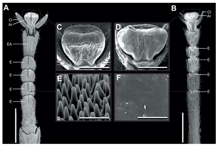
Figure 1.
Tarsal morphology. Scanning electron micrographs of the feet of Sungaya inexpectata (A,C,E) and Medauroidea extradentata (B,D,F). (A,B) Ventral overviews. (C,D) Arolia. (E,F) microstructure of the euplantulae. Ar, arolium; EA, accessory euplantula (5th euplantula); E, euplantula; Cl, claw. Scale bars: 1 mm (A,B), 300 µm (C,D), 3 µm (E,F). Figure from Büscher and Gorb 2019 [26] reproduced with permission.

