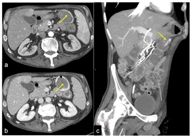
Figure 13.
Different radiologic pathways of bleeding. CTA multiphasic study in arterial (a) and venous (b) phases reveals intestinal bleeding with circular morphology (a,b, arrow) along the endoluminal wall of splenic flexure with enlargement and intensification in the venous phase. The MPR reconstruction of the arterial phase in the coronal-oblique plane (c) shows the whorled morphology of bleeding (arrow).

