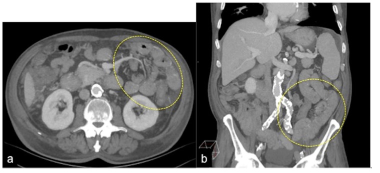
Figure 18.
Axial (a) and coronal (b) MIP reconstruction of the arterial phase in a patient of occult gastrointestinal bleeding. The MIP post-processing permits the intensification of the density of thin vascular structures in the wall (a,b, discontinuous circle) of the jejunum and proximal ileus as signs of small bowel telangectasia.

