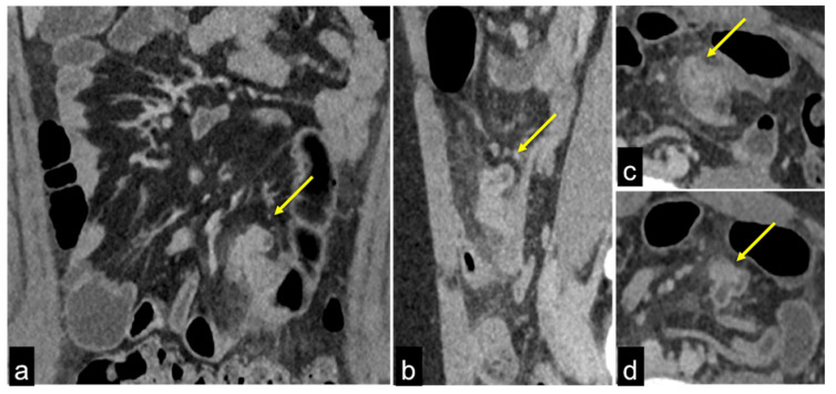
Figure 30.
Fifteen-year-old patient complaining of abdominal pain and anaemia. Enhanced CTA scan in portal venous phase in coronal (a), sagittal (b), axial (c,d) views. In the mesogastric region, there is a diverticular ileal structure (a–d, arrows) with increased wall enhancement at CTA venous phase, associated with adjacent fat stranding. Findings are consistent with Meckel diverticulitis.

