Abstract
Synthesis of thiazolidinone based on quinolone moiety was established starting from 4-hydroxyquinol-2-ones. The strategy started with the reaction of ethyl bromoacetate with 4-hydroxyquinoline to give the corresponding ethyl oxoquinolinyl acetates, which reacted with hydrazine hydrate to afford the hydrazide derivatives. Subsequently, hydrazides reacted with isothiocyanate derivatives to give the corresponding N,N-disubstituted thioureas. Finally, on subjecting the N,N-disubstituted thioureas with dialkyl acetylenedicarboxylates, cyclization occurred, and thiazolidinone derivatives were obtained in good yields. The two series based on quinolone moiety, one containing N,N-disubstituted thioureas and the other containing thiazolidinone functionalities, were screened for their in vitro urease inhibition properties using thiourea and acetohydroxamic acid as standard inhibitors. The inhibition values of the synthesized thioureas and thiazolidinones exhibited moderate to good inhibitory effects. The structure−activity relationship revealed that N-methyl quinolonyl moiety exhibited a superior effect, since it was proved to be the most potent inhibitor in the present series achieving (IC50 = 1.83 ± 0.79 µM). The previous compound exhibited relatively much greater activity, being approximately 12-fold more potent than thiourea and acetohydroxamic acid as references. Molecular docking analysis showed a good protein−ligand interaction profile against the urease target (PDBID: 4UBP), emphasizing the electronic and geometric effect of N,N-disubstituted thiourea.
Keywords: N,N-disubstituted thioureas; dialkyl acetylenedicarboxylates; thiazolidinones; in vitro urease inhibition properties; molecular docking
1. Introduction
Urease is a well-recognized enzyme that hydrolyzes urea to ammonia and carbon dioxide in living organisms. It is found in fungi, bacteria, plants, and vertebrates [1]. The amount of ammonia generated during hydrolysis tends to raise the pH, and an increase in medium pH is linked to the development of a variety of health problems in people who depend on colonization sites by urease-producing microorganisms [2,3]. It is known that the ureolytic activity of several microorganisms, e.g., Proteus mirabilis, is related to the formation of urinary tract stones, which can lead to chronic kidney and pelvic inflammation. In addition, urinary catheter obstruction in patients results in the colonization of urease-producing microorganisms, primarily P. mirabilis. Infectious bacteria that produce too much ammonia can cause ammonia encephalopathy or hepatic coma [4,5]. Another mechanism by which urease participates in pathogenic bacteria infection is the establishment of a microenvironment favorable to pathogen survival [6].
Due to the role of urease in such clinically significant complications, urease activity must be regulated using inhibitors [1,7]. Several classes of compounds have been identified as urease inhibitors [8]. Because of the association of ureases with several pathological conditions [9], the discovery of effective and safe urease inhibitors has been an important area of pharmaceutical research.
Thiosemicarbazide derivatives have been prominent precursors for synthesizing nitrogen and sulfur-containing heterocyclic compounds in recent decades due to their abundance of reactive centers [10]. In addition, thiosemicarbazones have also been evaluated as ribonucleotide reductase inhibitors and exhibit potential as anticancer drugs similar to methisazone and triapine [11,12,13,14,15]. These sulfur and nitrogen donor ligands and their coordination complexes have attracted significant attention due to their activity against the smallpox virus and protozoa influenza [16]. Many studies have recently been published on the efficacy of thiosemicarbazides and their hybrid derivatives in suppressing urease enzyme activity [17,18].
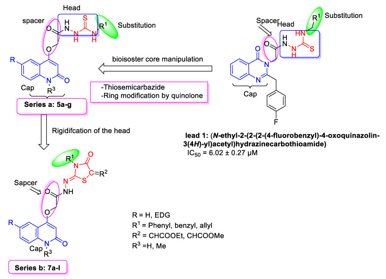
The current study is a part of our ongoing research into the synthesis of bioactive hybrid molecules, and a continuation of our previous work on the design of antibacterial urease inhibitors [19]. That previous work was involved the synthesis of 3-thiosemicarbazides derived by quinolin-2-one derivatives. The synthesized compounds were tested and screened in vitro against the urease-producing R. mucilaginosa and Proteus mirabilis strains. The results revealed that most of the tested compounds showed moderate-to-good activity [19]. Meanwhile, here we aim to synthesize another new series of quinolone-based 4-O-substituted-thiosemicarbazones and their 4-thiazolidinone derivatives, and explore them as antimicrobial and/or urease inhibitors (Figure 1).
Figure 1.
Rationale for designing the current compounds (series a and series b).
The intention to include quinolonyl moiety in our strategy was owing to its diverse range of biological properties, which include acetylcholinesterase inhibitor [20], antiallergenic [21], antimalarial [22], calcium-signaling inhibition [23] and antifungal [24] activities. Furthermore, quinolone hybrids have also been reported as potential candidates for antibacterial [25] and anticancer functions [26,27]. On the other hand, thiazolidinone ring has been linked to a variety of biological activities, including antibacterial [28], antitumor [29], antituberculous [30], and anti-inflammatory activities [31], and as potent urease inhibitors [32,33]. Furthermore, thiazolidinones are unique inhibitors of the bacterial enzyme MurB, a precursor involved in the biosynthesis of peptidoglycan as an essential component of both Gram-positive and Gram-negative bacterial cell wall [34,35,36].
2. Results and Discussion
2.1. Chemistry
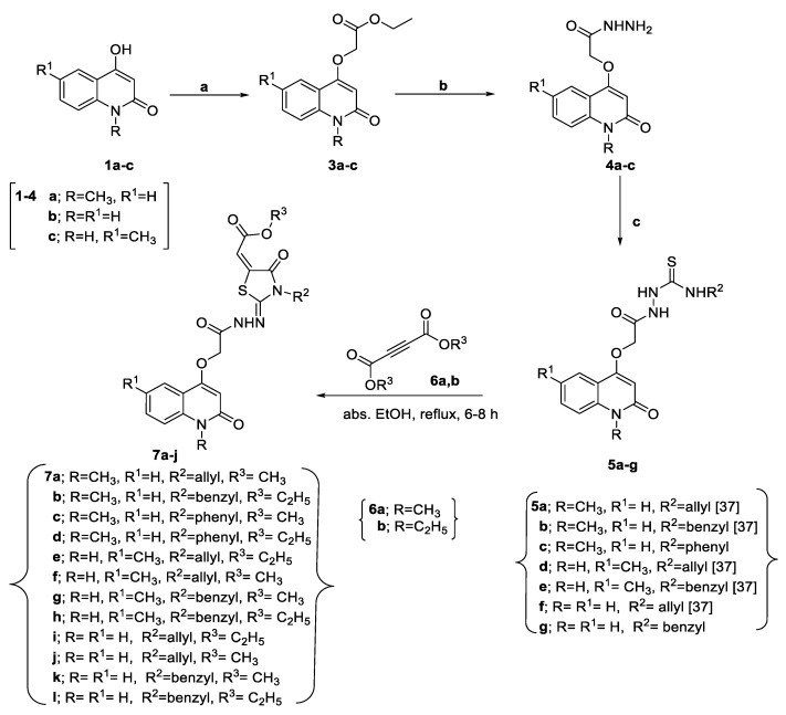
The reaction sequences for synthesizing 4-thiazolidinones-quinolone hybrids 7a–l starting from 4-hydroxyquinoline are outlined in Scheme 1. The synthesis of ethyl 2-((2-oxo-1,2-dihydroquinolin-4-yl)oxy)acetate derivatives 3a–c were obtained by refluxing ethyl bromoacetate (2) with 4-hydroxyquinoline 1a–c in dry acetone in the presence of anhydrous potassium carbonate. For the synthesis of new 4-oxothiazolidin-quinolone hybrids 7a–l, we planned to prepare the N,N-disubstituted thiourea derivatives 5a–g as precursors for functionalized 4-oxothiazolidine derivatives. To approach these targets, the reaction of compounds 4a–c and isothiocyanate derivatives in refluxing ethanol yielded the reported N,N-disubstituted thiourea derivatives 5a,b,d–f [37]. All the newly synthesized compounds gave satisfactory analyses for the proposed structures, which were confirmed based on their IR, NMR, mass spectra, and elemental analyses.
Scheme 1.
Synthesis of 4-oxothiazolidinquinolone 7a–l. Reagents and Conditions: (a) ethyl bromoacetate (2), anhydrous K2CO3, dry acetone, reflux 7–9 h; (b) hydrazine hydrate, EtOH, reflux 12–14 h; (c) isothiocyanate derivatives, EtOH, reflux, 4–6 h.
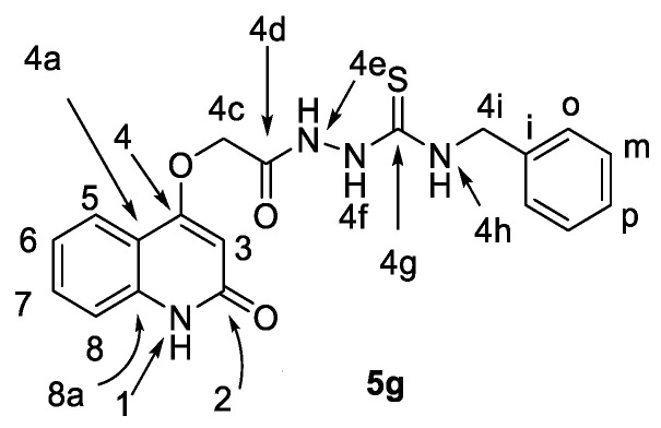
On the other hand, the structure of the newly prepared derivatives 5c and 5g were examined by elemental analyses, IR, and NMR in addition to mass spectra. For example, the 1H NMR spectrum of 5g showed a singlet at δN = 177.9 ppm assigned for N-4f. N-4f gives HMBC correlation with a singlet at δH 5.29 and 5.36 ppm, assigned as H-4c and benzylic H-4i, respectively. Additionally, distinctive are the benzylic (2C-m), (2C-o), and (C-4i) at δC = 128.48, 126.65, and 46.25 ppm, respectively. The distinctive carbons of 5g are shown in Figure 2; for full correlations, please see Figures S1–S19.
Figure 2.
Structure of compound 5g.
Interestingly, heterocyclization of 4-oxothiazolidine derivatives 7a–l was carried out when hydrazinecarbothioamides 5a–g were treated with dimethyl but-2-ynedioate (6a) and diethyl but-2-ynedioate (6b) in refluxing absolute ethanol for 6–8 h. The spectral and elemental data showed that series 7a–l underwent the reaction smoothly to give the respective 4-oxothiazolidin-quinolone hybrid structure. The 1H NMR spectra revealed the disappearance of two NH signals. Additionally, the appearance of a new signal at ~160–163 ppm in 13C NMR spectra for new carbonyl thiazolidine moiety augments the formation of the thiazolidinone hybrid. As a representative example, the 1H NMR spectrum of compound 7b (Table 1) showed quartet and triplet signals at δH = 4.27, 1.28 ppm for ethyl protons, as well as a singlet signal at δH = 6.91 ppm for (H-5a′); the benzylic protons (H-3a′) appeared as a singlet signal at δH = 4.69 ppm. Moreover, the 13C NMR spectrum (Table 1) showed signals at δC = 160.94 ppm representing additional carbonyl group C-4′, and another signal of thiazolidin-4-one at δC = 137.42 ppm is assigned as C-5′, which showed HMBC with δH = 6.91 and 4.69 ppm, assigned as H-5a′ and H-3a′, respectively. Further structural confirmation was also provided by analysis of the 1H-15N HSQC spectrum of 7b (Table 1 and Figures S20–S40), which showed a broad singlet at δH = 11.29 ppm, assigned as NH-4d, correlated with attached nitrogen, which appears at δN = 127.20 ppm.
Table 1.
NMR spectroscopic assignments of compound 7b.
| 1H NMR | COSY | Assignment | |
| 11.29 (s; 1H) | NH-4d | ||
| 8.10 (dd, J = 8.0, 1.0; 1H) | 7.68, 7.30 | H-5 | |
| 7.68 (ddd, J = 7.2, 7.2, 1.1; 1H) | 8.10, 7.53, 7.30 | H-7 | |
| 7.53 (d, J = 8.5; 1H) | 7.68, 7.30 | H-8 | |
| 7.30 (m; 6H) | 8.10, 7.68, 7.53 | H-o, m, p, 6 | |
| 6.91 (s; 1H) | H-5a′ | ||
| 6.12 (s; 1H) | H-3 | ||
| 5.03 (s; 2H) | H-4b | ||
| 4.69 (s; 2H) 4.27 (q, J = 7.1; 2H) 3.57 (s; 3H) 1.28 (t, J = 7.1; 3H) |
1.28 4.27 |
H-3a′ H-5c′ H-1a H-5d′ |
|
| 13C NMR | HSQC | HMBC | Assignment |
| 165.01 | 4.27 | C-5b′ | |
| 162.00 | 3.57 | C-2 | |
| 160.94 | 6.91 | C-4′ | |
| 160.00 | 8.10, 6.12, 5.03 | C-4 | |
| 145.89 | 4.69 | C-2′ | |
| 139.42 | 8.10, 7.68, 3.57 | C-8a | |
| 138.26 | 7.30 | C-i | |
| 137.42 | 6.91, 4.69 | C-5′ | |
| 131.57 | 7.68 | 8.10, 7.68, 7.53 | C-7 |
| 128.30 | 7.30 | 7.30, 7.30 | C-m |
| 127.40 | 7.30 | 7.30, 7.30, 4.69 | C-o |
| 126.91 | 7.30 | 7.30 | C-p |
| 123.24 | 8.10 | 7.68, 5.03 | C-5 |
| 121.43 | 7.30 | 7.53, 6.12 | C-6 |
| 116.97 | 6.91 | 6.91 | C-5a |
| 115.15 | 7.53 | C-4a | |
| 114.58 | 7.53 | 7.53, 7.30, 6.12, 3.57 | C-8 |
| 97.92 | 6.12 | C-3 | |
| 66.06 | 5.03 | 1.28 | C-4b |
| 61.63 | 4.27 | 7.30 | C-5c′ |
| 54.76 | 4.69 | 6.12 | C-3a′ |
| 28.67 | 3.57 | 3.57 | C-1a |
| 13.94 | 1.28 | 4.27, 1.28 | C-5d′ |
| 15N-NMR | HSQC | HMBC | Assignment |
| 137.9 127.2 |
11.29 |
7.53, 6.12, 3.57 |
N-1 N-4d |
2.2. Bioactivities
2.2.1. Antibacterial Screening
The Proteus mirabilis (P. mirabilis) strain was isolated from the urine of patients suffering from urinary tract infections. The strain was negative for hemolysis and motile and was urease positive. The test was performed using the cup-plate diffusion method [38] (Table 2). The results indicated that most tested compounds are weak or moderately active against P. mirabilis.
Table 2.
Urease inhibition IC50 (μM), anti-P. mirabilis, expressed as inhibition zone in mm and MICs (in μg/mL, given in brackets) of compounds 5a–g and 7a–l.
| Compd. | IC50 (µM) Urease | MIC (µM) P. mirabilis |
|---|---|---|
| 5a | 2.48 ± 0.18 | 22 (100) |
| 5b | 2.58 ± 0.10 | 20 (100) |
| 5c | 1.83 ± 0.79 | 27 (100) |
| 5d | 5.29 ± 0.36 | 15 (100) |
| 5e | 5.60 ± 0.84 | 9 (100) |
| 5f | 9.45 ± 0.08 | 12 (100) |
| 5g | 11.21 ± 0.27 | 10 (100) |
| 7a | 36.28 ± 0.10 | 9 (200) |
| 7b | 23.98 ± 0.88 | 9 (200) |
| 7c | 18.80 ± 1.72 | 8 (200) |
| 7d | 26.8 ± 1.11 | 6 (200) |
| 7e | 29.8 ± 1.31 | 11 (200) |
| 7f | 31.58 ± 0.15 | 10 (200) |
| 7g | 45.43 ± 0.19 | 7 (200) |
| 7h | 31.29 ± 0.36 | 9 (200) |
| 7i | 42.8 ± 1.51 | 11 (200) |
| 7j | 51.4 ± 0.91 | 6 (200) |
| 7k | 49.45 ± 0.09 | 8 (200) |
| 7l | 37.21 ± 0.28 | 9 (200) |
| lead I [21] | 6.02 ± 0.27 | |
| Thiourea | 22.8 ± 1.31 | |
| Acetohydroxamic acid | 21.03 ± 0.94 | |
| Ciprofloxacin | 8 (1.26) |
2.2.2. Urease Inhibition Activity
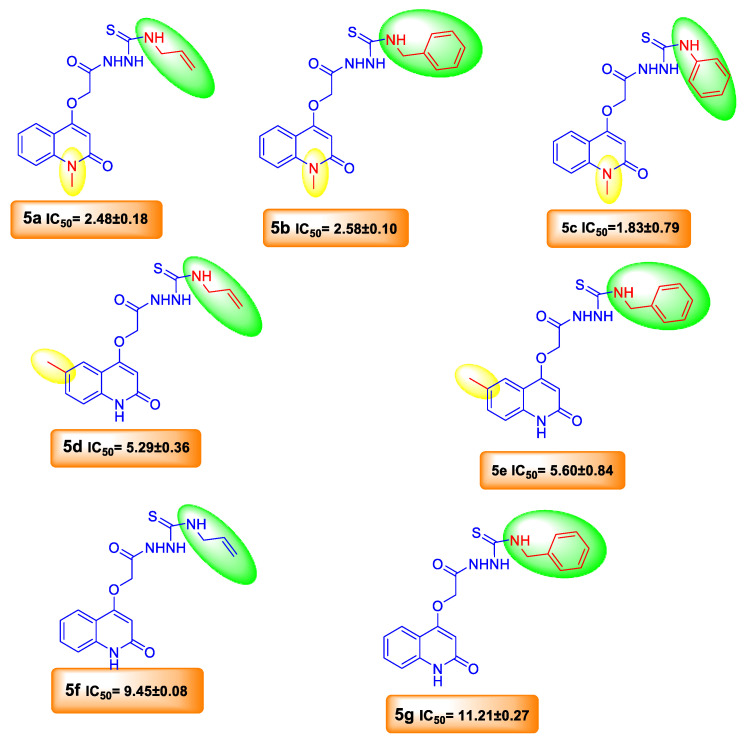
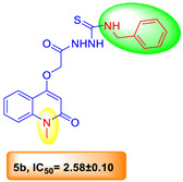
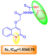
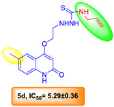
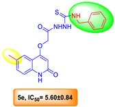
Two series of quinolone derivatives containing thiosemicarbazone and thiazole functionalities 5a–g and 7a–l were synthesized and then screened for their in vitro urease inhibition properties using thiourea and acetohydroxamic acid as standard inhibitors [39]. The inhibition values of the synthesized compounds 5a–g and 7a–l exhibited moderate to good inhibitory effects when compared to clinically used enzyme inhibitors, thiourea and acetohydroxamic acid (Table 2 and Figure 3). It is worth mentioning that quinolone-thiosemicarbazone hybrids 5a–g are effective molecules for urease inhibition ranking (IC50 = 1.83–11.21 µM) (Table 2 and Figure 3). Among the 5a–g series, compounds 5a–c, bearing a methyl group at position-1 on the quinolone skeleton, are the most active for urease inhibition (IC50 = 1.83–2.48 μM) when compared with the standard inhibitors thiourea and acetohydroxamic acid, which have IC50 values of 22.8 ± 1.31 and 21.03 ± 0.94 μM, respectively. Next, compounds 5d and 5e with p-methylquinolone moiety achieving IC50 = 5.29 ± 0.36 and 5.60 ± 0.84 μM were found to have superior activity compared to the standard inhibitors, thiourea and acetohydroxamic acid.
Figure 3.
Comparison of SAR of compounds 5a–g.
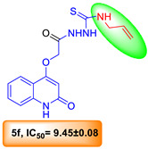
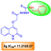
Meanwhile, 5f and 5g, with no substituents on quinolone moiety, showed the lowest activity (IC50 = 9.45 ± 0.08 and 11.21 ± 0.27 μM, respectively) compared to other members of the series, but were still more active than the standard inhibitors. It is clear that incorporating a methyl group at position-1 of the quinolone moiety enhances urease inhibitory activity. Overall, compound 5c bearing a methyl group at position-1 and thiosemicarbazide phenyl terminal was proved to be the most potent inhibitor in the present series, achieving (IC50 = 1.83 ± 0.79 uM), as it exhibited relatively much greater activity, being approximately 12-fold more potent than thiourea and acetohydroxamic acid as references. On the other hand, the quinolone-thiazole hybrids 7a–l were also screened in vitro for their human urease inhibitory potential. The results showed that most tested derivatives exhibited week to moderate urease inhibitory activities (IC50 = 18.80–45.43 µM).
Among the 7a–l series, compound 7c exhibited good activity compared with the standard inhibitors, thiourea, and acetohydroxamic acid, achieving IC50 = 18.80 ± 1.72 μM (Table 2). From the above discussion, it can be concluded that the positions of substituents at the quinolone moiety and the thiosemicarbazone chain play vital roles in urease inhibitory activity. However, in silico docking was performed, and is discussed in the following paragraphs to verify these interpretations.
2.3. Molecular Docking
A molecular docking study was performed to elucidate the in vitro activities of all synthesized compounds and to understand their binding with the protein and subsequently figure out the most important pharmacophoric features in this scaffold. The docking protocol was performed by the Openeye scientific program against bacillus pasteurii urease (PDB: ID: 4ubp) [40,41,42].
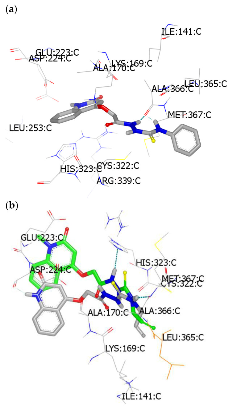
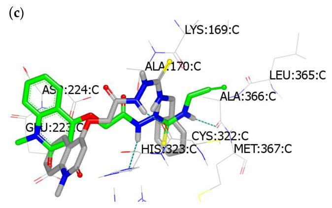
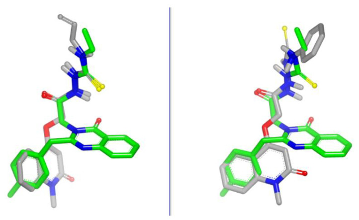
The compounds’ interaction and their binding mode and pose are illustrated in Table 3. By analyzing the compound pose, the methylation of NH of quinolinyl moiety was very important because it hindered the quinolonyl part from forming HB with the amino acid clefts. Compound 5c (N-phenylthiourea derivative) was the only compound whose thiourea functionality formed two HBs with the receptor in a chelation fashion (Figure 4a). To discover the great difference in activity between compounds 5a and 5d, overlay docking between these two ligands inside the receptor was performed. From Figure 4b, the quinolone ring in compound 5f formed extra HB with Asp: 224A and one HB with Ala: 366A. Comparing compound 5c with compound 5b (benzylthiourea), it is clear that both compounds adopted different poses and modes with the receptor. However, compounds 5a and 5b illustrated similar poses (Figure 4c). These results indicate the electronic and geometric effect of the N-substituted thiourea part.
Table 3.
Docking mode and consensus scores for compound 5a–g.
| Compound | Structure | Consensus Score | Binding Mode |
|---|---|---|---|
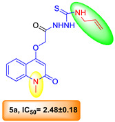
| 5a |
|
102 | Thiourea moiety forms HB Ala:366A; hydrazide forms HB with His:323A |
| 5b |
|
53 | Thiourea moiety forms HB Cys: 322A, |
| 5c |
|
65 | Thiourea moiety forms Two HBs with Ala:366A |
| 5d |
|
127 | Thiourea moiety forms HB with Leu: 365:A; NH of quinolinyl moiety forms HB with Asp:224A. |
| 5e |
|
109 | Thiourea moiety forms HB with Ala: 366A, NH of quinolinyl moiety forms HB with Asp: 224A. |
| 5f |
|
114 | Thiourea moiety forms HB with Ala: 366A, NH of quinolonyl moiety forms HB with Asp: 224A |
| 5g |
|
90 | Thiourea moiety forms HB with Asp:224A Leu:365:A; NH of quinolinyl moiety forms HB with Ala: 366A |
Figure 4.
Visualization of compounds docking with PDBID: 4uBP: (a) compound 5c; (b) compounds 5a and 5f; (c) compounds 5b and 5a.
The shape similarity for compounds 5a and 5c in comparison to lead 1 showed the quinolone ring overlaid with 4-F benzyl moiety (Figure 5).
Figure 5.
ROCS visualization of compound 5a (left, grey color) and compound 5c (right, grey color) overlaid with lead 1 (green color).
2.4. Structure–Activity Relationship (SAR)
Based on the compound activity and binding with the selected receptor, the series substituted thiourea has better activity than series b (thiazolidindione core). HB formation of the quinolonyl system is undesirable. Substitution on the phenyl of quinolone ring is ineffective. N-Phenylthiourea is the best derivative, and terminal substitution by alley or benzyl is very similar in terms of both activity and binding mode. These results open the gate for designing new derivatives bearing a substituted phenyl moiety. The latter is shown as in the case of compound 5c, since the incorporation of a methyl group at position-1 of the quinolone moiety and thiosemicarbazide phenyl terminal enhances the urease inhibitory activity. Thus, compound 5c exhibited relatively much greater activity, being approximately 12-fold more potent than thiourea and acetohydroxamic acid, as references. The same trend occurred in the case of thiazolidinone derivatives 7a–l, as the electronic effect of the methyl groups in the quinolinone molecule, together with the presence of phenyl group (N-phenylthiazolidine) in compound 7c (1,6-disubstituted-quinolinone-N-phenylthiazolidine), led the molecule to exhibit good activity compared with the standard inhibitors, thiourea, and acetohydroxamic acid.
3. Experimental Section
3.1. General Information
All reagents were purchased from Merck (St. Louis, MO, USA). The progress of all reactions was monitored with thin-layer chromatography (TLC) on Merck alumina-backed TLC plates and visualized under UV light. Spectra were measured in DMSO-d6 on a Bruker AV-400 spectrometer (400 MHz for 1H, 100 MHz for 13C, and 40.54 MHz for 15N, in the Chemistry Department, Florida Institute of Technology, 150 W University Blvd, Melbourne, FL 32901, USA. Chemical shifts are expressed in δ (ppm) versus internal tetramethylsilane (TMS) = 0 ppm for 1H and 13C, and external liquid ammonia = 0 ppm for 15N. Coupling constants are stated in Hz. Correlations were established using 1H-1H COSY, and 1H-13C and 1H-15N HSQC and HMBC experiments. All 15N signals were observed indirectly via HSQC or HMBC experiments. Chemical shifts (δ) are reported in parts per million (ppm) relative to tetramethylsilane (TMS) as the internal standard, and the coupling constants (J) are reported in Hertz (Hz). Splitting patterns are denoted as follows: singlet (s), doublet (d), multiplet (m), triplet (t), quartet (q), doublet of doublets (dd), doublet of triplets (dt), triplet of doublets (td), and doublet of quartet (dq). Melting points (mp’s) were determined with a Stuart melting point instrument in the Chemistry Department, Florida Institute of Technology, 150 W University Blvd, Melbourne, FL, USA, and are expressed in °C. Mass spectra were recorded on a Finnigan Fab 70 eV at Al-Azhar University, Egypt. Elemental analyses were carried out on a Perkin Elmer device at the Microanalytical Institute of Organic Chemistry, Karlsruhe Institute of Technology, Karlsruhe, Germany.
3.2. Starting Materials
4-Hydroxyquinoline derivatives 1a–c were prepared according to the literature [19,20]. Ethyl bromoacetate (2), isothiocyanate derivatives, dimethyl but-2-ynedioate (6a) and diethyl but-2-ynedioate (6b) were bought from Aldrich and used as received.
General method for the synthesis of compounds 3a–c [37]
General method for the synthesis of compounds 4a–c [37]
General method for the synthesis of compounds 5a–e
To a suspension of hydrazide derivatives 4a–c (1 mmol) in absolute ethanol (30 mL), the appropriate isothiocyanate derivatives (1 mmol) were added, and the mixture was heated at reflux on a boiling water bath for 4–6 h. The mixture was then left to cool, and the precipitate so formed was collected by filtration, washed with methanol, and recrystallized from ethanol to give the target compounds 5a–e.
N-Allyl-2-(2-((1-methyl-2-oxo-1,2-dihydroquinolin-4-yl)oxy)acetyl)hydrazinecarbo-thioamide (5a) [37]
Yield: 0.270 g (78%), m.p. 177–179 °C. IR (KBr) υmax/cm−1 3255, 3100.
N-Benzyl-2-(2-((1-methyl-2-oxo-1,2-dihydroquinolin-4-yl)oxy)acetyl)hydrazinecarbothioamide (5b) [37]
Yield: 0.281 g (71%), m.p. 190–192 °C. IR (KBr) υmax/cm−1 3249, 3150.
2-(2-((1-Methyl-2-oxo-1,2-dihydroquinolin-4-yl)oxy)acetyl)-N-phenylhydrazinecarbothioamide (5c)
Yield: 0.275 g (72%), m.p. 161–163 °C. IR (KBr) υmax/cm−1 3214, 3110. 1H NMR: 10.40 (bs, 1H; H-4e), 9.50 (bs, 1H; H-4f), 8.65 (b, 1H; H-4h), 8.11 (d, J = 7.9, 1H; H-5), 7.68 (dd, J = 7.7, 7.7, 1H; H-7), 7.53 (d, J = 8.5, 1H; H-8), 7.45 (dd, J = 8.0, 7.6, 2H; H-m), 7.30 (dd, J = 7.4, 7.4, 1H; H-6)), 7.25 (t, J = 7.4, 1H; H-p), 7.00 (dd, J = 8.5, 1.2, 2H; H-o), 6.04 (s, 1H; H-3), 4.86 (s, 2H; H-4c), 3.57 (s, 3H; H-1a). 13C NMR: 165.41 (C-4d), 162.05 (C-2), 160.29 (C-4), 147.67 (C-i), 139.41 (C-8), 138.60 (C-8a), 131.55 (C-7), 129.57 (2C-m), 125.41 (C-p), 123.30 (C-5), 121.45 (C-6), 120.74 (2C-o), 115.24 (C-8), 114.56 (C-4a), 97.54 (C-3), 66.20 (C-4c), 28.66 (C-1a). 15N NMR: 140.4 (N-1), 133.2 (N-4e/4f), 128.0 (N-4f/4e), 115.7 (N-4h). MS m/z (%): 382 (M+, 8), 257 (17), 132 (15), 65 (100). Anal. Calcd. for C19H18N4O3S (382.44): C, 59.67; H, 4.74; N, 14.65. Found: C, 59.82; H, 4.85; N, 14.83.
N-Allyl-2-(2-((6-methyl-2-oxo-1,2-dihydroquinolin-4-yl)oxy)acetyl)hydrazine-1-carbothioamide (5d) [37]
Yield: 0.290 g (80%), m.p. 174–176 °C. IR (KBr) υmax/cm−1 3289, 3165.
N-Benzyl-2-(2-((6-methyl-2-oxo-1,2-dihydroquinolin-4-yl)oxy)acetyl)hydrazinecarbothioamide (5e) [37]
Yield: 0.297 g (75%), m.p. 183–185 °C. IR (KBr) υmax/cm−1 3266, 3199.
N-Allyl-2-(2-((2-oxo-1,2-dihydroquinolin-4-yl)oxy)acetyl)hydrazinecarbothioamide (5f) [37]
Yield: 0.245 g (74%), m.p. 169–171 °C. IR (KBr) υmax/cm−1 3234, 3120.
N-Benzyl-2-(2-((2-oxo-1,2-dihydroquinolin-4-yl)oxy)acetyl)hydrazinecarbothioamide (5g)
Yield: 0.271 g (71%), m.p. 199–201 °C. IR (KBr) υmax/cm−1 3225, 3129. 1H NMR: 14.16 (s, 1H; H-4e/4f/4h), 11.39 (bs, 1H; H-1), 7.44 (dd, J = 7.6, 7.6, 1H; H-7), 7.22 (m, 4H; H-m, p, 8), 7.18 (d, J = 7.7, 2H; H- o), 6.96 (d, J = 7.9, 1H; H-5), 6.91 (dd, J = 7.5, 7.5, 1H; H-6), 6.03 (s, 1H; H-3), 5.36 (s, 2H; H-4i), 5.29 (s, 2H; H-4c). 13C NMR: 168.56 (C-4d), 162.83 (C-2), 161.00 (C-4), 147.36 (C-4g), 138.38 (C-8a), 135.50 (C-i), 130.90 (C-7), 128.48 (2C-m), 127.46 (C-p), 126.65 (2C-o), 122.21 (C-5), 121.16 (C-6), 114.87 (C-8), 113.84 (C-4a), 97.97 (C-3), 60.26 (C-4c), 46.25 (C-4i). 15N NMR: 282.4 (N-4e), 177.9 (N-4f), 144.2 (N-1). MS m/z (%): 382 (M+, 28), 280 (100), 188 (34), 47 (33). Anal. Calcd. for C19H18N4O3S (382.44): C, 59.67; H, 4.74; N, 14.65. Found: C, 59.85; H, 4.97; N, 14.83
General method for the synthesis of compounds 7a–l
To a solution of thiosemicarbazide 5a–g (1 mmol) in abs. ethanol (25 mL), DMAD (6a), and DEAD (6b) (0.17 gm, 1 mmol) were added, and the mixture was heated under reflux for 8–10 h. The mixture was then left to cool. The formed precipitate was filtered off, washed with hot ethanol, and recrystallized from methanol to give the target compounds 7a–l.
(2E)-Methyl 2-(3-allyl-2-(2-(2-((1-methyl-2-oxo-1,2-dihydroquinolin-4-yl)oxy)acetyl)-hydrazono)-4-oxothiazolidin-5-ylidene)acetate (7a)
Yield: 0.342 g (75%), m.p. 233–235 °C. IR (KBr) υmax/cm−1 1735, 1630, 1051. 1H NMR: 7.81 (d, J = 7.8, 1H; H-5), 7.66 (dd, J = 8.2, 7.4, 1H; H-7), 7.53 (d, J = 8.5, 1H; H-8), 7.26 (dd, J = 7.5, 7.5, 1H; H-6), 6.77 (s, 1H; H-5a′), 6.31 (s, 1H; H-3), 5.99 (ddt, Jd = 17.0, 10.2, Jt = 5.2, 1H; H-3b′), 5.52 (s, 2H; H-4b), 5.18 (d, J = 10.3, 1H; H-3c′), 5.00 (d, J = 17.4, 1H; H-3c′), 4.86 (d, J = 4.2, 2H; H-3a′), 3.78 (s, 3H; H-5c′), 3.57 (s, 3H; H-1a), 3.48 (s; NH-1). 13C NMR: 164.84 (C-5b′), 163.11 (C-4′), 162.00 (C-2), 159.84 (C-4), 151.49 (C-4c), 148.25 (C-5a′), 139.42 (C-8a), 132.11 (C-3b′), 131.57 (C-7), 122.78 (C-5a′), 122.56 (C-5), 121.60 (C-6), 117.80 (C-3c′), 115.10 (C-8), 114.74 (C-4a), 97.95 (C-3), 60.52 (C-4b), 52.37 (C-5c′), 46.69 (C-3a′), 28.67 (C-1a). 15N NMR: 329.1 (N-4e), 177.1 (N-4d), 137.6 (N-1). N-3′ n/o. MS m/z (%): 456 (M+, 38), 392 (62), 148 (38), 44 (100). Anal. Calcd. for C21H20N4O6S (456.47): C, 55.26; H, 4.42; N, 12.27. Found C, 55.47; H, 4.59; N, 12.58.
(2E)-Methyl 2-(3-allyl-2-(2-(2-((1-methyl-2-oxo-1,2-dihydroquinolin-4-yl)oxy)acetyl)-hydrazono)-4-oxothiazolidin-5-ylidene)acetate (7b)
Yield: 0.405 g (77%), m.p. 243–245 °C. IR (KBr) υmax/cm−1 1738, 1640, 1054. 1H NMR: 11.29 (s, 1H; NH-4d), 8.10 (dd, J = 8.0, 1.0, 1H; H-5), 7.68 (ddd, J = 7.2, 7.2, 1.1, 1H; H-7), 7.53 (d, J = 8.5, 1H; H-8), 7.30 (m, 6H; H-o, m, p, 6), 6.91 (s, 1H; H-5a′), 6.12 (s, 1H; H-3), 5.03 (s, 2H; H-4b), 4.69 (s, 2H; H-3a′), 4.27 (q, J = 7.1, 2H; H-5c′), 3.57 (s, 3H; H-1a), 1.28 (t, J = 7.1, 3H; H-5d′). 13C NMR: 165.61 (C-4c), 165.01 (C-5b′), 162.00 (C-2), 160.94 (C-4′), 160.00 (C-4), 145.89 (C-2′), 139.42 (C-8a), 138.26 (C-i), 137.42 (C-5′), 131.57 (C-7), 128.30 (2C-m), 127.40 (2C-o), 126.91 (C-p), 123.24 (C-5), 121.43 (C-6), 116.97 (C-5a′), 115.15 (C-4a), 114.58 (C-8), 97.92 (C-3), 66.06 (C-4b), 61.63 (C-5c′), 54.76 (C-3a′), 28.67 (C-1a), 13.94 (C-5d′). 15N NMR: 137.9 (N-1), 127.2 (N-4d). N-3′, 4e n/o. MS m/z (%): 520 (M+, 77), 236 (93), 200 (100), 40 (71). Anal. Calcd. for C26H24N4O6S (520.56): C, 59.99; H, 4.65; N, 10.76. Found C, 60.12; H, 4.82; N, 10.89.
(2E)-Methyl 2-(2-(2-(2-((1-methyl-2-oxo-1,2-dihydroquinolin-4-yl)oxy)acetyl)-hydrazono)-4-oxo-3-phenylthiazolidin-5-ylidene)acetate (7c)
Yield: 0.400 g (81%), m.p. 229–231 °C. IR (KBr) υmax/cm−1 1740, 1634, 1060. 1H NMR: 11.47 (b, 1H; NH-4d), 8.15 (d, J = 8.0, 1H; H-5), 7.69 (ddd, J = 8.5, 7.1, 1.4, 1H; H-7), 7.54 (d, J = 8.4, 1H; H-8), 7.45 (dd, J = 8.0, 7.6, 2H; H-m), 7.31 (dd, J = 7.8, 7.3, 1H; H-6), 7.25 (t, J = 7.4, 1H; H-p), 7.00 (dd, J = 8.3, 0.9, 2H; H-o), 6.97 (s, 1H; H-5a′), 6.11 (s, 1H; H-3), 5.09 (s, 2H; H-4b), 3.77 (s, 3H; H-5c′), 3.57 (s, 3H; H-1a). 13C NMR: 165.60 (C-4c), 165.06 (C-5b′), 162.00 (C-2), 160.92 (C-4′), 160.03 (C-4), 147.57 (C-i), 146.44 (C-2′), 139.43 (C-8a), 137.56 (C-5′), 131.60 (C-7), 129.58 (2C-m), 125.43 (C-p), 123.28 (C-5), 121.48 (C-6), 120.73 (2C-o), 117.65 (C-5a′), 115.15 (C-8), 114.61 (C-4a), 97.93 (C-3), 66.06 (C-4b), 52.71 (C-5c′), 28.68 (C-1a). 15N NMR: 137.7 (N-1). MS m/z (%): 492 (M+, 29), 204 (100), 145 (49), 40 (22). Anal. Calcd. for C24H20N4O6S (492.50): C, 58.53; H, 4.09; N, 11.38. Found C, 58.67; H, 4.27; N, 11.56.
(2E)-Ethyl 2-(2-(2-(2-((1-methyl-2-oxo-1,2-dihydroquinolin-4-yl)oxy)acetyl)-hydrazono)-4-oxo-3-phenylthiazolidin-5-ylidene)acetate (7d)
Yield: 0.399 g (78%), m.p. 223–225 °C. IR (KBr) υmax/cm−1 1736, 1639, 1077. 1H NMR: 11.47 (bs, 1H; NH-4d), 8.15 (dd, J = 8.0, 1.3, 1H; H-5), 7.68 (ddd, J = 8.5, 7.2, 1.4, 1H; H-7), 7.53 (d, J = 8.5, 1H; H-8), 7.45 (dd, J = 8.0, 7.6, 2H; H-m), 7.31 (ddd, J = 7.9, 7.3, 0.6, 1H; H-6), 7.25 (t, J = 7.4, 1H; H-p), 7.00 (dd, J = 8.5, 1.2, 2H; H-o), 6.94 (s, 1H; H-5a′), 6.11 (s, 1H; H-3), 5.09 (s, 2H; H-4b), 4.22 (q, J = 7.1, 2H; H-5c′), 3.57 (s, 3H; H-1a), 1.24 (t, J = 7.1, 3H; H-5d′). 13C NMR: 165.60 (C-4c), 165.06 (C-5b′), 162.00 (C-2), 160.92 (C-4′), 160.03 (C-4), 147.67 (C-i), 146.45 (C-2′), 139.42 (C-8a), 137.53 (C-5′), 131.60 (C-7), 129.57 (2C-m), 125.41 (C-p), 123.28 (C-5), 121.48 (C-6), 120.74 (2C-o), 117.65 (C-5a′), 115.15 (C-8), 114.60 (C-4a), 97.92 (C-3), 66.06 (C-4b), 61.71 (C-5c′), 28.68 (C-1a), 13.87 (C-5d′). 15N NMR: 265.2 (N-3′), 137.7 (N-1), 127.4 (N-4d). N-4e n/o. MS m/z (%): 506 (M+, 54), 316 (58), 181 (100), 45 (26). Anal. Calcd. for C25H22N4O6S (506.53): C, 59.28; H, 4.38; N, 11.06. Found C, 59.39; H, 4.62; N, 11.31.
(E)-Ethyl 2-((Z)-3-allyl-2-(2-(2-((6-methyl-2-oxo-1,2-dihydroquinolin-4-yl)oxy)acetyl)-hydrazono)-4-oxothiazolidin-5-ylidene)acetate (7e)
Yield: 0.375 g (79%), m.p. 239–241 °C. IR (KBr) υmax/cm−1 1744, 1640, 1074.1H NMR: 11.35 (b, 1H; NH-1), 11.13 (bs, 1H; NH-4d), 7.71 (s, 1H; H-5), 7.37 (d, J = 8.3, 1H; H-7), 7.20 (d, J = 8.4, 1H; H-8), 6.81 (s, 1H; H-5a′), 5.89 (ddt, Jd = 17.2, 9.9, Jt = 5.2, 1H; H-3b′), 5.83 (s, 1H; H-3), 5.19 (d, J = 10.4, 1H; H-3c′), 5.15 (d, J = 17.4, 1H; H-3c′), 4.88 (s, 2H; H-4b), 4.43 (m, 2H; H-3a′), 4.27 (q, J = 7.1, 2H; H-5c′), 2.37 (s, 3H; H-6a), 1.27 (t, J = 6.7, 3H; H-5d′). 13C NMR: 165.32 (C-5b′), 163.27 (C-4c, 4′), 162.86 (C-2), 161.62 (C-4), 151.5 (C-2′), 140.17 (C-5′), 136.66 (C-8a), 132.24 (C-7), 130.73 (C-3b′), 130.37 (C-6), 122.07 (C-5), 117.64 (C-3c′), 115.58 (C-5a′), 115.10 (C-8), 114.24 (C-4a), 97.56 (C-3), 66.15 (C-4b), 61.57 (C-5c′), 44.60 (C-3a′), 20.48 (C-6a), 13.96 (C-5d′). 15N NMR: 157.0 (N-4d), 143.8 (N-1). MS m/z (%): 470 (M+, 18), 338 (53), 106 (100), 40 (16). Anal. Calcd. for C22H22N4O6S (470.50): C, 56.16; H, 4.71; N, 11.91. Found C, 56.37; H, 4.89; N, 12.07.
(E)-Methyl 2-((Z)-3-allyl-2-(2-(2-((6-methyl-2-oxo-1,2-dihydroquinolin-4-yl)oxy)-acetyl)hydrazono)-4-oxothiazolidin-5-ylidene)acetate (7f)
Yield: 0.342 g (75%), m.p. 232–233 °C. IR (KBr) υmax/cm−1 1780, 1666, 1090. 1H NMR: 11.35 (s, 1H; NH-1), 11.15 (bs, 1H; NH-4d), 7.71 (s, 1H; H-5), 7.37 (d, J = 8.2, 1H; H-7), 7.20 (d, J = 8.3, 1H; H-8), 6.83 (s, 1H; H-5a′), 5.89 (ddt, Jd = 17.1, 10.2, Jt = 5.0, 1H; H-3b′), 5.83 (s, 1H; H-3), 5.19 (d, J = 11.2, 1H; H-3c′), 5.15 (d, J = 17.1, 1H; H-3c′), 4.88 (s, 2H; H-4b), 4.43 (m, 2H; H-3a′), 3.81 (s,3H; H-5c′), 2.37 (s, 3H; H-6a). 13C NMR: 165.78 (C-5b′), 163.28 (C-4c), 163.08 (C-4′), 162.86 (C-2), 161.62 (C-4), 151.28 (C-2′), 140.22 (C-5′), 136.66 (C-8a), 132.25 (C-7), 130.73 (C-3b′), 130.37 (C-6), 122.05 (C-5), 117.66 ( C-3c′), 115.32 (C-8), 115.10 (C-5a′), 114.22 (C-4a), 97.56 (C-3), 66.13 (C-4b), 52.63 (C-5c′), 44.64 (C-3a′), 20.48 (C-6a). 15N NMR: 157.0 (N-4d), 143.4 (N-1); N-4e, 3′ n/o. MS m/z (%): 456 (M+, 22), 360 (46), 216 (100), 43 (45). Anal. Calcd. for C21H20N4O6S (456.47): C, 55.26; H, 4.42; N, 12.27. Found C, 55.49; H, 4.60; N, 12.53.
(2E)-Methyl 2-(3-benzyl-2-(2-(2-((6-methyl-2-oxo-1,2-dihydroquinolin-4-yl)oxy)acetyl)-hydrazono)-4-oxothiazolidin-5-ylidene)acetate (7g)
Yield: 0.355 g (70%), m.p. 241–243 °C. IR (KBr) υmax/cm−1 1799, 1694, 1041. 1H NMR: 11.36 (s, 1H; NH-1), 11.25 (s, 1H; NH-4d), 7.77 (s, 1H; H-5), 7.31 (m, 6H; H-7, o, m, p), 7.23 (d, J = 8.3, 1H; H-8), 6.86 (s, 1H; H-5a′), 5.81 (s, 1H; H-3), 5.90 (s, 2H; H-3a′), 4.99 (s, 2H; H-4b), 3.81 (s, 3H; H-5c′), 2.34 (s, 3H; H-6a). 13C NMR: 165.6 (C-5b′), 165.4 (C-4c), 162.7 (C-4′) 161.1 (C-2), 160.9 (C-4), 145.8 (C-2′), 138.2 (C-5′), 137.5 (C-i), 136.6 (C-8a), 132.2 (C-7), 130.2 (C-6), 128.3 (5C-o, m, p), 126.9 (C-5), 122.0 (C-5a′), 116.7 (C-8), 114.1 (C-4a), 98.2 (C-3), 66.0 (C-4b), 54.7 (C-5c′), 52.6 (C-3a′), 20.47 (C-6a). MS m/z (%): 506 (M+, 22), 377 (33), 283 (100), 57 (20). Anal. Calcd. for C25H22N4O6S (506.53): C, 59.28; H, 4.38; N, 11.06. Found C, 59.64; H, 4.51; N, 11.34.
(E)-Ethyl 2-((Z)-3-benzyl-2-(2-(2-((6-methyl-2-oxo-1,2-dihydroquinolin-4-yl)oxy)acetyl)-hydrazono)-4-oxothiazolidin-5-ylidene)acetate (7h)
Yield: 0.390 g (75%), m.p. 233–235 °C. IR (KBr) υmax/cm−1 1756, 1660, 1083. 1H NMR: 11.35 (s, 1H; NH-1), 11.17 (s, 1H; NH-4d), 7.71 (s, 1H; H-5), 7.3 (m, 6H; H-7, o, m, p), 7.20 (d, J = 8.3, 1H; H-8), 6.83 (s, 1H; H-5a′), 5.83 (s, 1H; H-3), 5.01 (s, 2H; H-3a′), 4.88 (s, 2H; H-4b), 4.27 (q, J = 6.9, 2H; H-5c′), 2.37 (s, 3H; H-6a), 1.27 (t, J = 7.1, 3H; H-5d′). 13C NMR: 165.6 (C-5b′), 163.7 (C-4c), 163.3 (C-4′), 162.5 (C-2), 162.4 (C-4), 151.6 (C-2′), 137.7 (C-i), 137.41 (C-5′), 136.8 (C-8a), 132.7 (C-7), 130.7 (C-6), 128.6 (5C-o, m, p), 122.4 (C-5), 116.0 (C-5a′), 115.4 (C-8), 114.9 (C-4a), 98.0 (C-3), 66.4 (C-4b), 62.0 (C-5c′), 46.1 (C-3a′), 20.9 (C-6a), 14.4 (C-5d′). MS m/z (%): 520 (M+, 17), 428 (14), 91 (100), 40 (39). Anal. Calcd. for C26H24N4O6S (520.56): C, 59.99; H, 4.65; N, 10.76. Found C, 60.13; H, 4.81; N, 10.98.
(2E)-Ethyl 2-(3-allyl-4-oxo-2-(2-(2-((2-oxo-1,2-dihydroquinolin-4-yl)oxy)acetyl)hydra-zono)thiazolidin-5-ylidene)acetate (7i)
Yield: 0.360 g (78%), m..p. 210–212 °C. IR (KBr) υmax/cm−1 1741, 1639, 1041. 1H NMR: 11.44 (b, 1H), 11.15 (b, 1H; NH-1, 4d), 7.92 (d, J = 7.9, 1H; H-5), 7.54 (dd, J = 7.8, 7.5, 1H; H-7), 7.30 (d, J = 8.2, 1H; H-8), 7.20 (dd, J = 7.6, 7.5, 1H; H-6), 6.80 (s, 1H; H-5a′), 5.88 (ddt, Jd = 17.3, 10.3, Jt = 5.2, 1H; H-3b′), 5.85 (s, 1H; H-c), 5.28 (d, J = 17.2, 1H; H-3c′), 5.20 (m, 1H; H-3c′), 4.90 (s, 2H; H-4b), 4.43 (m, 2H; H-3a′), 4.27 (q, J = 7.1, 2H; H-5c′), 1.27 (t, J = 6.9, 3H; H-5d′). 13C NMR: 165.32 (C-5b′), 163.25 (C-4′), 163.08 (C-2′), 162.97 (C-2), 161.77 (C-4), 151.54 (C-4c), 140.12 (C-5′), 138.64 (C-8a), 131.09 (C-7), 130.72 (C-3b′), 122.63 (C-5), 121.34 (C-6), 117.63 (C-3c′), 115.57 (C-5a′), 115.14 (C-8), 114.34 (C-4a), 97.61 (C-3), 66.19 (C-4b), 61.57 (C-5c′), 44.61 (C-3a′), 13.97 (C-5d′). 15N NMR: 156.8, (N-1) 144.2 (N4d). N-3′, 4e n/o. MS m/z (%): 456 (M+, 19), 320 (30), 129 (100), 40 (13). Anal. Calcd. for C21H20N4O6S (456.47): C, 55.26; H, 4.42; N, 12.27. Found C, 55.43; H, 4.57; N, 12.45.
(2E)-Methyl 2-(3-allyl-4-oxo-2-(2-(2-((2-oxo-1,2-dihydroquinolin-4-yl)oxy)acetyl)-hydrazono)thiazolidin-5-ylidene)acetate (7j)
Yield: 0.330 g (73%), m.p. 244–246 °C. IR (KBr) υmax/cm−1 1794, 1680, 1084. 1H NMR: 11.43 (bs, 1H; NH-1), 11.21 (b, 1H; NH-4d), 7.92 (d, J = 7.9, 1H; H-5), 7.54 (dd, J = 7.7, 7.6, 1H; H-7), 7.30 (d, J = 8.2, 1H; H-8), 7.20 (dd, J = 7.6, 7.4, 1H; H-6), 6.83 (s, 1H; H-5a′), 5.90 (ddt, Jd = 16.7, 10.9, Jt = 5.4, 1H; H-3b′), 5.85 (s, 1H; H-3), 5.19 (d, J = 11.4, 1H; H-3c′), 5.17 (d, J = 16.7, 1H; H-3c′), 4.90 (s, 2H; H-4b), 4.44 (m, 2H; H-3a′), 3.81 (s, 3H; H-5c′). 13C NMR: 165.78 (C-5b′), 163.28 (C-2′, 4′), 162.98 (C-2), 161.78 (C-4), 151.34 (C-4c), 140.27 (C-5′), 138.64 (C-8a), 131.27 (C-7), 131.09 (C-3b′), 122.62 (C-5), 121.34 (C-6), 117.65 (C-3c′), 115.30 (C-8), 115.14 (C-5a′), 114.35 (C-4a), 97.61 (C-3), 66.23 (C-4b), 52.63 (C-5c′), 44.65 (C-3a′). 15N NMR: 144.2 (N-1); N-3′, N-4d, N-4e n/o. MS m/z (%): 442 (M+, 33), 360 (63), 283 (100), 41 (13). Anal. Calcd. for C20H18N4O6S (442.45): C, 54.29; H, 4.10; N, 12.66. Found C, 54.51; H, 4.28; N, 12.92
(2E)-Methyl 2-(3-benzyl-4-oxo-2-(2-(2-((2-oxo-1,2-dihydroquinolin-4-yl)oxy)acetyl)-hydrazono)thiazolidin-5-ylidene)acetate (7k)
Yield: 0.375 g (76%), m.p. 251–253 °C. IR (KBr) υmax/cm−1 1781, 1683, 1092. 1H NMR: 11.42 (bs, 1H; NH-1), 11.19 (b, 1H; NH-4d), 7.91 (d, J = 7.9, 1H; H-5), 7.44 (dd, J = 7.6, 7.5, 1H; H-7), 7.32 (m, 4H; H-o, m), 7.32 (m, 1H; H-p), 7.29 (d, J = 8.5, 1H; H-8), 7.20 (dd, J = 7.4, 6.8, 1H; H-6), 6.80 (s, 1H; H-5a′), 5.83 (s, 1H; H-3), 5.11 (s, 2H; H-4b), 4.95 (s, 2H; H-3a′), 4.30 (s, 3H; H-5c′). 13C NMR: 165.77 (C-5b′), 163.38 (C-4c), 162.98 (C-2), 161.45 (2C-4, 4′), 151.48 (C-2′), 138.64 (2C-8a, i), 135.35 (C-5′), 131.08 (C-7), 128.50 (C-p), 128.33 (2C-o),, 127.98 (2C-m), 127.73 (C-5), 127.42 (C-6), 121.34 (C-5a′), 115.14 (2C-8, 4a), 97.64 (C-3), 66.16 (C-3a′), 61.59 (C-5c′), 52.61 (C-4b). MS m/z (%): 492 (M+, 25), 375 (20), 343 (100), 147 (38). Anal. Calcd. for C24H20N4O6S (492.51): C, 58.53; H, 4.09; N, 11.38. Found C, 58.64; H, 4.28; N, 11.56.
(E)-Ethyl 2-((Z)-3-benzyl-4-oxo-2-(2-(2-((2-oxo-1,2-dihydroquinolin-4-yl)oxy)acetyl)-hydrazono)thiazolidin-5-ylidene)acetate (7l)
Yield: 0.370 g (72%), m.p. 249–251 °C. IR (KBr) υmax/cm−1 1790, 1689, 1088. 1H NMR: 11.44 (bs, 1H; NH-1), 11.22 (b, 1H; NH-4d), 7.92 (d, J = 7.9, 1H; H-5), 7.54 (dd, J = 7.6, 7.5, 1H; H-7), 7.39 (m, 4H; H-o, m), 7.34 (m, 1H; H-p), 7.30 (d, J = 8.5, 1H; H-8), 7.20 (dd, J = 7.4, 6.8, 1H; H-6), 6.82 (s, 1H; H-5a′), 5.86 (s, 1H; H-3), 5.01 (s, 2H; H-4b), 4.90 (s, 2H; H-3a′), 4.27 (q, J = 7.0, 2H; H-5c′), 1.27 (t, J = 7.0, 3H; H-5d′). 13C NMR: 165.31 (C-5b′), 163.38 (C-4c), 162.99 (C-2), 161.75 (2C-4, 4′), 151.48 (C-2′), 138.64 (2C-8a, i), 135.35 (C-5′), 131.09 (C-7), 128.49 (C-p), 127.96 (2C-o), 127.73 (2C-m), 122.64 (C-5), 121.34 (C-6), 115.82 (C-5a′), 115.14 (2C-8, 4a), 97.62 (C-3), 66.16 (C-3a′), 61.59 (C-5c′), 45.83 (C-4b), 13.96 (C-5d′). 15N NMR: 144.2 (N-1). MS m/z (%): 506 (M+, 57), 372 (37), 232 (45), 113 (100). Anal. Calcd. for C25H22N4O6S (506.53): C, 59.28; H, 4.38; N, 11.06. Found C, 59.45; H, 4.57; N, 11.32.
3.3. Biology
Urease Inhibitory Activity
In vitro screening and inhibitory studies on urease (Jack bean urease) were determined using the colored Berthelot phenols method, which measures the liberation of ammonia from the reaction [43]. Briefly, the assay mixture containing 1 unit of the enzyme was added to 650 µL of buffer solution (50 mmol phosphate buffer Ph 6.7, 400 mmol sodium salicylate, 10 mmol sodium nitroprusside, and 2 mmol EDTA/L) and mixed with 10 µL of different concentration 0.1–100 Mm of the tested compounds in DMF as a solvent. DMF was tested alone and showed no inhibitory effect on the enzyme. After 15 min incubation at room temperature, 10 µL of 50 mg/L urea solution was added. The mixture was incubated for 0.5 h in a water bath at 37 °C to allow the hydrolysis process.
After complete urea hydrolysis and ammonia liberation, the reaction was stopped by adding 200 µL of the hypochlorite reagent (150 mmol/L sodium hydroxide, 140 mm/L sodium hypochlorite). The liberated ammonia was allowed to complex with the hypochlorite and salicylate for 25 min at 30 °C. The absorbance was measured at 578 nm using a UV/VIS Spectrophotometer (Optizen POP, 5U4608, Daejeon, Korea), and experiments were performed in triplicate in a final volume of 1 mL. All results were compared with thiourea, a standard inhibitor of urease. The percentage inhibition was calculated as the difference of absorbance values with and without the test compounds. The concentration that provokes an inhibition halfway between the minimum and maximum response of each compound (relative IC50) was determined by monitoring the inhibition effect of various concentrations of compounds in the assay.
4. Molecular Docking Study
A docking study was performed for the target compounds using the Openeye scientific program (academic license 2021). The coordinate for the protein structure was obtained from the Protein Data Bank (PDB ID: 4ubp). The compound conformers were energy minimized using the Omega application. The docking step was operated by the Fred application, and the results were visualized by the Vida command.
5. Conclusions
Thiazolidinone derivatives were achieved starting from 4-hydroxyquinolin-2-ones, which were subjected to ethyl bromoacetate to afford the corresponding ethyl oxoquinolinyl acetates. The latter species reacted with hydrazine hydrate to afford the hydrazide derivatives. Then, the hydrazide derivatives reacted with isothiocyanate derivatives to give the corresponding N,N-disubstituted thioureas. On subjecting the N,N-disubstituted thioureas with dialkyl acetylenedicarboxylates, the thiazolidinone derivatives was obtained in good yields. The two series based on quinolone ring, one bearing N,N-substituted thiourea, and the other bear thiazolidinone ring were designed with different substituents at different positions. The N,N-disubstituted thiourea scaffold with N-methyl quinolone system exhibited the most potent urease inhibitor activity. Besides the study of the previous results dealing with the results of urease inhibition activity of 4-O-substituted-thiosemicarbazones and derived by quinolin-2-ones, it can be concluded that the quinolinone moiety plays an important role in the mechanism of urease inhibition process.
Acknowledgments
We acknowledge support from the KIT-Publication Fund of the Karlsruhe Institute of Technology. Stefan Bräse is grateful for support from the DFG-funded cluster program “3D Matter Made to Order” under Germany’s Excellence Strategy-2082/1-390761711. The NMR spectrometer at the Florida Institute of Technology was purchased with assistance from the U.S. National Science Foundation (CHE 03-42251).
Supplementary Materials
The following are available online at https://www.mdpi.com/article/10.3390/molecules27207126/s1, they are as: NMR spectra of compound 5g (Figures S1–S19), compound 7b (Figures S20–S39), compound 7a (Figures S40–S57), compound 7c (Figures S58–S75), compound 7d (Figures S76–S92), compound 7e (Figures S93–S116), compound 7f (Figures S117–S139), compound 7g (Figures S140–S159), compound 7h (Figures S160–S171), compound 7j (Figures S172–S189), compound 7k (Figures S190–S205) and compound 7l (Figures S206–S228).
Author Contributions
Y.A.M.M.E.: Writing, editing; A.A.A.: Conceptualization, Writing, editing; M.A.E.-A.: Revising; H.M.F.: Experimental; A.B.B.: Editing; S.B.: Writing, editing; M.R.: Writing, Editing. All authors have read and agreed to the published version of the manuscript.
Institutional Review Board Statement
Not applicable.
Informed Consent Statement
Not applicable.
Data Availability Statement
Not applicable.
Conflicts of Interest
The authors declare no conflict of interest.
Sample Availability
Not available.
Funding Statement
This research received no external funding.
Footnotes
Publisher’s Note: MDPI stays neutral with regard to jurisdictional claims in published maps and institutional affiliations.
References
- 1.Kosikowska P., Berlicki Ł. Urease inhibitors as potential drugs for gastric and urinary tract infections: A patent review. Expert Opin. Ther. Pat. 2011;21:945–957. doi: 10.1517/13543776.2011.574615. [DOI] [PubMed] [Google Scholar]
- 2.Mobley H., Hausinger R. Microbial ureases: Significance, regulation, and molecular characterization. Microbiol. Mol. Biol. Rev. 1989;53:85–108. doi: 10.1128/mr.53.1.85-108.1989. [DOI] [PMC free article] [PubMed] [Google Scholar]
- 3.Ali B., Khan K.M., Hussain S., Hussain S., Ashraf M., Riaz M., Wadood A., Perveen S. Synthetic nicotinic/isonicotinic thiosemicarbazides: In vitro urease inhibitory activities and molecular docking studies. Bioorg. Chem. 2018;79:34–45. doi: 10.1016/j.bioorg.2018.04.004. [DOI] [PubMed] [Google Scholar]
- 4.Maroney M.J., Ciurli S. Nonredox Nickel Enzymes. Chem. Rev. 2014;114:4206–4228. doi: 10.1021/cr4004488. [DOI] [PMC free article] [PubMed] [Google Scholar]
- 5.Coker C., Poore C.A., Li X., Mobley H.L. Pathogenesis of Proteus mirabilis urinary tract infection. Microbes Infect. 2000;2:1497–1505. doi: 10.1016/S1286-4579(00)01304-6. [DOI] [PubMed] [Google Scholar]
- 6.Preininger C., Wolfbeis O.S. Disposable cuvette test with integrated sensor layer for enzymatic determination of heavy metals. Biosens. Bioelectron. 1996;11:981–990. doi: 10.1016/0956-5663(96)87657-3. [DOI] [Google Scholar]
- 7.Beraldo H., Gambino D. The wide pharmacological versatility of semicarbazones, thiosemicarba-zones and their metal complexes. Mini Rev. Med. Chem. 2004;4:31–39. doi: 10.2174/1389557043487484. [DOI] [PubMed] [Google Scholar]
- 8.Kafarski P., Taka M. Recent advances in design of new urease inhibitors: A review. J. Adv. Res. 2018;13:101–112. doi: 10.1016/j.jare.2018.01.007. [DOI] [PMC free article] [PubMed] [Google Scholar]
- 9.Svane S., Sigurdarson J.J., Finkenwirth F., Eitinger T., Karring H. Inhibition of urease activity by different compounds provides insight into the modulation and association of bacterial nickel import and ureolysis. Sci. Rep. 2020;10:8503. doi: 10.1038/s41598-020-65107-9. [DOI] [PMC free article] [PubMed] [Google Scholar]
- 10.Hassan A.A., Shawky A.M. Thiosemicarbazides in heterocyclization. J. Heterocycl. Chem. 2011;48:495–516. doi: 10.1002/jhet.553. [DOI] [Google Scholar]
- 11.Kalinowski D.S., Yu Y., Sharpe P.C., Islam M., Liao Y.-T., Lovejoy D.B., Kumar N., Bernhardt P.V., Richardson D.R. Design, Synthesis, and Characterization of Novel Iron Chelators: Structure−Activity Relationships of the 2-Benzoylpyridine Thiosemicarbazone Series and Their 3-Nitrobenzoyl Analogues as Potent Antitumor Agents. J. Med. Chem. 2007;50:3716–3729. doi: 10.1021/jm070445z. [DOI] [PubMed] [Google Scholar]
- 12.Abid M., Azam A. Synthesis and antiamoebic activities of 1-N-substituted cyclised pyrazoline analogues of thiosemicarbazones. Biorg. Med. Chem. 2005;13:2213–2220. doi: 10.1016/j.bmc.2004.12.050. [DOI] [PubMed] [Google Scholar]
- 13.Hameed A., Khan K.M., Zehra S.T., Ahmed R., Shafiq Z., Bakht S.M., Yaqub M., Hussain M., de León A.D.L.V., Furtmann N. Synthesis, biological evaluation and molecular docking of N-phenyl thiosemicarbazones as urease inhibitors. Bioorg. Chem. 2015;61:51–57. doi: 10.1016/j.bioorg.2015.06.004. [DOI] [PubMed] [Google Scholar]
- 14.Hameed A., Yaqub M., Hussain M., Hameed A., Ashraf M., Asghar H., Naseer M.M., Mahmood K., Muddassar M., Tahir M.N. Coumarin-based thiosemicarbazones as potent urease inhibitors: Synthesis, solid state self-assembly and molecular docking. RSC Adv. 2016;6:63886–63894. doi: 10.1039/C6RA12827K. [DOI] [Google Scholar]
- 15.Shehzad M.T., Imran A., Njateng G.S.S., Hameed A., Islam M., Al-Rashida M., Uroos M., Asari A., Shafiq Z., Iqbal J. Benzoxazinone-thiosemicarbazones as antidiabetic leads via aldose reductase inhibition: Synthesis, biological screening and molecular docking study. Bioorg. Chem. 2019;87:857–866. doi: 10.1016/j.bioorg.2018.12.006. [DOI] [PubMed] [Google Scholar]
- 16.Refat M.S., El-Deen I.M., Zein M.A., Adam A.M.A., Kobeasy M.I. Spectroscopic, Structural and Electrical Conductivity Studies of Co(II), Ni(II) and Cu(II) Complexes derived from 4- Acetylpyridine with Thiosemicarbazide. Int. J. Electrochem. Sci. 2013;8:9894–9917. [Google Scholar]
- 17.Menteşe E., Emirik M., Sökmen B.B. Design, molecular docking and synthesis of novel 5,6-dichloro-2-methyl-1H-benzimidazole derivatives as potential urease enzyme inhibitors. Bioorg. Chem. 2019;86:151–158. doi: 10.1016/j.bioorg.2019.01.061. [DOI] [PubMed] [Google Scholar]
- 18.Menteşe E., Akyüz G., Emirik M., Baltaş N. Synthesis, in vitro urease inhibition and molecular docking studies of some novel quinazolin-4(3H)-one derivatives containing triazole, thiadiazole and thiosemicarbazide functionalities. Bioorg. Chem. 2019;83:289–296. doi: 10.1016/j.bioorg.2018.10.031. [DOI] [PubMed] [Google Scholar]
- 19.Elbastawesy M.A., El-Shaier Y.A., Ramadan M., Brown A.B., Aly A.A., Abuo-Rahma G.E.-D.A. Identification and molecular modeling of new quinolin-2-one thiosemicarbazide scaffold with antimicrobial urease inhibitory activity. Mol. Divers. 2021;25:13–27. doi: 10.1007/s11030-019-10021-0. [DOI] [PubMed] [Google Scholar]
- 20.Martínez-Grau A., Marco J. Friedländer reaction on 2-amino-3-cyano-4H-pyrans: Synthesis of derivatives of 4H-pyran [2,3-b] quinoline, new tacrine analogues. Bioorg. Med. Chem. Lett. 1997;7:3165–3170. doi: 10.1016/S0960-894X(97)10165-2. [DOI] [Google Scholar]
- 21.Kamperdick C., Van N.H., van Sung T., Adam G. Bisquinolinone alkaloids from Melicope ptelefolia. Phytochemistry. 1999;50:177–181. doi: 10.1016/S0031-9422(98)00500-7. [DOI] [Google Scholar]
- 22.Isaka M., Tanticharoen M., Kongsaeree P., Thebtaranonth Y. Structures of cordypyridones A-D, antimalarial N-hydroxy- and N-methoxy-2-pyridones from the insect pathogenic fungus Cordyceps nipponica. J. Org. Chem. 2001;66:4803–4808. doi: 10.1021/jo0100906. [DOI] [PubMed] [Google Scholar]
- 23.Koizumi F., Fukumitsu N., Zhao J., Chanklan R., Miyakawa T., Kawahara S., Iwamoto S., Suzuki M., Kakita S., Rahayu E.S. YCM1008A, a Novel Ca2-Signaling Inhibitor, Produced by Fusarium sp. YCM1008. J. Antibiot. 2007;60:455–458. doi: 10.1038/ja.2007.58. [DOI] [PubMed] [Google Scholar]
- 24.Othman E.S., Hassanin H.M., Mostafa M.A. Synthesis of Pyrano[3,2-c]quinoline-3-carboxaldehyde and 3-(Ethoxymethylene)-pyrano[3,2-c]quinolinone and Their Chemical Behavior toward Some Nitrogen and Carbon Nucleophiles. J. Heterocycl. Chem. 2019;56:1598–1604. doi: 10.1002/jhet.3539. [DOI] [Google Scholar]
- 25.Magesh C.J., Makesh S.V., Perumal P.T. Highly diastereoselective inverse electron demand (IED) Diels–Alder reaction mediated by chiral salen–AlCl complex: The first, target-oriented synthesis of pyranoquinolines as potential antibacterial agents. Bioorg. Med. Chem. Lett. 2004;14:2035–2040. doi: 10.1016/j.bmcl.2004.02.057. [DOI] [PubMed] [Google Scholar]
- 26.Fouda A.M. Halogenated 2-amino-4H-pyrano[3,2-h]quinoline-3-carbonitriles as antitumor agents and structure–activity relationships of the 4-, 6-, and 9-positions. Med. Chem. Res. 2017;26:302–313. doi: 10.1007/s00044-016-1747-z. [DOI] [Google Scholar]
- 27.El-Agrody A.M., Abd-Rabboh H.S., Al-Ghamdi A.M. Synthesis, antitumor activity, and structure–activity relationship of some 4H-pyrano[3,2-h]quinoline and 7H-pyrimido[4′,5′:6,5]pyrano[3,2-h]quinoline derivatives. Med. Chem. Res. 2013;22:1339–1355. doi: 10.1007/s00044-012-0142-7. [DOI] [Google Scholar]
- 28.Deep A., Kumar P., Narasimhan B., Ramasamy K., Mani V., Kumar P., Mishra R., Bakar Abdul Majeed A. Synthesis, antimicrobial, anticancer evaluation of 2-(aryl)-4- thiazolidinone derivatives and their QSAR studies. Curr. Top. Med. Chem. 2015;15:990–1002. doi: 10.2174/1568026615666150317221849. [DOI] [PubMed] [Google Scholar]
- 29.Szychowski K.A., Kaminskyy D.V., Leja M.L., Kryshchyshyn A.P., Lesyk R.B., Tobiasz J., Wnuk M., Pomiane K.T., Gmiński J. Anticancer properties of 5Z-(4-fluorobenzylidene)-2-(4-hydroxyphenyl-amino)-thiazol-4-one. Sci. Rep. 2019;9:1–16. doi: 10.1038/s41598-019-47177-6. [DOI] [PMC free article] [PubMed] [Google Scholar]
- 30.Kumar A.S., Kudva J., Bharath B., Ananda K., Sadashiva R., Kumar S.M., Revanasiddappa B., Kumar V., Rekha P., Naral D. Synthesis, structural, biological and in silico studies of new 5-arylidene-4-thiazolidinone derivatives as possible anticancer, antimicrobial and antitubercular agents. New J. Chem. 2019;43:1597–1610. doi: 10.1039/C8NJ03671C. [DOI] [Google Scholar]
- 31.Mahdi M.F., Raauf A., Kadhim F. Synthesis, characterization and preliminary pharmacological evaluation of new non-steroidal anti-inflammatory pyrazoline derivatives. J. Nat. Sci. Res. 2015;5:21–28. doi: 10.5155/eurjchem.6.4.461-467.1321. [DOI] [Google Scholar]
- 32.Rahim F., Zaman K., Ullah H., Taha M., Wadood A., Javed M.T., Rehman W., Ashraf M., Uddin R., Uddin I. Synthesis of 4-thiazolidinone analogs as potent in vitro anti-urease agents. Bioorg. Chem. 2015;63:123–131. doi: 10.1016/j.bioorg.2015.10.005. [DOI] [PubMed] [Google Scholar]
- 33.Khomami A., Rahimi M., Tabei A., Saniee P., Mahboubi A., Foroumadi A., Koopaei N.N., Almasirad A. Synthesis and Docking Study of Novel 4-Thiazolidinone Derivatives as Anti-Gram-positive and Anti-H. pylori Agents. Mini Rev. Med. Chem. 2019;19:239–249. doi: 10.2174/1389557518666181017142630. [DOI] [PubMed] [Google Scholar]
- 34.Bonde C.G., Gaikwad N.J. Synthesis and preliminary evaluation of some pyrazine containing thiazolines and thiazolidinones as antimicrobial agents. Biorg. Med. Chem. 2004;12:2151–2161. doi: 10.1016/j.bmc.2004.02.024. [DOI] [PubMed] [Google Scholar]
- 35.Aridoss G., Balasubramanian S., Parthiban P., Kabilan S. Synthesis, stereochemistry and antimicrobial evaluation of some N-morpholinoacetyl-2,6-diarylpiperidin-4-ones. Eur. J. Med. Chem. 2007;42:851–860. doi: 10.1016/j.ejmech.2006.12.005. [DOI] [PubMed] [Google Scholar]
- 36.El-Gaby M.S., Ali G.A.E.-H., El-Maghraby A.A., Abd El-Rahman M.T., Helal M.H. Synthesis, characterization and in vitro antimicrobial activity of novel 2-thioxo-4-thiazolidinones and 4, 4′-bis (2-thioxo-4-thiazolidinone-3-yl) diphenylsulfones. Eur. J. Med. Chem. 2009;44:4148–4152. doi: 10.1016/j.ejmech.2009.05.005. [DOI] [PubMed] [Google Scholar]
- 37.Aly A.A., Abd El-Aziz M., Elshaier Y.A.M.M., Brown A.A., Fathy H.M., Bräse S., Nieger M., Ramadan M. Regioselective formation of new 3-S-alkylated-1,2,4-triazole-quinolones. J. Sulfur Chem. 2021;43:215–231. doi: 10.1080/17415993.2021.2006659. [DOI] [Google Scholar]
- 38.Benini S., Rypniewski W.R., Wilson K.S., Miletti S., Ciurli S., Mangani S. The complex of Bacillus pasteurii urease with acetohydroxamate anion from X-ray data at 1.55 Å resolution. J. Biol. Inorg. Chem. 2000;5:110–118. doi: 10.1007/s007750050014. [DOI] [PubMed] [Google Scholar]
- 39.Weatherburn M.W. Phenol-hypochlorite reaction for determination of ammonia. Anal. Chem. 1967;39:971–974. doi: 10.1021/ac60252a045. [DOI] [Google Scholar]
- 40.OpenEye Scientific Software; Santa Fe, NM, USA: [(accessed on 10 May 2022)]. Fast Rigid Exhaustive Docking (FRED) Receptor, Version 2.2.5. Available online: http://www.eyesopen.com. [Google Scholar]
- 41.OpenEye Scientific Software; Santa Fe, NM, USA: [(accessed on 10 May 2022)]. OMEGA, Version 2.5.1.4. Available online: http://www.eyesopen.com. [Google Scholar]
- 42.OpenEye Scientific Software; Santa Fe, NM, USA: [(accessed on 10 May 2022)]. VIDA, Version 4.1.2. Available online: http://www.eyesopen.com. [Google Scholar]
- 43.Sharaf M., Arif M., Hamouda H.I., Khan S., Abdalla M., Shabana S., Rozan H.E., Khan T.U., Chi Z., Liu C. Preparation, urease inhibition mechanisms, and anti-Helicobacter pylori activities of hesperetin-7-rhamnoglucoside. Curr. Res. Microb. Sci. 2022;3:100103. doi: 10.1016/j.crmicr.2021.100103. [DOI] [PMC free article] [PubMed] [Google Scholar]
Associated Data
This section collects any data citations, data availability statements, or supplementary materials included in this article.
Supplementary Materials
Data Availability Statement
Not applicable.


