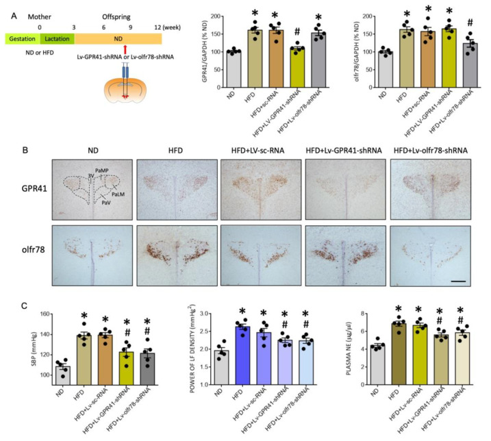
Figure 6.
Percentage change in the expression of GPR41 and olfr78 mRNA (A), representative photomicrographs of GPR41- or olfr78-immunoreactive cells (brown color) in the PVN (B), and SBP, plasma NE levels and power density of LF component of SBP signals (C) in adult ND or HFD offspring, alone or with bilateral microinjection into the PVN of lentiviral vectors encoding shRNA targeting GPR41 (Lv-GPR41-shRNA; 1 × 10⁵ IFU), olfr78 (Lv-olfr78-shRNA; 1 × 10⁵ IFU) or scramble shRNA (Lv-sc-RNA) control, performed at the age of 8 weeks. Also presented in (A) is the schematic diagram of the experimental design. Arrow indicates the age (8 weeks) when the lentiviral vector is microinjected into the RVLM. Data are presented as mean ± SD, n = 5 in each group. * p < 0.05 vs. ND group, and # p < 0.05 vs. HFD group in the post hoc Tukey’s multiple-range test. 3V, third ventricle; PaLM, paraventricular nucleus, lateral magnocellularis; PaMP, paraventricular nucleus median pavicellularis; PaV, paraventricular nucleus ventrolaris. Scale bar in (B): 500 μm.

