Abstract
Five domains of antigenic importance were previously mapped on the nucleocapsid protein (N) of the porcine reproductive and respiratory syndrome virus (PRRSV), and a domain comprised of the 11 C-terminal-most amino acids (residues 112 to 123) was shown to be essential for binding of N-specific conformation-dependent monoclonal antibodies (MAbs). In the present study, the importance of individual residues within this C-terminal domain for antigenicity was investigated using eight different mutant constructs of N expressed in HeLa cells. Single amino acid substitutions were introduced into the C-terminal domain of the N protein, and the significance of individual amino acids for MAb reactivity was determined by immunoprecipitation. None of the MAbs tested recognized the mutant with a leucine-to-proline substitution at residue 114 (L114P), while V112P, R113P, R113D, I115P, and R116P reduced MAb binding significantly. Conversely, substitution of amino acids at positions 118 (T118S) and 121 (P121A) had little effect on MAb binding. Secondary-structure predictions indicate that amino acids 111 to 117 form a beta-strand. In view of the fact that replacement of beta-strand-forming amino acids with proline elicited the greatest effect on MAb binding, it appears that secondary structure in the C terminus of the N protein is an important determinant of conformational epitope formation. While the crystal structure of the PRRSV N protein remains to be determined, results from these studies broaden our understanding of the secondary structures that make up the PRRSV N protein and shed some light on how they may relate to function.
Since its emergence in the late 1980s, the porcine reproductive and respiratory syndrome virus (PRRSV) has spread widely throughout pig-producing countries, imposing a considerable economic burden on the swine industry worldwide (1). Clinical signs associated with the syndrome vary greatly. In general, symptoms are more apparent in young pigs and are often associated with respiratory illness leading to secondary infections, while sows suffer primarily from reproductive problems (22).
PRRSV is a small, enveloped RNA virus belonging to the family Arteriviridae in the order Nidovirales (3). The PRRSV genome is a nonsegmented, plus-strand RNA molecule that is capped and polyadenylated (23). The full-length genomic sequence for both the North American (2, 18, 27) and the European (15) genotypes of PRRSV has been determined. The nonstructural proteins responsible for genome replication are encoded in two large open reading frames (ORFs 1a and 1b) that comprise the 5′-terminal two-thirds of the genome. The structural proteins are translated from a 3′-coterminal nested set of subgenomic mRNAs that are synthesized via a discontinuous mechanism of transcription (25). ORFs 2a, 2b (24), 3, and 4 (encoding GP2a, GP2b, GP3, and GP4) are thought to encode minor envelope proteins, ORFs 5 (encoding GP5) and 6 (encoding M) encode major envelope proteins, and ORF 7 encodes the nucleocapsid protein (N) (5).
The N protein is a highly abundant protein that experiences relatively little amino acid variability (11, 13). The early immunological response generated in PRRSV-infected pigs is directed mainly to the N protein, and this response, which can be detected as early as 1 week postinfection (12), declines at a much lower rate than that directed to the major structural proteins M and GP5 (30). Since the majority of antibodies produced during PRRSV infection in pigs are specific for the N protein, for which major antigenic determinants are highly conserved, the N protein has been targeted as a suitable candidate for the detection of virus-specific antibodies and diagnosis of the disease. Indeed, recombinant N protein expressed either in insect cells (9, 16) or in Escherichia coli (8) has been used as an antigen in the development of indirect and competitive enzyme-linked immunosorbent assays, respectively, for detection of serum antibodies against PRRSV. These methods are relatively inexpensive, sensitive, and easy to perform and therefore represent a feasible economic alternative to the present methods that rely upon whole-virus antigen (14). Therefore, knowledge of the antigenic makeup of the PRRSV N protein would be beneficial in the development of more effective detection methods.
In a previous study, a series of nine N protein deletion mutants expressed in HeLa cells and a collection of N-specific monoclonal antibodies (MAbs) were used to identify antigenically important domains on the PRRSV N protein (26). Of the five domains identified, the C-terminal-most domain appeared to be critical for correct folding of the N protein, as the mutant construct with a deletion of 11 amino acids from its C terminus (designated C-11) was unrecognizable by any of the conformation-dependent MAbs examined. In light of the fact that the majority of N-specific MAbs produced in mice and in pigs are conformational (7, 10, 19), we wanted to investigate the structural nature of the C-terminal domain of the N protein in greater detail by introducing specific mutations to assess the role of individual C-terminal amino acids in MAb binding.
MATERIALS AND METHODS
Cells, viruses, and antibodies.
HeLa cells were maintained at 37°C and 5% CO2 in Dulbecco's modified Eagle's medium supplemented with 10% heat-inactivated fetal bovine serum (FBS) (CanSera, Rexdale, Ontario, Canada). MARC-145 cells were maintained in Dulbecco's modified Eagle's medium supplemented with 4% heat-inactivated FBS. To prepare the recombinant vaccinia virus vTF7-3, HeLa cells were infected at a multiplicity of infection (MOI) of 1 PFU/cell. At approximately 36 h postinfection, concomitant with the appearance of severe cytopathic effects, cells were scraped into the media and subjected to two cycles of freeze-thawing to release virus particles. Cellular debris was removed by centrifugation at 1,500 × g for 10 min, and the clarified supernatant was used as crude virus stock. To prepare the PRRSV isolate PA8 (27), MARC-145 cells were infected with an MOI of 5 PFU/cell. At 2 days postinfection, cells and supernatant were collected and centrifuged for 10 min at 1,500 × g. The clarified supernatant was used as crude virus stock. E. coli strains XL1-Blue (Stratagene, La Jolla, Calif.) and DH5α were used as hosts for generating N gene mutations after PCR mutagenesis and for general-purpose cloning, respectively.
Generation of mouse ascites for the PRRSV-specific MAbs utilized in this study is described elsewhere (19, 20, 28, 29). Ascites fluid for MAbs 2D6, 1D2, 2G7, and 7C10 was kindly provided by D. Deregt at the Animal Disease Research Institute, Lethbridge, Alberta, Canada.
Construction of N gene mutants.
cDNA cloning of the N gene from PRRSV isolate PA8 to produce pGEM3zf-ORF7 was described previously (26). The N gene excised with BamHI from pGEM3zf-ORF7 was subcloned into the BamHI site of pCITE-2c (Novagen, Madison, Wis.) downstream of the T7 promoter producing pCITE-ORF7. This construct was used as the parental plasmid for the generation of N gene mutants by oligonucleotide-directed PCR mutagenesis according to the QuickChange site-directed mutagenesis protocol (Stratagene). The desired amino acid replacements were incorporated into PCR-amplified fragments using the primer pairs listed in Table 1. PCR amplifications were carried out using 15 ng of pCITE-ORF7 plasmid DNA; 300 ng of forward and reverse primer; 1 mM concentrations each of dCTP, dGTP, dATP, and dTTP; 1× PCR buffer [10 mM KCl, 10 mM (NH4)2SO4, 20 mM Tris-HCl (pH 8.8), 2 mM MgSO4, 0.1% Triton X-100]; and 2.5 U of Pfu DNA polymerase (Stratagene). The samples were subjected to 12 cycles of amplification under the following conditions: denaturation at 94°C for 30 s, primer annealing at 55°C for 1 min, and primer extension at 68°C for 8 min. Upon incorporation of the oligonucleotide primers, a mutated plasmid containing staggered nicks was generated. Following PCR cycling, the products were digested with DpnI to remove the methylated and hemimethylated parental plasmid DNA template. E. coli XL1-Blue cells were transformed by heat shock with 1 μl of the PCR-DpnI digest mixture containing the mutated plasmids. XL1-Blue competent cells were used because of their ability to repair nicks in the mutated plasmid in vivo. Random colonies were selected and plasmid DNA was prepared using a QIAprep spin miniprep kit (Qiagen, Santa Clarita, Calif.). The presence of desired mutations was verified by nucleotide sequencing in both directions. Plasmid DNA for transfection experiments was prepared using a plasmid midi kit (Qiagen) according to the manufacturer's recommended procedures.
TABLE 1.
Oligonucleotide sequences used to generate C-terminal point mutants
| Mutant | Primer pair | Primer sequencea | Locationb |
|---|---|---|---|
| V112P | V112P-Fwd | 5′-TACGCATCATACTccGCGCCTGATC-3′ | 321–345 |
| V112P-Rv | 5′-GATCAGGCGCggAGTATGATGCGTA-3′ | ||
| R113D | R113D-Fwd | 5′-ATCATACTGTGgaCCTGATCCGCGT-3′ | 326–350 |
| R113D-Rv | 5′-ACGCGGATCAGGtcCACATGATGAT-3′ | ||
| R113P | R113P-Fwd | 5′-TCATACTGTGCcaCTGATCCGCGT-3′ | 327–350 |
| R113P-Rv | 5′-ACGCGGATCAGtgGCACAGTATGA-3′ | ||
| L114P | L114P-Fwd | 5′-ATACTGTGCGCCctATCCGCGTCA-3′ | 329–352 |
| L114P-Rv | 5′-TGACGCGGATagGGCGCACAGTA-3′ | ||
| I115P | I115P-Fwd | 5′-TACTGTGCGCCTGcctCGCGTCAC-3′ | 330–353 |
| I115P-Rv | 5′-GTGACGCGaggCAGGCGCACAGTA-3′ | ||
| R116P | R116P-Fwd | 5′-TGCGCCTGATCCctGTCACAGCAT-3′ | 335–358 |
| R116P-Rv | 5′-TGCTGTGACagGGATCAGGCGCA-3′ | ||
| T118S | T118S-Fwd | 5′-TGATCCGCGTCtCAGCATCACCC-3′ | 341–363 |
| T118S-Rv | 5′-GGGTGATGCTGaGACGCGGATCA-3′ | ||
| P121A | P121A-Fwd | 5′-CACAGCATCAgCCTCAGCATGATG-3′ | 351–374 |
| P121A-Rv | 5′-CATCATGCTGAGGcTGATGCTGTG-3′ |
Lowercase letters represent mutated nucleotides.
Location (in nucleotides) of primer sequence relative to the N gene sequence
Protein expression and radiolabeling.
HeLa cells grown to 90% confluence in 100-mm-diameter dishes were infected at an MOI of 5 PFU/cell with vaccinia virus vTF7-3 and allowed to adsorb for 1 h at 37°C with occasional rocking. Five milliliters of fresh medium containing 10% FBS was added, and incubation continued for an additional 1 h at 37°C. Transfection solution consisting of 8 μg of plasmid DNA, 30 μl of LipofectACE (Gibco BRL, Burlington, Ontario, Canada), and 800 μl of OPTI-MEM (Gibco BRL) was incubated at room temperature for 30 min prior to overlay on cells. At 2 h postinfection, medium was removed and the transfection solution, diluted with 6.5 ml of OPTI-MEM, was added to the cells. The transfection solution was removed at 10 h postinfection, and cells were labeled for 16 h with 50 μCi of Easy Tag EXPRESS protein labeling mix ([35S]methionine and [35S]cysteine; specific activity, 407 MBq/ml; New England Nuclear, Boston, Mass.)/ml in methionine-free Eagle's minimal essential medium (Sigma, Oakville, Ontario, Canada) supplemented with 2% FBS. The cells were harvested, washed twice with cold phosphate-buffered saline, and resuspended in 600 μl of lysis buffer (0.1% Triton X-100, 0.5% NP-40, 10 mM Tris-HCl [pH 7.4]) per dish. After incubation for 10 min on ice, cell lysates were centrifuged at 14,000 rpm for 30 min in a microcentrifuge. The supernatant containing the cytoplasmic fraction was collected for immunoprecipitation experiments.
Immunoprecipitation and SDS-polyacrylamide gel electrophoresis analysis.
Thirty-microliter aliquots of labeled cell lysates (equivalent to approximately 105 cells) were adjusted with radioimmunoprecipitation assay (RIPA) buffer (1% Triton X-100, 1% sodium deoxycholate, 150 mM NaCl, 50 mM Tris-HCl [pH 7.4], 10 mM EDTA, 0.1% sodium dodecyl sulfate [SDS]) to a final volume of 100 μl and incubated for 2 h at room temperature with 1 μl of MAb ascites fluid. The immune complexes were adsorbed to 10 mg of protein A Sepharose CL-4B beads (Amersham Pharmacia Biotech, Baie d'Urfe, Quebec, Canada) for 16 h at 4°C in 800 μl of RIPA buffer containing a final concentration of 0.3% SDS. The precipitates, collected by microcentrifugation in an Eppendorf 5415C microcentrifuge at 6,000 rpm for 2 min, were washed twice with RIPA buffer and once with wash buffer (50 mM Tris-HCl [pH 7.4], 150 mM NaCl). Pellets were resuspended in 20 μl of SDS sample buffer (10 mM Tris-HCl [pH 6.8], 25% glycerol, 10% SDS, 10% β-mercaptoethanol, 0.12% [wt/vol] bromophenol blue) and heated for 5 min at 95°C. After microcentrifugation in an Eppendorf 5415C microcentrifuge at 10,000 rpm for 5 min, the supernatant was analyzed by SDS polyacrylamide gel electrophoresis on 12% polyacrylamide gels. The gels were dried and exposed to X-ray film at −70°C.
RESULTS
Construction and expression of mutant proteins.
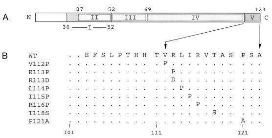
Secondary-structure predictions indicate that amino acids 111 to 117 of the N protein form a strong beta-strand, while the extreme C-terminal amino acids form a coil structure (6). To examine the contribution of the beta-strand to MAb binding, single amino acid substitutions were introduced to replace individual residues in the C-terminal domain of the PRRSV N protein. Valine at position 112 and isoleucine at position 115, both of which are strong beta-strand formers, were replaced with proline, a strong beta-strand breaker, to construct mutants V112P and I115P, respectively. Arginine at position 113 was replaced with aspartic acid (R113D), effectively reversing the charge and permitting the role of charge in MAb binding to be analyzed. Arginine at position 113, leucine at position 114, and arginine at position 116 were each replaced with proline in order to examine their effects on beta-strand structure and ultimately their involvement in MAb binding (R113P, L114P and R116P, respectively). Two additional amino acids downstream of the putative beta-strand were mutated to generate T118S and P121A. Threonine at position 118 was replaced with serine (T118S) to assess the effect of a relatively conservative change in this region, while proline at position 121 was replaced with alanine (P121A) to examine the significance of chain flexibility. N-gene mutants were generated by oligonucleotide-directed PCR mutagenesis using the primer pairs listed in Table 1 to alter amino acid codons. A total of eight mutants were constructed (Fig. 1), and the mutant proteins were expressed in HeLa cells using the T7-based vaccinia virus expression system.
FIG. 1.
(A) Illustration of five antigenically important domains localized on the PRRSV N protein. Boxes flanked by numbers indicate amino acid positions of the antigenic domains identified in the previous study (26). Domains: I, amino acids 30 to 52; II, 37 to 52; III, 52 to 69; IV, 69 to 112; V, 112 to 123. (B) Illustration of amino acid substitutions made in the C terminus of the PRRSV N protein. Substitutions are indicated at their respective positions below the wild-type (WT) sequence. Dots represent identical amino acids, and numbers indicate amino acid positions. The desired amino acid replacements were introduced into PCR-amplified fragments using the primer pairs listed in Table 1.
The specificity of MAbs was initially confirmed using the authentic viral N protein as well as the recombinant N protein expressed in HeLa cells, and the confirmed N-specific MAbs were subsequently tested for their C terminus dependency using the C-11 deletion construct (Table 2). A total of 10 MAbs (ISU15B, ISU15C, ISU15E, ISU15Fd, ISU15Fd-11, ISU15Fd-32, K9-3, K9-5, K9-9, and 2D6) were identified as C terminus dependent, and thus a total of 19 MAbs were used for this study, including the 9 MAbs previously characterized (SDOW17, VO17, EP147, SR30, MR40, JP25, 1D2, 2G7, and 7C10) (26).
TABLE 2.
MAb reactivities with C-terminal mutants of the PRRSV N protein
| MAb | Fig. 2 panel | Reactivitya with
|
Reference | ||||||||||
|---|---|---|---|---|---|---|---|---|---|---|---|---|---|
| PA8 Nb | Rec Nc | C-11d | V112P | R113P | R113D | L114P | I115P | R116P | T118S | P121A | |||
| ISU15A | A | + | + | + | + | + | + | + | + | + | + | + | 28 |
| ISU15B | B | + | + | − | − | − | + | − | − | − | + | + | 28 |
| ISU15C | C | + | + | − | − | − | − | − | − | + | + | − | 28 |
| ISU15E | D | + | + | − | − | − | − | − | − | − | + | + | 28 |
| K9-5 | D | + | + | − | − | − | − | − | − | − | + | + | 29 |
| MR40 | D | + | + | − | − | − | − | − | − | − | + | + | 20 |
| K9-3 | E | + | + | − | − | − | − | − | − | − | − | + | 29 |
| ISU15Fd-11 | F | + | + | − | − | − | − | − | + | − | + | + | 29 |
| ISU15Fd-32 | G | + | + | − | − | − | − | − | +/− | +/− | +/− | + | 29 |
| ISU15Fd | H | + | + | − | − | − | − | − | − | +/− | +/− | + | 29 |
| SDOW17 | I | + | + | − | − | − | +/− | − | +/− | +/− | +/− | + | 19 |
| K9-9 | I | + | + | − | − | − | +/− | − | +/− | +/− | +/− | + | 29 |
| SR30 | J | + | + | − | − | + | + | − | − | − | +/− | + | 20 |
| VO17 | K | + | + | − | + | + | + | − | − | − | + | + | 19 |
| JP25 | L | + | + | − | − | + | + | − | − | − | + | + | 20 |
| EP147 | M | + | + | − | − | − | − | − | − | − | + | − | 19 |
| 1D2 | M | + | + | − | − | − | − | − | − | − | + | − | D. Deregte |
| 7C10 | M | + | + | − | − | − | − | − | − | − | + | − | D. Deregt |
| 2D6 | N | + | + | − | + | − | − | − | − | − | + | +/− | D. Deregt |
| 2G7 | N | + | + | − | + | − | − | − | − | − | + | +/− | D. Deregt |
+, positive (N protein detected after 48 h of exposure); +/−, weakly positive (N protein detected after 1 week of exposure); −, negative (N protein undetectable after 1 week of exposure).
Authentic N protein from PRRSV-infected cell lysates.
Recombinant N protein expressed from vaccinia virus.
N protein with a deletion of the 11 C-terminal-most amino acids.
Animal Disease Research Institute.
Immunoreactivitiy of the beta-strand mutants with C-terminus dependent MAbs.
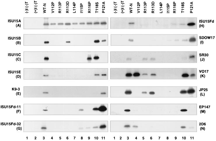
The immunoreactivities of individual MAbs with the beta-strand mutant proteins were determined by immunoprecipitation to assess the interaction of antibody with antigen expressed in its natural environment under the normal physiological conditions of the eukaryotic cell. Expression of the mutant proteins was first confirmed by immunoprecipitation with the C-terminus independent MAb ISU15A (Table 2). MAb ISU15A detected all eight mutant proteins with equal affinity, indicating that the mutants were efficiently expressed in this system and that the levels of protein expression were comparable (Fig. 2A). Experiments were repeated a minimum of two times to validate the results. Antibody binding was scored as follows: an interaction was considered positive if the N protein was detectable after 48 h of exposure, weakly positive if the N protein was detectable after 1 week of exposure, and negative if the N protein was undetectable even after 1 week of exposure. Thirteen different binding patterns were discerned from these immunoprecipitation studies. MAb ISU15B displayed a unique reactivity profile with the mutant proteins since replacement of arginine at position 113 with proline (R113P) abolished MAb binding (Fig. 2B, lane 5) while reversal of the amino acid charge from positive to negative by replacing arginine with aspartic acid (R113D) had no effect (Fig. 2B, lane 6). In almost every other case, with the exception of a very weak signal for MAbs SDOW17 and K9-9 (Fig. 2I, lane 6), replacement of arginine at position 113 with proline or aspartic acid affected MAb binding in a similar manner. MAbs ISU15C (Fig. 2C, lane 11), EP147, 7C10, and 1D2 (Fig. 2M, lane 11) were the only MAbs that did not detect the P121A mutant. MAbs ISU15E, K9-5, and MR40 (Fig. 2D) appear to be particularly sensitive to mutations in the putative beta-strand domain, as only the T118S and P121A mutants were detected. MAb K9-3 was unusual in that it was the only MAb unable to detect the T118S mutant (Fig. 2E, lane 10). ISU15Fd-11 was the only MAb capable of detecting the I115P mutant with high affinity (Fig. 2F, lane 8); however, a weak signal was seen for MAbs ISU15Fd-32, SDOW17, and K9-9 (Fig. 2G and I, lanes 8). Finally, VO17 (Fig. 2K, lane 4), 2D6, and 2G7 (Fig. 2N, lane 4) were the only MAbs able to detect the V112P mutant. Occasionally, point mutations reduced, but did not completely abolish, MAb binding, and these were scored as weakly positive (Fig. 2G, lanes 8 through 10; 2H, lanes 9 and 10; 2I, lanes 6, 8, 9, and 10; 2J, lane 10, and 2N, lane 11). However, in a few cases, particularly with ISU15Fd-11, ISU15Fd, SDOW17, and SR30, point mutations actually enhanced binding affinity (Fig. 2F, H, I, and J, respectively, lanes 11). In the case of enhanced binding, the P121A mutant appeared to be favored. Since the MAbs displayed variable affinity for the PA8 N protein, scores were determined relative to the individual MAb's affinity for the wild-type N protein. The immunoreactivity scores are summarized in Table 2. Interestingly, none of the MAbs tested were able to recognize the L114P mutant and very few detected the neighboring mutants I115P and R116P. Conversely, mutation of T118S and P121A had very little negative effect on MAb binding. Sixty-eight percent (13 out of 19) of the MAbs tested displayed a unique reactivity profile with the mutants, suggesting that while all of the MAbs examined were C terminus dependent, they differ with respect to their epitope specificities.
FIG. 2.
Immunoprecipitation of N protein point mutants. (A) ISU15A; (B) ISU15B; (C) ISU15C; (D) ISU15E (representative of MAbs K9-5 and MR40); (E) K9-3; (F) ISU15Fd-11; (G) ISU15Fd-32; (H) ISU15Fd; (I) SDOW17 (representative of K9-9); (J) SR30; (K) VO17; (L) JP25; (M) EP147 (representative of 1D2 and 7C10); (N) 2D6 (representative of 2G7), [I(−)T(−)], uninfected and untransfected; [I(+)T(−)], vTF7-3 infected and untransfected; [I(+)T(+)], vTF7-3 infected and pCITE-2c transfected.
DISCUSSION
The present study was conducted in order to characterize the involvement of the 11 C-terminal-most amino acids in conformation-dependent MAb binding and, in doing so, contribute to our understanding of the antigenic structure of the PRRSV N protein. The importance of the C terminus was highlighted in a previous study, where deletion of as little as 11 amino acids from the C terminus of the PRRSV N protein (North American isolate PA8) abolished binding of conformation-dependent MAbs (26). The C terminus has also been recognized as being an important determinant of discontinuous epitope formation in the N protein of the European lineage of PRRSV. In this case, deletion of either 23 or 31 amino acids from the C terminus of N proteins derived from European PRRSV isolates destroyed binding of conformation-dependent MAbs (17, 21). These findings support the notion that the C terminus of the N protein is critical for the generation of discontinuous epitopes. Moreover, C-terminus-dependent epitopes appear to be significant in vivo, as conformation-dependent MAbs were the only MAbs able to efficiently compete with PRRSV-positive porcine sera in a competitive enzyme-linked immunosorbent assay (21). Therefore, C-terminus-dependent epitopes may be among the most dominant of the PRRSV N protein, at least in the natural host.
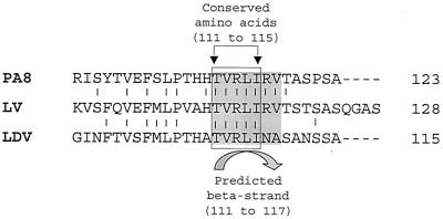
A structural role for the C terminus was alluded to in a recent report aimed at identifying antigenic determinants on the Lelystad virus (LV) N protein. In this case, to circumvent the structural changes associated with deleting C-terminal amino acids, a chimera between the N protein of LV and lactate dehydrogenase elevating virus (LDV), another member of the family Arteriviridae, was constructed (17). In the chimeric protein, C-terminal amino acids 111 to 128 of the LV N protein (the European N protein is 128 amino acids long, whereas the North American N protein is 123 amino acids) were replaced by the corresponding amino acids of the LDV N protein. Of those substituted amino acids, only residues which make up the putative beta-strand domain are identical to the LV N protein sequence (Fig. 3). Interestingly, amino acids 110 to 118 (North American numbering scheme) comprise one of the four invariable regions common to the N protein of both North American and European genotypes of PRRSV (11). Replacement with LDV C-terminal amino acids was able to restore conformation-dependent MAb binding. Given that only those C-terminal amino acids that make up the putative beta-strand are identical between the LV and LDV N proteins, this result suggests that this region of the C terminus plays an important role in determining N protein conformation. These observations correlate well with our results in which the most dramatic effect on C-terminus dependent MAb binding was observed when amino acids comprising the beta-strand were mutated. Therefore, it is likely that these extreme C-terminal amino acids have been conserved due to their requirement for nucleocapsid function and/or structure, thus substantiating the importance of this region of the N protein.
FIG. 3.
Illustration of the sequence conservation in the C terminus of N proteins from PA8 virus, LV, and LDV. Vertical lines designate amino acids common to all three viruses. The box defines the stretch of absolutely conserved amino acids, while the shaded area inside highlights amino acids comprising the predicted beta-strand. Amino acid position numbers in descriptions above and below the diagram apply to the PA8 sequence only. GenBank accession numbers: PA8, AF066068; LV, U03040; LDV, AF092283.
We found that C-terminal amino acids comprising the beta-strand exert the greatest influence on N protein structure, as determined by binding of C-terminus-dependent MAbs to mutant proteins where in which C-terminal amino acids were individually mutated. Although actual epitopes are still unknown for the majority of MAbs examined in this study, information described by Yang et al. (28) facilitates interpretation of ISU15B and ISU15C binding activities with the C-terminal point mutants. In their studies using 70 different PRRSV isolates, single amino acid substitutions responsible for abolishing binding of MAbs ISU15B and ISU15C were found in the C terminus. Substitution of what is typically a serine at positions 120 and 122 for proline was shown to correlate with the destruction of ISU15B and ISU15C specific epitopes, respectively. Furthermore, these amino acid substitutions correlate to changes in the secondary structure of the C terminus, as predicted by Chou and Fasman (4). In our studies, MAbs ISU15B and ISU15C both displayed C terminus dependency. We were able to mutate threonine at position 118 and proline at position 121 on either side of serine 120, the amino acid found to be necessary for ISU15B epitope formation, without disrupting ISU15B binding (Table 2). Therefore, serine at position 120 may be a critical residue for epitope formation. Interestingly, while the LV N protein amino acid sequence is only 63% identical to that of North American strains, ISU15B is able to detect the LV N protein and sequence analysis indicates that the LV N protein possesses a serine at the equivalent to position 120. For MAb ISU15C, mutation of proline at position 121 adjacent to serine 122, the residue shown to be essential for ISU15C epitope formation, abolishes MAb binding while mutation of threonine at position 118 does not, suggesting that the extreme C-terminal amino acids may participate in ISU15C-specific epitope formation. The combination of results from these and previous mutational studies of the PRRSV N protein suggests that the requirement of the C terminus for conformation-dependent MAb binding correlates with the proper formation of the predicted beta-sheet structure formed by amino acids 111 to 117. Crystallographic studies will provide a better understanding of the structure-function relationship of the N protein, and such studies are currently in progress.
ACKNOWLEDGMENTS
This work was supported by the Ontario Ministry of Agriculture, Food, and Rural Affairs (OMAFRA), Ontario Pork, and Vetrepharm Animal Health.
REFERENCES
- 1.Albina E. Epidemiology of porcine reproductive and respiratory syndrome virus (PRRS): an overview. Vet Microbiol. 1997;55:309–316. doi: 10.1016/s0378-1135(96)01322-3. [DOI] [PubMed] [Google Scholar]
- 2.Allende R, Lewis T L, Lu Z, Rock D L, Kutish G F, Ali A, Doster A R, Osorio F A. North American and European porcine reproductive and respiratory syndrome viruses differ in non-structural protein coding regions. J Gen Virol. 1999;80:307–315. doi: 10.1099/0022-1317-80-2-307. [DOI] [PubMed] [Google Scholar]
- 3.Cavanagh D. Nidovirales: a new order comprising Coronaviridae and Arteriviridae. Arch Virol. 1997;142:629–633. [PubMed] [Google Scholar]
- 4.Chou P Y, Fasman G D. Beta-turns in proteins. J Mol Biol. 1977;115:135–175. doi: 10.1016/0022-2836(77)90094-8. [DOI] [PubMed] [Google Scholar]
- 5.Conzelmann K, Visser N, van Woensel P, Thiel H. Molecular characterization of porcine reproductive and respiratory syndrome virus, a member of the arterivirus group. Virology. 1993;193:329–229. doi: 10.1006/viro.1993.1129. [DOI] [PMC free article] [PubMed] [Google Scholar]
- 6.Cuff J A, Clamp M E, Siddiqui A S, Finlay M, Barton G, Jpred J. A consensus secondary structure prediction server. Bioinformatics. 1998;14:892–893. doi: 10.1093/bioinformatics/14.10.892. [DOI] [PubMed] [Google Scholar]
- 7.Dea S, Gagnon C A, Mardassi H, Milane G. Antigenic variability among North American and European strains of porcine reproductive and respiratory syndrome virus as defined by monoclonal antibodies to the matrix protein. J Clin Microbiol. 1995;34:1488–1493. doi: 10.1128/jcm.34.6.1488-1493.1996. [DOI] [PMC free article] [PubMed] [Google Scholar]
- 8.Dea S, Wilson L, Therrien D, Cornaglia E. Competitive ELISA for detection of porcine reproductive and respiratory syndrome virus using recombinant E. coli-expressed nucleocapsid protein as antigen. J Virol Methods. 2000;87:109–122. doi: 10.1016/s0166-0934(00)00158-0. [DOI] [PubMed] [Google Scholar]
- 9.Denac H, Moser C, Tratschin J D, Hofmann M A. An indirect ELISA for the detection of antibodies against porcine reproductive and respiratory syndrome virus using recombinant nucleocapsid protein as antigen. J Virol Methods. 1997;65:169–181. doi: 10.1016/s0166-0934(97)02186-1. [DOI] [PubMed] [Google Scholar]
- 10.Drew T W, Meulenberg J J M, Sands J J, Paton D. Production, characterization and reactivity of monoclonal antibodies to the porcine reproductive and respiratory syndrome virus. J Gen Virol. 1995;76:1361–1369. doi: 10.1099/0022-1317-76-6-1361. [DOI] [PubMed] [Google Scholar]
- 11.Le Gall A, Legeay O, Bourhy H, Arnauld C, Albina E, Jestin A. Molecular variation in the nucleocapsid protein gene (ORF7) of the porcine reproductive and respiratory syndrome virus (PRRSV) Virus Res. 1998;54:9–21. doi: 10.1016/s0168-1702(97)00146-9. [DOI] [PubMed] [Google Scholar]
- 12.Loemba H D, Mounir S, Mardassi H, Archambault D, Dea S. Kinetics of humoral immune response to the major structural proteins of the porcine reproductive and respiratory syndrome virus. Arch Virol. 1996;141:751–761. doi: 10.1007/BF01718333. [DOI] [PMC free article] [PubMed] [Google Scholar]
- 13.Meng X-J, Paul P S, Halbur P G, Lum M A. Phylogenetic analysis of the putative M (ORF 6) and N (ORF 7) genes of porcine reproductive and respiratory syndrome virus (PRRSV): implication for the existence of two genotypes of PRRSV in the U.S.A. and Europe. Arch Virol. 1995;140:745–755. doi: 10.1007/BF01309962. [DOI] [PMC free article] [PubMed] [Google Scholar]
- 14.Mengeling W L, Lager K M. A brief review of procedures and potential problems associated with the diagnosis of porcine reproductive and respiratory syndrome. Vet Res. 2000;31:61–69. doi: 10.1051/vetres:2000108. [DOI] [PubMed] [Google Scholar]
- 15.Meulenberg J J M, Hulst M M, de Meijer E J, Moonen P J M, den Besten A, De Kluyver E P, Wensvoort G, Moormann R J M. Lelystad virus, the causative agent of porcine epidemic abortion and respiratory syndrome (PEARS), is related to LDV and EAV. Virology. 1993;192:62–72. doi: 10.1006/viro.1993.1008. [DOI] [PMC free article] [PubMed] [Google Scholar]
- 16.Meulenberg J J M, Bende R J, Pol J M A, Wensvoort G, Moormann R J M. Nucleocapsid protein of Lelystad virus: expression by recombinant baculovirus, immunological properties, and suitability for detection of serum antibodies. Clin Diagn Lab Immunol. 1995;2:652–656. doi: 10.1128/cdli.2.6.652-656.1995. [DOI] [PMC free article] [PubMed] [Google Scholar]
- 17.Meulenberg J J M, van Nieuwstadt A P, van Essen-Zandbergen A, Bos-de-Ruijter J N A, Langeveld J P M, Meloen R H. Localization and fine mapping of antigenic sites on the nucleocapsid protein of porcine reproductive and respiratory syndrome virus with monoclonal antibodies. Virology. 1998;252:106–114. doi: 10.1006/viro.1998.9436. [DOI] [PubMed] [Google Scholar]
- 18.Nelson C J, Murtaugh M P, Faaberg K S. Porcine reproductive and respiratory syndrome virus comparison: divergent evolution on two continents. J Virol. 1999;73:270–280. doi: 10.1128/jvi.73.1.270-280.1999. [DOI] [PMC free article] [PubMed] [Google Scholar]
- 19.Nelson E A, Christopher-Hennings J, Drew T, Wensvoort G, Collins J E, Benfield D A. Differentiation of US and European isolates of porcine reproductive and respiratory syndrome virus by monoclonal antibodies. J Clin Microbiol. 1993;31:3184–3189. doi: 10.1128/jcm.31.12.3184-3189.1993. [DOI] [PMC free article] [PubMed] [Google Scholar]
- 20.Nelson E A, Nelson J K, Christopher-Hennings J, Yoon K-J, Magar R, Benfield D A. Proceedings of the 14th International Pig Veterinary Society Congress, Bologna, Italy. 1996. Reactivity of North American PRRSV isolates with a monoclonal antibody panel; p. 88. [Google Scholar]
- 21.Rodriguez M J, Sarraseca J, Jarcia J, Sanz A, Plana-Duran J, Casal I. Epitope mapping of the nucleocapsid protein of European and North American isolates of porcine reproductive and respiratory syndrome virus. J Gen Virol. 1997;78:2269–2278. doi: 10.1099/0022-1317-78-9-2269. [DOI] [PubMed] [Google Scholar]
- 22.Rossow K D. Porcine reproductive and respiratory syndrome. Vet Pathol. 1998;35:1–20. doi: 10.1177/030098589803500101. [DOI] [PubMed] [Google Scholar]
- 23.Sagripanti J L, Zandonmeni R O, Weimann R. The cap structure of simian hemorrhagic fever virion RNA. Virology. 1986;151:146–150. doi: 10.1016/0042-6822(86)90113-3. [DOI] [PubMed] [Google Scholar]
- 24.Snijder E J, van Tol H, Pedersen K W, Raamsman M J, de Vries A A. Identification of a novel structural protein of arteriviruses. J Virol. 1999;73:6335–6345. doi: 10.1128/jvi.73.8.6335-6345.1999. [DOI] [PMC free article] [PubMed] [Google Scholar]
- 25.Van Marle G, Dobbe J C, Gultyaev A P, Luytjes W, Spaan W J, Snijder E J. Arterivirus discontinuous mRNA transcription is guided by base pairing between sense and antisense transcription-regulating sequences. Proc Natl Acad Sci USA. 1999;96:12056–12061. doi: 10.1073/pnas.96.21.12056. [DOI] [PMC free article] [PubMed] [Google Scholar]
- 26.Wootton S K, Nelson E A, Yoo D. Antigenic structure of the nucleocapsid protein of porcine reproductive and respiratory syndrome virus. Clin Diagn Lab Immunol. 1998;5:773–779. doi: 10.1128/cdli.5.6.773-779.1998. [DOI] [PMC free article] [PubMed] [Google Scholar]
- 27.Wootton S K, Rogan D, Yoo D. Full-length sequence of a Canadian porcine reproductive and respiratory syndrome virus (PRRSV) isolate. Arch Virol. 2000;145:2297–2323. doi: 10.1007/s007050070022. [DOI] [PMC free article] [PubMed] [Google Scholar]
- 28.Yang L, Yoon Y-J, Li Y, Lee J-H, Zimmermann J J, Frey M L, Harmon K M, Platt K B. Antigenic and genetic variations of the 15 kD nucleocapsid protein of porcine reproductive and respiratory syndrome virus isolates. Arch Virol. 1999;144:525–546. doi: 10.1007/s007050050523. [DOI] [PubMed] [Google Scholar]
- 29.Yang L, Frey M L, Yoon Y-J, Zimmermann J J, Platt K B. Categorization of North American porcine reproductive and respiratory syndrome viruses: epitope profiles of the N, M, GP5 and GP3 proteins and susceptibility to neutralization. Arch Virol. 2000;145:1599–1619. doi: 10.1007/s007050070079. [DOI] [PubMed] [Google Scholar]
- 30.Yoon K-J, Zimmerman J J, Swenson S L, McGinley M J, Eernisse K A, Brevik A, Rhinehart L L, Frey M L, Hill H T, Platt K B. Characterization of the humoral immune response to porcine reproductive and respiratory syndrome (PRRS) virus infection. J Vet Diagn Investig. 1995;7:305–312. doi: 10.1177/104063879500700302. [DOI] [PubMed] [Google Scholar]


