Abstract
The in vivo pathogenicity of canine parvovirus (CPV) type 2c (strain V203) and of CPV type 2a (strain V154) against cats was investigated. Our results indicate that both types of CPV have the potential to induce disease in cats.
Canine parvovirus (CPV) and feline panleukopenia virus (FPLV) are members of the feline parvovirus (FPV) subgroup and are classified as autonomous parvoviruses of the family Parvoviridae (23). CPV type 2 (CPV-2) was first observed in dogs in 1978, and this virus subsequently became globally distributed such that it is now endemic in populations of domestic and wild canids (16, 22). The origin of CPV-2 has not yet been identified, although various hypotheses explaining its derivation and sudden emergence have been proposed. The most widely accepted hypothesis for its emergence is that CPV is derived from FPLV in cats or from FPLV-like viruses in wild animals by natural genetic mutation. Genetic analyses of parvovirus DNA obtained from a number of wild carnivore isolates might support the latter hypothesis (26, 30, 31).
Since the emergence of CPV-2, new antigenic types of this virus (which can be distinguished using specific monoclonal antibodies) have arisen (18, 19). These antigenic variants have been designated CPV type 2a (CPV-2a) and type 2b (CPV-2b). CPV-2a was first isolated in 1979, while CPV-2b was not isolated until 1984 (19). CPV-2a and -2b replaced the original CPV-2 worldwide in a relatively short period in dogs.
CPV strains can replicate in both canine and feline cells in culture, whereas FPLV strains can replicate efficiently only in feline cells (9, 13, 27). Recently, Truyen et al. (28, 29) reported that approximately 5% of FPV isolates from domestic cats from Germany and the United States were either CPV-2a or -2b and, furthermore, CPV-2a and -2b could replicate in feline tissues, while the original CPV-2 could not. CPV-2a and -2b infections in large felids were recently observed by Steinel et al. (24). These observations indicate that the host range of CPV-2a and -2b has now expanded into domestic cats and the wild felids. In addition, we have demonstrated that CPV-2a and -2b are prevalent in cat populations in southeast Asia (7). We isolated several CPV strains from peripheral blood mononuclear cells (PBMCs) of apparently healthy Vietnamese leopard cats (Felis bengalensis) which had high titers of virus-neutralizing (VN) antibodies (8). Intriguingly, among the strains of CPV designated LCPV, three viral strains (V139, V140, and V203) were regarded as new antigenic types which were less reactive to conventional anti-CPV monoclonal antibodies than FPLV, CPV-2, and CPV-2a and -2b (7). Sequence analyses of the VP2 genes of these viruses revealed that they were closely related to either CPV-2a or -2b but commonly possessed a specific amino acid substitution at residue 300 in the VP2 capsid protein. Therefore, we named these new antigenic type strains CPV type 2c (CPV-2c) (7).
The pathogenicities of CPV-2a and -2b in cats are not fully understood. Mochizuki et al. (14) isolated a strain of CPV-2a from a cat showing clinical signs typical of feline panleukopenia, suggesting that it had some pathogenic potential in cats. However, we reported that two strains of CPV-2a, originating from either Japanese domestic cats or domestic dogs, did infect cats but could replicate only poorly. Clinical signs were not observed in infected animals, with the exception of transient leukopenia, which was induced by subcutaneous infection only (21). Similarly, Chalmers et al. (2) reported that cats which were orally infected with CPV-2b did not develop overt clinical signs. These preliminary results suggested the pathogenicities of CPV-2a and -2b to be relatively low. In the study presented here, we performed in vivo experiments designed to investigate the pathogenicity and biological characteristics of both CPV-2c (strain V203) and a strain of CPV-2a (recently isolated from Vietnamese wild and domestic cats, respectively).
Crandell feline kidney cells (3) and a feline T-lymphoblastoid (FL74) cell line (25) were grown in Dulbecco's modified Eagle's medium supplemented with 10% fetal calf serum. CPV-2a strain V154 was isolated from PBMCs of a Vietnamese domestic cat (12), CPV-2c strain V203 was isolated from PBMCs of a Vietnamese leopard cat (8), and FPLV strain no. 311 (14) was isolated from the feces of a Japanese domestic cat. These isolates were used in experimental infections. FPLV strain TU-1 (10) and LCPV strain V203 were used in VN tests. Stock viruses were prepared using Crandell feline kidney cells and were titrated on FL74 cells, as described previously (6). Virus titers were expressed as the 50% tissue culture infective dose (TCID50).
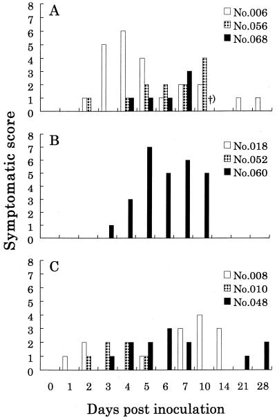
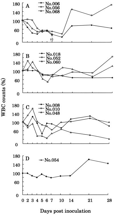
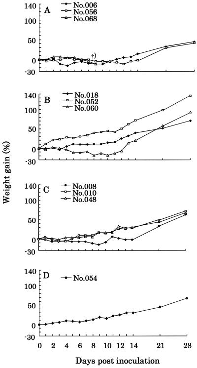
Eight-week-old specific pathogen-free kittens were purchased from Harlan-Sprague-Dawley Inc. (Indianapolis, Ind.), and these animals were used in experimental infections. Each group of three cats was separated into a different isolation room, and each individual animal was placed in a separate cage. Animals were inoculated orally with 107 TCID50 of either FPLV no. 311, CPV-2a V154, or CPV-2c V203. One cat was kept under the same conditions without virus inoculation as an uninfected control. The clinical condition of each animal was monitored daily. The total number of white blood cells (WBC) was measured with a Celltac Automatic Analyzer (Nihon Kohden Co., Inc., Tokyo, Japan) according to the manufacturer's instructions. In general, cats infected with parvovirus show a great variety of symptoms, although leukopenia is a consistent feature (20). A scoring system was devised from such a background. We had the evaluators score the animals without knowledge of which was the treatment group, and each group of cats was scored separately. When any symptom related to parvovirus infection was observed, scores were registered according to the point system shown in Table 1. Cumulative scores for individual cats are presented in Fig. 1. Changes in WBC counts and body weight are presented in Fig. 2 and 3, respectively. No cat showed fevers caused from virus infection during this experiment period although abnormal hypothermia was observed in some animals. In the group of cats inoculated with FPLV strain no. 311, all animals showed prominent leukopenia and poor growth (Fig. 2A and 3A) and registered moderate to severe scores (Fig. 1A). One cat (no. 068) died 9 days postinoculation (d.p.i.). In the group inoculated with CPV-2a strain V154, one cat (no. 060) developed symptoms frequently associated with parvovirus infection (Fig. 1B), including leukopenia and weight loss (Fig. 2B and 3B), although the other two animals remained asymptomatic. In the group inoculated with CPV-2c strain V203, all cats showed clinical signs, although the symptoms were relatively milder than those observed in the FPLV no. 311-inoculated group (Fig. 1C). Two cats (no. 008 and 048) were found to display leukopenia (Fig. 2C). The uninfected control did not show any symptoms through the experiment period.
TABLE 1.
Scoring system for symptoms of FPV infectiona
| Symptom | Daily score |
|---|---|
| Body temp (°C) | |
| 37.6 | 1 |
| 37.7–39.4 | 0 |
| 39.5–39.9 | 1 |
| 40.0–40.4 | 2 |
| 40.5 | 3 |
| WBC countb | |
| 59–40 | 1 |
| 39–20 | 2 |
| 20 | 3 |
| Diarrhea | |
| Mucoid | 1 |
| Fluid | 2 |
| Dysenteric | 3 |
| Loss of appetite | 1 |
| Vomiting | 1 |
| Depression | 1 |
| Dehydration | |
| Mild | 1 |
| Moderate | 2 |
| Severe | 3 |
When any of the indicated symptoms related to parvovirus were observed, scores were registered according to the point system outlined here.
The WBC counts (per microliter) of each cat are shown as the percentages of WBCs determined immediately prior to and after inoculation.
FIG. 1.
Symptomatic scores obtained for cats inoculated with FPVs. FPLV no. 311 (A), CPV-2a V154 (B), and CPV-2c V203 (C) strains were orally inoculated. †), cat no. 068 died at 9 d.p.i.
FIG. 2.
WBC counts of cats inoculated with FPVs. The WBC counts (per microliter) are shown as the percentages of WBCs determined immediately prior to and after inoculation. Shown are the orally inoculated groups of FPLV no. 311 (A), CPV-2a V154 (B), and CPV-2c V203 (C) strains and the uninfected control (D). †), cat no. 068 died at 9 d.p.i.
FIG. 3.
Body weights of cats inoculated with FPVs. The body weight of each cat is shown as the percentage of weight gain, compared with the weight determined immediately prior to infection. Shown are the orally inoculated groups of FPLV no. 311 (A), CPV-2a V154 (B), and CPV-2c V203 (C) strains and the uninfected control (D). †), cat no. 068 died at 9 d.p.i.
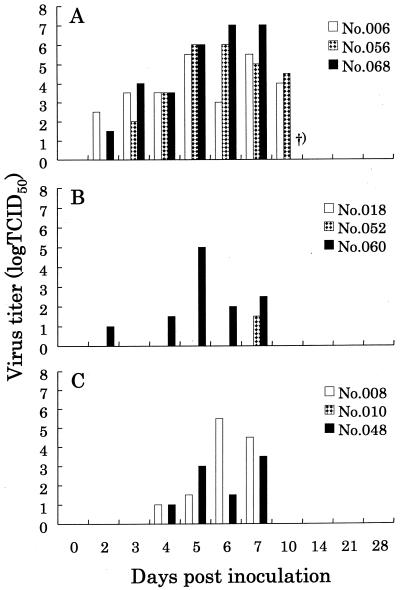
To test viral shedding into feces, rectal swabs were collected at appropriate time intervals. Each swab was transferred into a sterile tube containing 2.0 ml of phosphate-buffered saline, and the tube was mixed vigorously. The phosphate-buffered saline was subsequently centrifuged at 3,000 × g for 10 min, and the supernatant was filtrated through Millipore filters (pore size, 200 nm). One hundred microliters of the filtrate was subjected to virus titration using FL74 cells, as described previously (6). In all cats inoculated with FPLV no. 311, shedding of virus particles into feces was observed from 2 to 10 d.p.i. (Fig. 4A). In both CPV-2a V154- and CPV-2c V203-inoculated animals, two of the three cats were found to have shed viruses in their feces (Fig. 4B and C, respectively). Virus shedding had also ceased by 10 d.p.i. for these two experimental groups. In the uninfected control cat, virus shedding into feces was never observed during the experiments.
FIG. 4.
Virus titers in the feces of cats inoculated with FPVs. FPLV no. 311 (A), CPV-2a V154 (B), and CPV-2c V203 (C) strains were orally inoculated. †), cat no. 068 died at 9 d.p.i.
In the past, viruses in the feces of infected animals have commonly been titrated as a means of monitoring virus proliferation in cats. It is generally considered that viruses shed in feces reflect virus proliferation in cats. However, in addition to the epithelial cells of the intestine, lymphoid tissues are also a major target for FPVs in both dogs and cats (1, 5). Recently, we found that FPVs can frequently be isolated even in the presence of high VN antibodies in infected cats (12). This was surprising because virus shedding into feces stops once the immune response develops (17). To determine how long FPVs are present in the PBMCs of infected cats, we performed virus isolations from PBMCs as described previously (12). We first performed PCR, using VP2 targeting primers of sequence F1 (5′-AGATAGTAATAATACTATGCCATTT-3′) and R2 (5′-TTTTGAATCCAATCTCCTTCTGGAT-3′) to detect viral DNA amongst DNA isolated from PBMCs. Viral DNA was not detected in any of the samples taken at the start of PBMC cultivation; however, some of the cultures (3 to 9 days after cultivation) showed severe cytophathic effect (CPE), such as cell rounding and nuclear disintegration. To confirm the presence of FPVs in the cultures and to isolate viruses, the cultures showing CPE were cocultured with MYA-1 cells (an interleukin-2-dependent feline T-lymphoblastoid cell line) (ATCC CRL-2417) (11). The cocultured MYA-1 cells showed similar CPE 1 to 3 days after cocultivation. The presence of FPVs was subsequently confirmed in all cocultured PBMC samples, by both PCR and indirect immunofluorescence assays. Cultures not showing CPE after 2 weeks were also subjected to these two assays; however, none were found to be positive. In the group of cats infected with FPLV no. 311, the virus was isolated from PBMCs at 1 to 3 or 4 weeks postinoculation (w.p.i.) in the two animals which survived (Table 2). In CPV-2a V154-inoculated cats, the virus was isolated from PBMCs in only one animal (no. 052) at 2 and 3 w.p.i., although no clinical symptoms were observed in this individual (Fig. 2). We failed to isolate virus from PBMCs of cat no. 060, although this animal developed clinical signs of infection, including virus shedding in feces. In the CPV-2c V203-inoculated animals, virus was successfully isolated at 1 and 2 w.p.i. from all three cats (Table 2).
TABLE 2.
Virus isolation from PBMCs
| Cat | Virus isolation at the following w.p.i.:
|
||||
|---|---|---|---|---|---|
| 0a | 1 | 2 | 3 | 4 | |
| FPLV no. 311 | |||||
| 006 | − | + | + | + | + |
| 056 | − | + | + | + | − |
| 068 | − | + | NAb | NA | NA |
| CPV-2a V154 | |||||
| 018 | − | − | − | − | − |
| 052 | − | − | + | + | − |
| 060 | − | − | − | − | − |
| CPV-2c V203 | |||||
| 008 | − | + | + | − | − |
| 010 | − | + | + | − | − |
| 048 | − | + | + | − | − |
| Uninoculated, 054 | − | − | − | − | − |
Blood samples were collected at the time of virus inoculation.
Not applicable; cat no. 068 died 9 d.p.i.
The VN antibody titers against FPLV strain TU-1 and CPV-2c strain V203 were determined weekly as described previously (6). In the cats inoculated with FPLV no. 311, high VN antibody titers were induced against FPLV TU-1, while relatively low titers were induced against CPV-2c V203. In the CPV-2a V154-inoculated cats, elevation of VN antibody titers against both FPLV TU-1 and CPV-2c V203 was obvious in two of the three cats, while the third animal (no. 018) failed to develop any detectable VN antibodies. In cats inoculated with LCPV V203, VN antibodies against both FPLV TU-1 and CPV-2c V203 were detected at high titers in all three animals (Table 3).
TABLE 3.
FPV neutralizing antibody titers
| Cat | Titer against FPLV TU-1/titer against CPV-2c V203 at the following w.p.i.:
|
||||
|---|---|---|---|---|---|
| 0a | 1 | 2 | 3 | 4 | |
| FPLV no. 311 | |||||
| 006 | <10/<10 | 320/40 | 2,560/160 | 2,560/160 | 2,560/160 |
| 056 | <10/<10 | 160/80 | 2,560/160 | 2,560/320 | 2,560/160 |
| 068 | <10/<10 | 40/40 | NAb | NA | NA |
| CPV-2a V154 | |||||
| 018 | <10/<10 | <10/<10 | <10/<10 | <10/<10 | <10/<10 |
| 052 | <10/<10 | <10/<10 | 80/160 | 80/320 | 320/320 |
| 060 | <10/<10 | 20/80 | 320/640 | 1,280/640 | 1,280/640 |
| CPV-2c V203 | |||||
| 008 | <10/<10 | 160/320 | 320/1,280 | 640/1,280 | 640/1,280 |
| 010 | <10/<10 | 40/320 | 320/640 | 320/640 | 1,280/1,280 |
| 048 | <10/<10 | 20/320 | 160/1,280 | 1,280/1,280 | 2,560/1,280 |
| Uninoculated, 054 | <10/<10 | <10/<10 | <10/<10 | <10/<10 | <10/<10 |
Blood samples were collected at the time of virus inoculation.
Not applicable; cat 068 died 9 d.p.i.
In the study presented here, we show diverse pathogenicity of CPV-2a V154 for individual cats. One animal (no. 060) manifested moderate symptoms, and shed viruses were detected in its feces. One cat (no. 018) of the three inoculated with CPV-2a strain V154 showed no evidence of infection, i.e., no detectable VN antibodies or virus shedding into feces, and virus was absent in PBMCs. Contrary to the results obtained in CPV-2a V154-inoculated animals, all cats inoculated with CPV-2c V203 developed mild diseases typical of FPV infection. These data indicate that CPV-2a V154 and CPV-2c V203 have the potential to cause diseases in cats involving some variation of symptoms. It seemed that there were some differences in disease virulence between CPV-2a V154 and CPV-2c V203, although the numbers of cats tested in this experiment were too small to draw any conclusions.
After VN antibodies were induced, virus shedding into feces ceased in all FPV-infected cats. However, FPVs could be isolated from the PBMCs of infected animals even after VN antibodies had been induced. Notably, in one cat (no. 006) infected with FPLV no. 311, virus could still be isolated from PBMCs until 4 w.p.i. (when we stopped blood sampling). This phenomenon is probably distinct from high levels of virus that can circulate during the initial viremia. It is possible that this was due to the very low amount of virus in the PBMCs, since FPV DNA could not be detected by PCR at the start of PBMC cultivation. Furthermore, a very small amount of FPV DNA was retained in quiescent lymphocytes under VN antibodies in cats, and viral replication occurred with the proliferation of the lymphocytes in vitro after concanavalin A stimulation. It was reported that parvoviruses persisted in lungs and kidneys over 50 weeks in recovered cats (4). The small amount of virus in PBMCs might be transmitted through the blood circulation in these organs.
The virulence of CPV-2c in leopard cats is unknown; however, we suspect that the virulence in leopard cats is similar to that observed in domestic cats. While the pathogenicity of these viruses is apparently mild, other secondary pathogens might induce severe disease in leopard cats whose immune systems are compromised by these viruses. Vaccination of wild cats in zoos against FPVs has recently been recommended (24). However, the use of vaccines made from FPLV might not be very effective against the novel antigenic strains of CPV, since the VN antibody titers recorded against CPV-2c V203 were relatively low in cats inoculated with FPLV no. 311. Although the prevalence of the new antigenic strains of CPV in domestic as well as nondomestic cat populations is still unclear, the development and application of vaccines using either CPV-2a, -2b, or the novel strains should be considered for the prevention of CPV infection in felids.
CPV-2c is shed into feces, and this could represent a source of viruses which could potentially infect other susceptible cats, including domestic cats. CPV-2c strains have a specific amino acid substitution in their VP2 protein, yielding an Asp residue at position 300 (7). This substitution has previously been suggested to reduce the ability of the viruses to infect dog cells and increase the stability of the viruses in the environment (15). In addition, this substitution seemed to be involved in an adaptation of the CPV-2c strains to cats. This viral phenotype might possess an advantage over CPV-2a and -2b, allowing it to spread more efficiently in cat populations. In the present study we confirmed that viruses recovered from feces still retained the Asp substitution (data not shown), suggesting that the novel viruses can be transmitted to domestic cats without reversion of this substitution. Therefore, if the new viruses described here enter the domestic cat population, they could potentially replace the original FPVs. Further epidemiological and virological surveillance of the new antigenic strains of CPV are clearly needed for controlling parvovirus diseases in wild and domestic cats, as well as in dogs.
Acknowledgments
We are grateful to C. Kuwahara, Y. Sanada, and S. Ishiguro (Kyoritsu Shoji Co., Ibaraki, Japan) for their excellent technical assistance. We thank M. Hattori (Kyoto University, Kyoto, Japan) for providing recombinant human interleukin-2-producing Ltk− IL-2.23 cells. We are also grateful to J. Martin (Imperial College, London, United Kingdom) for valuable suggestions and help in preparing this paper.
This study was supported in part by grants from the Ministry of Education, Science, Sports and Culture of Japan. K. Nakamura and E. Sato are supported by Research Fellowships from the Japanese Society for the Promotion of Science for Young Scientists.
REFERENCES
- 1.Carlson J H, Scott F W, Duncan J R. Feline panleukopenia. III. Development of lesions in the lymphoid tissues. Vet Pathol. 1978;15:383–392. doi: 10.1177/030098587801500314. [DOI] [PubMed] [Google Scholar]
- 2.Chalmers W S K, Truyen U, Greenwood N M, Baxendale W. Efficacy of feline panleucopenia vaccine to prevent infection with an isolate of CPV2b obtained from a cat. Vet Microbiol. 1999;69:41–45. doi: 10.1016/s0378-1135(99)00085-1. [DOI] [PubMed] [Google Scholar]
- 3.Crandell R A, Fabricant C G, Nelson-Rees W A. Development, characterization, and viral susceptibility of a feline (Felis catus) renal cell line (CRFK) In Vitro (Rockville) 1973;9:176–185. doi: 10.1007/BF02618435. [DOI] [PubMed] [Google Scholar]
- 4.Csiza C K, Scott F W, De Lahunta A, Gillespie J H. Immune carrier state of feline panleukopenia virus-infected cats. Am J Vet Res. 1971;32:419–426. [PubMed] [Google Scholar]
- 5.Goto H, Hosokawa S, Ichijo S, Shimizu K, Morohoshi Y, Nakano K. Experimental infection of feline panleukopenia virus in specific pathogen-free cats. Jpn J Vet Sci. 1983;45:109–112. doi: 10.1292/jvms1939.45.109. [DOI] [PubMed] [Google Scholar]
- 6.Ikeda Y, Miyazawa T, Kurosawa K, Naito R, Hatama S, Kai C, Mikami T. New quantitative methods for detection of feline parvovirus (FPV) and virus neutralizing antibody against FPV using a feline T lymphoid cell line. J Vet Med Sci. 1998;60:973–974. doi: 10.1292/jvms.60.973. [DOI] [PubMed] [Google Scholar]
- 7.Ikeda Y, Mochizuki M, Naito R, Nakamura K, Miyazawa T, Mikami T, Takahashi E. Predominance of canine parvovirus (CPV) in unvaccinated cat populations and emergence of new antigenic types of CPVs in cats. Virology. 2000;278:13–19. doi: 10.1006/viro.2000.0653. [DOI] [PubMed] [Google Scholar]
- 8.Ikeda Y, Miyazawa T, Nakamura K, Naito R, Inoshima Y, Tung K-C, Lee W-M, Chen M-C, Kuo T-F, Lin J A, Mikami T. Serosurvey for selected virus infections of wild carnivores in Taiwan and Vietnam. J Wildlife Dis. 1999;35:578–581. doi: 10.7589/0090-3558-35.3.578. [DOI] [PubMed] [Google Scholar]
- 9.Ikeda Y, Shinozuka J, Miyazawa T, Kurosawa K, Izumiya Y, Nishimura Y, Nakamura K, Cai J-S, Fujita K, Doi K, Mikami T. Apoptosis in feline panleukopenia virus-infected lymphocytes. J Virol. 1998;72:6932–6936. doi: 10.1128/jvi.72.8.6932-6936.1998. [DOI] [PMC free article] [PubMed] [Google Scholar]
- 10.Konishi S, Mochizuki M, Ogata M. Studies on feline panleukopenia. I. Isolation and properties of virus strains. Jpn J Vet Sci. 1975;37:439–449. doi: 10.1292/jvms1939.37.5_247. [DOI] [PubMed] [Google Scholar]
- 11.Miyazawa T, Furuya T, Itagaki S, Tohya Y, Takahashi E, Mikami T. Establishment of a feline T-lymphoblastoid cell line highly sensitive for replication of feline immunodeficiency virus. Arch Virol. 1989;108:131–135. doi: 10.1007/BF01313750. [DOI] [PubMed] [Google Scholar]
- 12.Miyazawa T, Ikeda Y, Nakamura K, Naito R, Mochizuki M, Tohya Y, Vu D, Mikami T, Takahashi E. Isolation of feline parvovirus from peripheral blood mononuclear cells of cats in northern Vietnam. Microbiol Immunol. 1999;43:609–612. doi: 10.1111/j.1348-0421.1999.tb02447.x. [DOI] [PubMed] [Google Scholar]
- 13.Mochizuki M, Hashimoto T. Growth of feline panleukopenia virus and canine parvovirus in vitro. Jpn J Vet Sci. 1986;48:841–844. doi: 10.1292/jvms1939.48.841. [DOI] [PubMed] [Google Scholar]
- 14.Mochizuki M, Horiuchi M, Hiragi H, San Gabriel M C, Yasuda N, Uno T. Isolation of canine parvovirus from a cat manifesting clinical signs of feline panleukopenia. J Clin Microbiol. 1996;34:2101–2105. doi: 10.1128/jcm.34.9.2101-2105.1996. [DOI] [PMC free article] [PubMed] [Google Scholar]
- 15.Parker J S L, Parrish C R. Canine parvovirus host range is determined by the specific conformation of an additional region of the capsid. J Virol. 1997;71:9214–9222. doi: 10.1128/jvi.71.12.9214-9222.1997. [DOI] [PMC free article] [PubMed] [Google Scholar]
- 16.Parrish C R. Emergence, natural history, and variation of canine, mink, and feline parvoviruses. Adv Virus Res. 1990;38:403–450. doi: 10.1016/S0065-3527(08)60867-2. [DOI] [PMC free article] [PubMed] [Google Scholar]
- 17.Parrish C R. Parvoviruses: cats, dogs and mink. In: Webster R G, Granoff A, editors. Encyclopedia of virology. London, England: Academic Press; 1994. pp. 1061–1067. [Google Scholar]
- 18.Parrish C R, O'Connell P H, Evermann J F, Carmichael L E. Natural variation of canine parvovirus. Science. 1985;230:1046–1048. doi: 10.1126/science.4059921. [DOI] [PubMed] [Google Scholar]
- 19.Parrish C R, Aquadro C F, Strassheim M L, Evermann J F, Sgro J-Y, Mohammed H O. Rapid antigenic-type replacement and DNA sequence evolution of canine parvovirus. J Virol. 1991;65:6544–6552. doi: 10.1128/jvi.65.12.6544-6552.1991. [DOI] [PMC free article] [PubMed] [Google Scholar]
- 20.Pedersen N C. Feline panleukopenia virus. In: Appel M J, editor. Virus infections of carnivores. Amsterdam, The Netherlands: Elsevier Science Publishers B. V.; 1985. pp. 247–254. [Google Scholar]
- 21.Sakamoto M, Ishiguro S, Mochizuki M. Experimental infection of canine parvoviruses in cats. J Jpn Vet Med Assoc. 1999;52:305–309. . (In Japanese.) [Google Scholar]
- 22.Siegl G. Canine parvovirus. Origin and significance of a “new” pathogen. In: Berns K, editor. The Parvoviruses. New York, N.Y: Plenum; 1984. pp. 363–388. [Google Scholar]
- 23.Siegl G, Bates R C, Berns K I, Carter B J, Kelly D C, Kurstak E, Tattersall P. Characteristics and taxonomy of Parvoviridae. Intervirology. 1985;23:61–73. doi: 10.1159/000149587. [DOI] [PubMed] [Google Scholar]
- 24.Steinel A, Munson L, van Vuuren M, Truyen U. Genetic characterization of feline parvovirus sequences from various carnivores. J Gen Virol. 2000;81:345–350. doi: 10.1099/0022-1317-81-2-345. [DOI] [PubMed] [Google Scholar]
- 25.Theilen G H, Kawakami T G, Rush J D, Munn R J. Replication of cat leukaemia virus in cell suspension cultures. Nature. 1969;222:589–590. doi: 10.1038/222589b0. [DOI] [PubMed] [Google Scholar]
- 26.Truyen U. Emergence and recent evolution of canine parvovirus. Vet Microbiol. 1999;69:47–50. doi: 10.1016/s0378-1135(99)00086-3. [DOI] [PubMed] [Google Scholar]
- 27.Truyen U, Parrish C R. Canine and feline host ranges of canine parvovirus and feline panleukopenia virus: distinct host cell tropisms of each virus in vitro and in vivo. J Virol. 1992;66:5399–5408. doi: 10.1128/jvi.66.9.5399-5408.1992. [DOI] [PMC free article] [PubMed] [Google Scholar]
- 28.Truyen U, Platzer G, Parrish C R. Antigenic type distribution among canine parvoviruses in dogs and cats in Germany. Vet Rec. 1996;138:365–366. doi: 10.1136/vr.138.15.365. [DOI] [PubMed] [Google Scholar]
- 29.Truyen U, Evermann J F, Vieler E, Parrish C R. Evolution of canine parvovirus involved loss and gain of feline host range. Virology. 1996;215:186–189. doi: 10.1006/viro.1996.0021. [DOI] [PubMed] [Google Scholar]
- 30.Truyen U, Müller T, Heidrich R, Tackmann K, Carmichael L E. Survey on viral pathogens in wild red foxes (Vulpes vulpes) in Germany with emphasis on parvoviruses and analysis of a DNA sequence from a red fox parvovirus. Epidemiol Infect. 1998;121:433–440. doi: 10.1017/s0950268898001319. [DOI] [PMC free article] [PubMed] [Google Scholar]
- 31.Truyen U, Gruenberg A, Chang S-F, Obermaier B, Veijalainen P, Parrish C R. Evolution of the feline-subgroup parvoviruses and the control of canine host range in vivo. J Virol. 1995;69:4702–4710. doi: 10.1128/jvi.69.8.4702-4710.1995. [DOI] [PMC free article] [PubMed] [Google Scholar]


