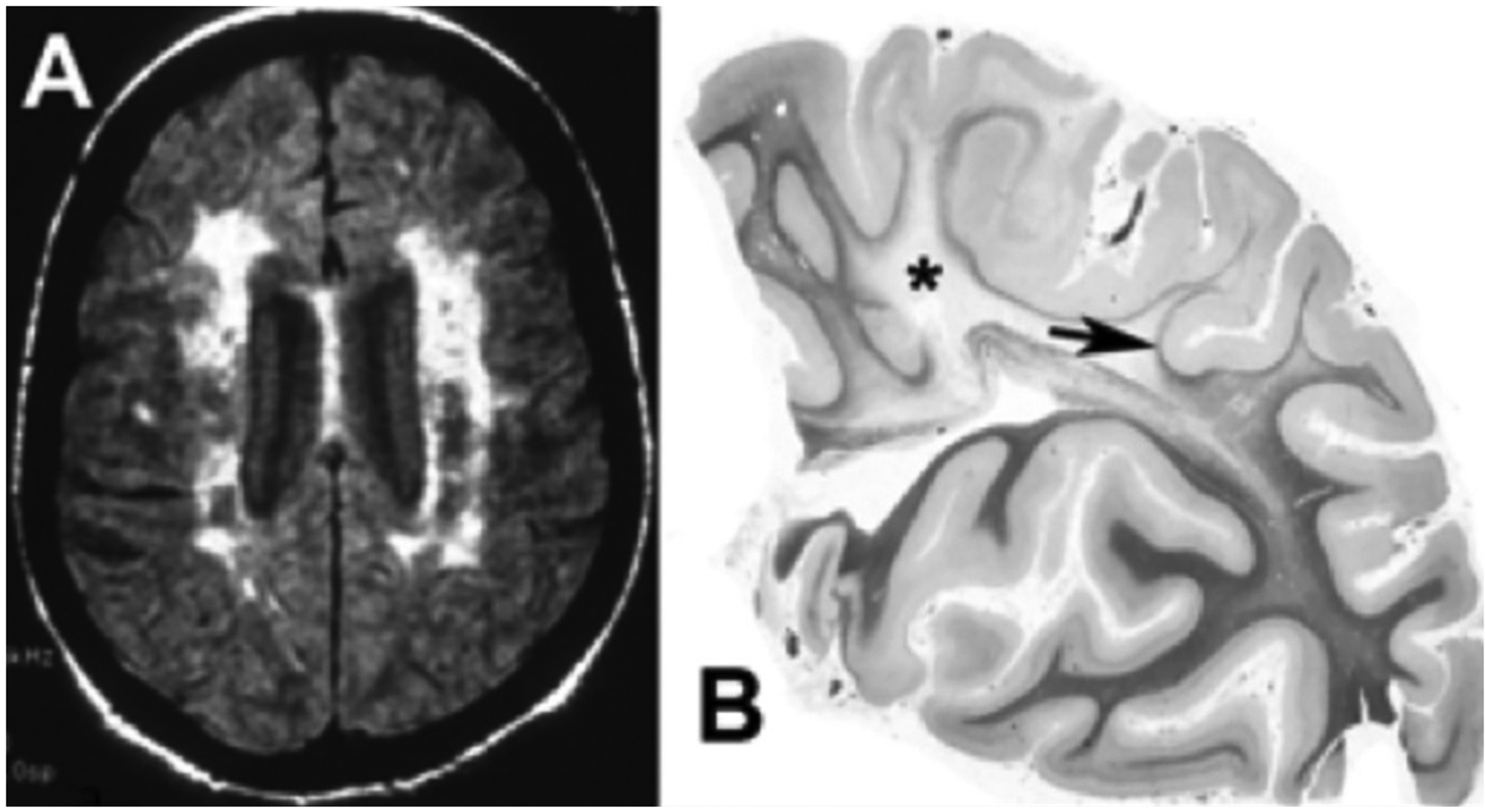
Figure 2:

A) FLAIR MRI from a patient with Binswanger’s disease showing extensive white matter involvement in the subcortical region. There is relative preservation of the cortex and lack of ventricular enlargement. B) A hemisphere section from a patient with long-standing hypertension. In this myelin stain, there is loss of myelination (Asterix) in the centrum semiovale extending into the sulci and sparing U-fibers (arrow). (Figure 2B is from46 with permission. Copyright © 2001, Wolters Kluwer Health).
