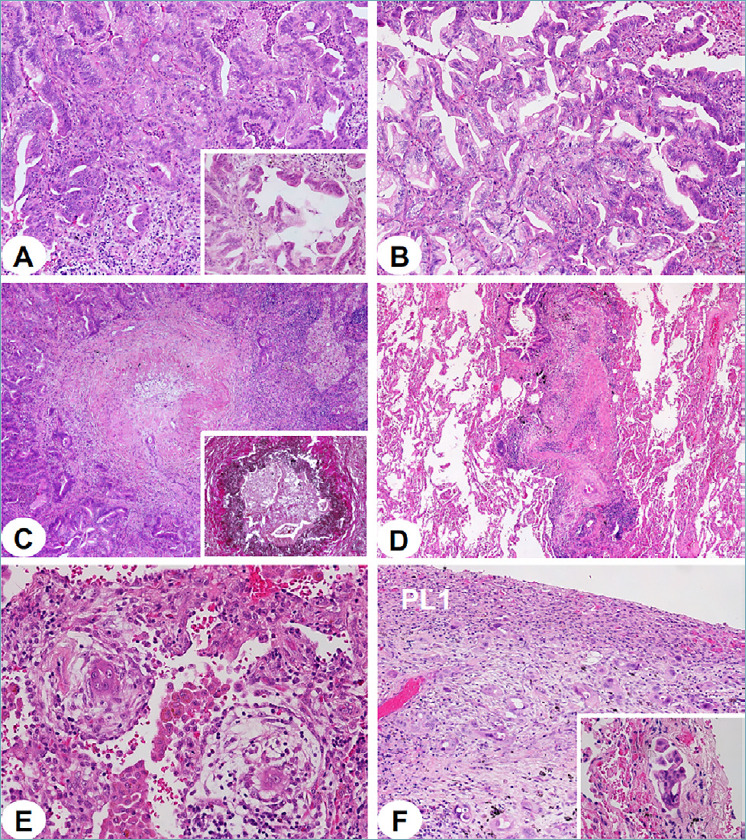
Figure 2.

(A-F). In the lung, invasive mucinous adenocarcinoma featured acinar and complex glandular patterns with labyrinthine growth appearance (A), lepidic-like growth (A, inset), goblet to columnar mucinous cells, basally located nuclei and gastrointestinal-type histologic appearance (B). Massive and multifocal carcinomatous thromboembolism was readily observed in all branching pulmonary arteries (elastin stain shows internal and external layers, inset) (C) and massively permeated their terminal branches along accompanying centrilobular bronchioles, thereby realizing a centrifugal-type colonization route (D). Even interstitial vascular channels were diffusely engulfed by cancer cells (E). Visceral pleural layer was infiltrated at the level PL1, with many tumor emboli accumulating with lymphatic vessels (F, inset).
