Figure 5.


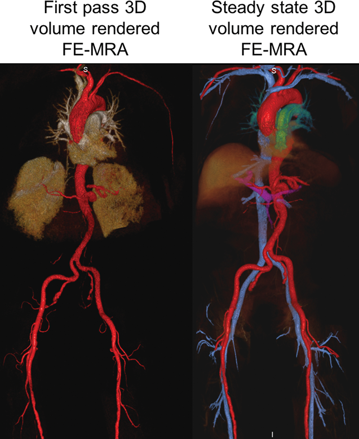
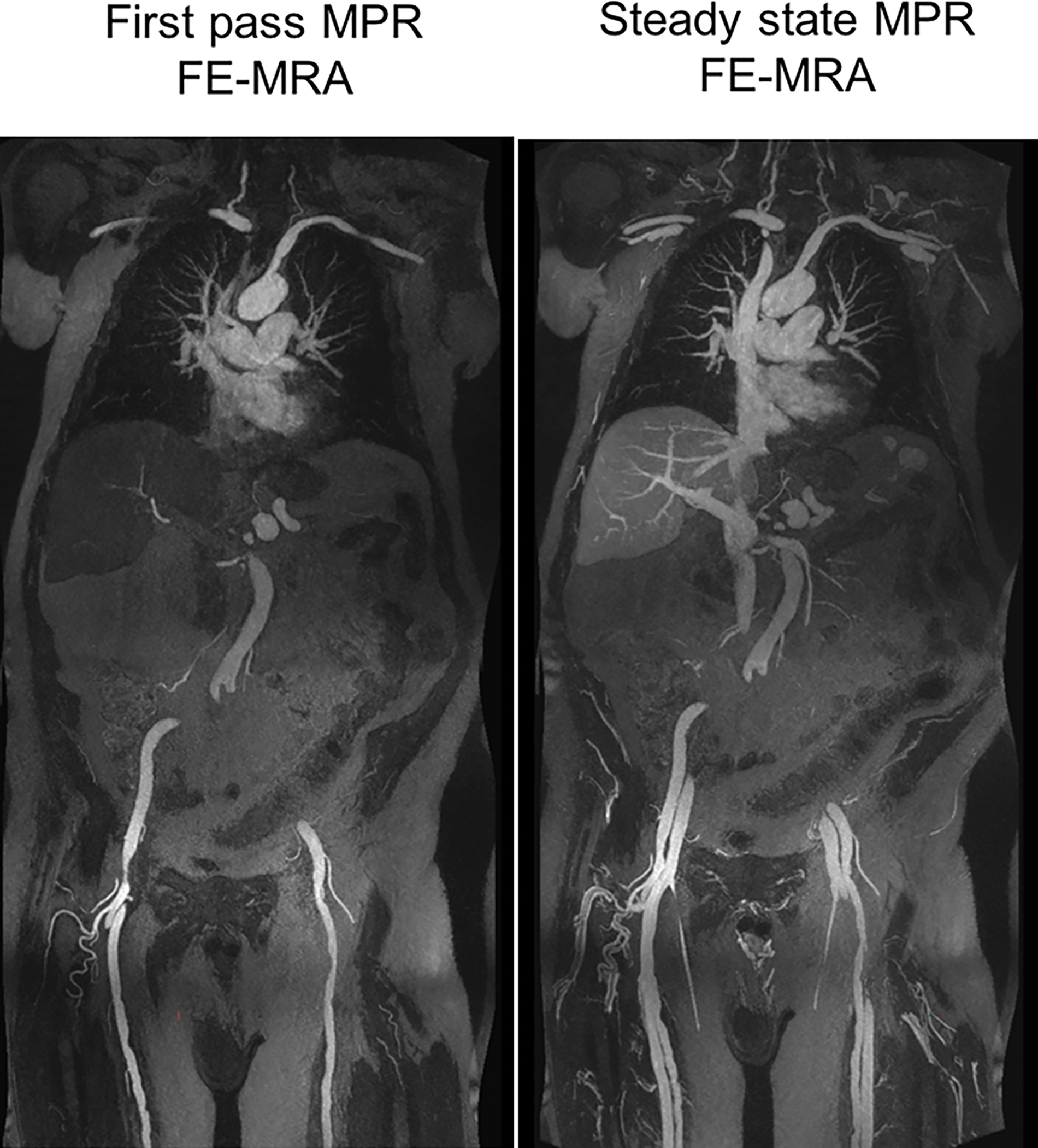
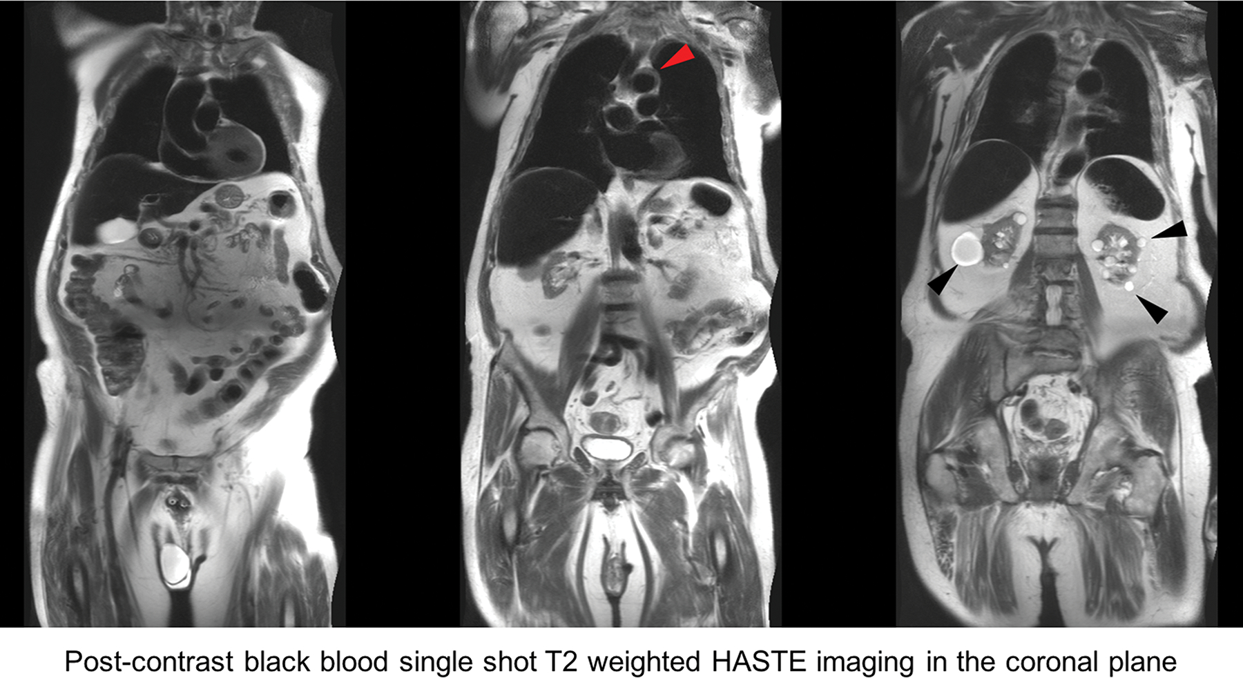
FE-MRA at 3.0T. 84-year-old male with severe AS and moderate renal impairment (creatinine of 1.7 mg/dL, stage 3b) presented for TAVR evaluation. First-pass and steady state (a) 3D volume rendered and (b) multiplanar reformat (MPR) FE-MRA of the entire aorta and pelvic access vessels show significant tortuosity. There is comparable luminal vascular enhancement between first-pass vs steady state imaging. (c) Post-contrast black blood HASTE imaging depicts atheromatous plaque morphology (red arrowhead) at the transverse aortic arch and incidental multiple renal cysts (black arrowhead). This patient had successful transfemoral placement of a 26mm Edwards Sapien valve.
