Abstract
BACKGROUND
Several studies have proved the safety and feasibility of robot-assisted percutaneous coronary intervention (PCI) in reducing the occupational hazards of interventionists while achieving precision medicine. However, an independently developed robot-assisted system for PCI in China has not yet emerged. This study aimed to evaluate the safety and feasibility of a robot-assisted system for elective PCI in China.
METHODS
This preclinical trial included 22 experimental pigs and preliminarily supported the safety and feasibility of the ETcath200 robot-assisted system for PCI. Then, eleven patients with coronary heart disease who met the inclusion criteria and had clinical indications for elective PCI were enrolled. PCI was performed using a robot-assisted system. The primary outcomes were clinical success (defined as visual estimated residual stenosis < 30% after PCI and no major adverse cardiovascular events during hospitalization and within 30 days after PCI) and technical success (defined as the ability to use the robot-assisted system to complete PCI successfully without conversion to the traditional manual PCI).
RESULTS
Eleven patients were included in this clinical trial. A drug-eluting stent with a diameter of 3 mm (interquartile range: 2.75–3.5 mm) and a length of 26 mm (interquartile range: 22–28 mm) was deployed in all patients. The clinical success rate was 100%, with no PCI-related complications and no in-hospital or 30-day major adverse cardiovascular events, and the technical success rate was 100%.
CONCLUSIONS
The results strongly suggest that the use of the independently developed robot-assisted system in China for elective PCI is feasible, safe, and effective.
Percutaneous coronary intervention (PCI) is one of the most important methods to treat coronary heart disease (CHD). Traditional manual PCI (M-PCI) can significantly improve the prognosis of patients with CHD. However, high radiation doses,[1] orthopedic injuries sustained by interventionists,[2] and many other issues cannot be ignored. At the same time, difficulties in precisely deploying devices in M-PCI often lead to a poor prognosis.[3]
Corindus (Siemens Healthineers Company, Waltham, MA, USA) proposed the concept of a robot-assisted system in 2011, focusing on solving the above problems. Numerous studies have demonstrated the safety and feasibility of robot-assisted PCI (R-PCI),[4–6] which can reduce interventionists’ exposure to radiation, thereby decreasing the occurrence of radiation-related occupational diseases compared to those with M-PCI.[2,3] In addition, robot-assisted systems can accurately measure the length of lesions and guide the deployment of devices, thereby achieving precision medicine.[7]
However, independent development of robot-assisted systems for PCI in China started relatively late and have not yet been utilized in clinical practice. This is the first study to test an independently developed robot-assisted system from China for PCI and the first to report on the preclinical and clinical efficacy of a Chinese robot-assisted system. The primary objective of this study was to evaluate the safety and feasibility of a robot-assisted system for elective PCI.
METHODS
ETcath200 Robot-assisted System
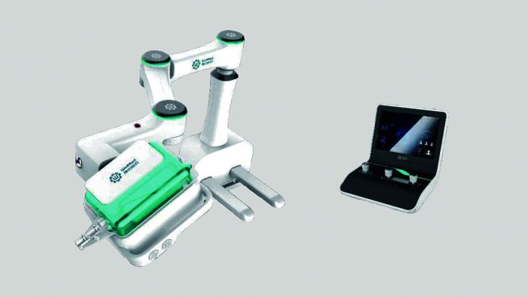
The robot-assisted system was developed by Beijing WeMed Medical Equipment Co., Ltd., Beijing, China and named the ETcath200 robot-assisted system. The ETcath200 robot-assisted system (Figure 1) consisted of control and executive units.
Figure 1.
ETcath200 robot-assisted system.
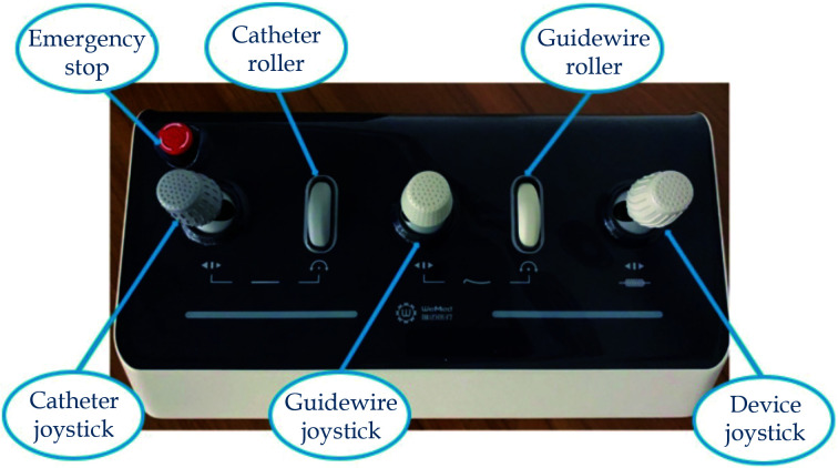
The control unit (Figure 2) included a control box, touch screen, and control cabinet. The control and executive units were connected using a wire. The control unit was located in the control room, and the executive unit was beside the operating room bed. Interventionists could remotely control the movement of the guiding catheter, guidewire, balloon, or stent in the control room through three joysticks on the control unit. They could also achieve axial movement (forward/backward) and rotational movement of the guidewire and simultaneously push and rotate the guidewire. Balloon or stent catheter advancement and retraction were controlled precisely, and the robot recorded the distance traveled by the device to accurately measure the length of the target lesion. Additionally, the emergency stop button was configured to deal with emergencies, including equipment failure.
Figure 2.
Control unit of ETcath200 robot-assisted system.
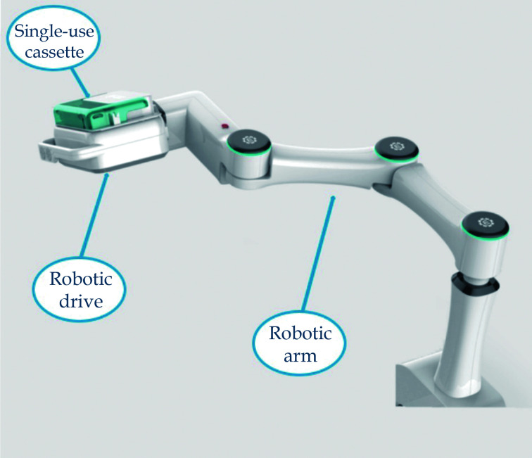
The executive unit (Figure 3) was used for the manipulation of catheters, guidewires, balloons, or stents at the bedside in the operating room. For the executive unit, structures such as a robotic drive which actually execute the movements of devices, a sterile single-use cassette, and a robotic arm were used. During the PCI procedure, a sterile single-use cassette was installed on the guidewire actuator in a pluggable manner, and its internal components were automatically matched with the guidewire actuator. The device was compatible with common components of interventional procedures, including guidewires, rapid exchange balloon dilatation catheters, and stent delivery systems. It was also equipped with a multi-dimensional guidewire resistance detection system, which converted the resistance the guidewire and balloon/stent encountered during the interventional procedure into a pressure curve. The cassettes were sterilized with ethylene oxide, and a new sterile cassette was used for each procedure.
Figure 3.
Executive unit of ETcath200 robot-assisted system.
Foot pedals for cine and fluoroscopy, contrast media injection, guidewire, balloon or stent catheter replacement, to mention a few, were controlled by bedside assistants and technicians.
All interventionists who operated the robot-assisted system used the coronary vessel simulator to conduct comprehensive and scientific operation practice before the procedures.
Preclinical Trial
The preclinical trial of the robot-assisted system was performed according to the Institutional Animal Care and Use Committee guidelines. A total of 22 Chinese experimental pigs were purchased from Jurong Kangrong poultry industry Co., Ltd, Jiangsu, China which were without coronary stenosis. The pigs, which were fed a standard laboratory diet with free access to food and water and maintained at 22 ± 1 °C and 65%–70% humidity under a 12-h light/12-h dark cycle, weighted among 35–45 kg and their age matched with their weight. The preclinical trial aimed to preliminary investigate the safety and feasibility of using the robot-assisted system for PCI. Of the 22 animals, two animals were pre-experimental animals. The remaining 20 animals were randomly divided into two groups: one underwent R-PCI (n = 10) and the other underwent M-PCI (n = 10). A stent was deployed in each animal’s left anterior descending coronary artery. The evaluation indicators included technical success (PCI could be successfully completed with the robot-assisted system, and there was no need to convert to M-PCI when the guidewire or balloon/stent catheter was unable to cross the blood vessel or the catheter was poorly supported), immediate PCI complications (vessel dissection, perforation, to mention a few), contrast volume, fluoroscopy time, interventionists’ cumulative radiation dose, and PCI procedure time. Angiography was performed to observe any procedure-related complications 28 days after the surgery.
Clinical Trial
This was a single-arm, single-center, open-label, prospective trial. Patients referred to the 12th Ward of Beijing Anzhen Hospital, Capital Medical University, Beijing, China, who met the indications for elective PCI between October 8, 2021 and October 21, 2021, were included. All patients were followed up for 30 days to evaluate clinical and technical endpoints. All patients understood the PCI procedure and provided informed consent. The trial protocol was approved by the Institutional Review Board of Beijing Anzhen Hospital, Capital Medical University, Beijing, China (No.2022162X).
Study Population
Patients with unstable angina and/or having undergone prior coronary computed tomography angiography/coronary angiography with a definitive diagnosis of CHD and indications for elective PCI were included in the trial. The inclusion criteria were as follows: (1) age ≥ 18 years, no gender restrictions; (2) patients with CHD and clinical indications for PCI; (3) voluntary participation and signed informed consent; and (4) meeting the following angiographic criteria: (a) de novo lesions; (b) reference vessel diameter of 2.5–4.0 mm, target lesion length ≤ 40 mm; (c) target lesions that could be completely covered by the stent; and (d) the degree of stenosis in the target lesion > 50%.
The clinical exclusion criteria were as follows: (1) acute myocardial infarction (MI) experienced within one week; (2) stroke occurrence within six months, or a history of major gastrointestinal bleeding in the past six months, or determination by the investigator that the patient had a bleeding disorder; (3) the target vessel having received a previous stent treatment, and the target lesion located within 5 mm of the proximal end of the treated lesion; (4) severe calcification at the proximal end of the target lesion or within the lesion; (5) thrombosis in the target vessel lumen; (6) unprotected left main coronary artery disease; (7) allergy to any component of the drug or research device necessary for surgery, such as aspirin, clopidogrel, heparin, contrast agent, ticagrelor, bivalirudin, and paclitaxel; (8) pregnant or breastfeeding women; (9) having participated in clinical trials of other drugs or medical devices during the same period; and (10) other reasons due to which the researcher believed that the patient was not suitable for inclusion in the trial, such as history of definite neurological or mental disorders.
Endpoints
The primary outcomes were clinical and technical success. Clinical success was defined as visual estimated residual stenosis < 30% after R-PCI and no major adverse cardiovascular events (MACEs) during hospitalization or within 30 days after the procedure. MACEs were defined as a combination of nonfatal stroke, nonfatal MI, and cardiovascular death. All patients were followed up by telephone at 30 days to evaluate postoperative 30-day MACEs. Technical success was defined as the availability of a robot-assisted system to complete PCI successfully without conversion to M-PCI in the event of a guidewire or balloon/stent catheter that was unable to cross the vessel or was poorly supported by the catheter.
Secondary outcomes included the total procedure time (defined as the time from the beginning of the procedure to the time when the guide catheter was withdrawn), PCI procedure time (defined as the time from the insertion of the guide catheter to the time when the guide catheter was withdrawn), fluoroscopy time, contrast volume, air kerma dose, and dose-area product. Reference vessel diameter and minimum lumen diameter are acquired based on a visual estimate of interventionists.
Statistical Analysis
Data were analyzed using the Stata 17.0 (Stata Corporation, College Station, TX, USA). Use normality tests to check whether data are normal or skewed distributions. Continuous variables with normal distributions were summarized using mean ± SD. Continuous variables with skewed distributions were summarized using the median [interquartile range (IQR)]. Categorical variables were summarized as counts (percentages). Two-sided P-value < 0.05 were considered statistically significant. The cardiac enzyme (creatine kinase, creatine kinase-myocardial band, high-sensitivity troponin I, and myoglobin) values before and 24 h after the procedure were analyzed using the Wilcoxon paired nonparametric test due to the small sample size (n = 11) and inability to satisfy the normal distribution.
RESULTS
Preclinical Trial
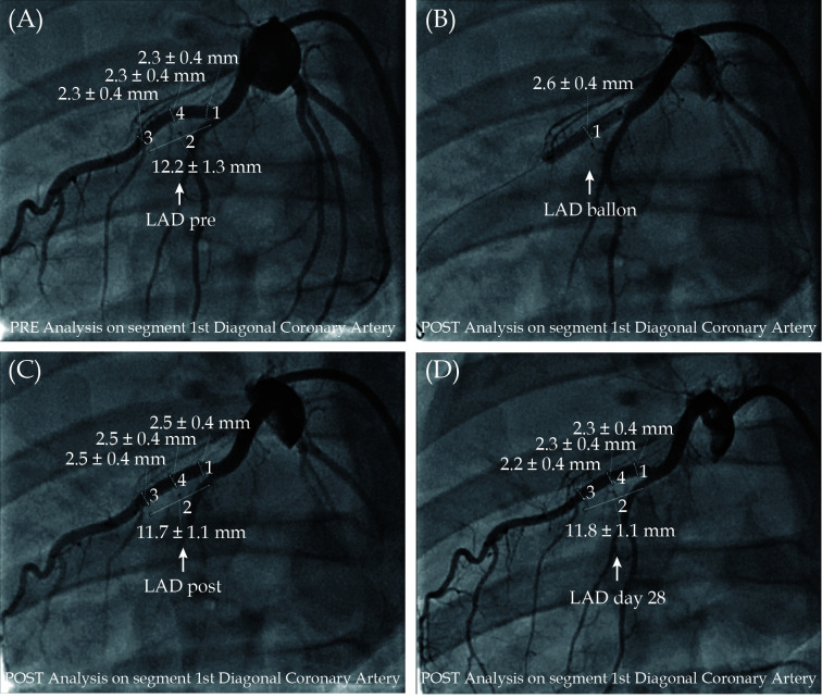
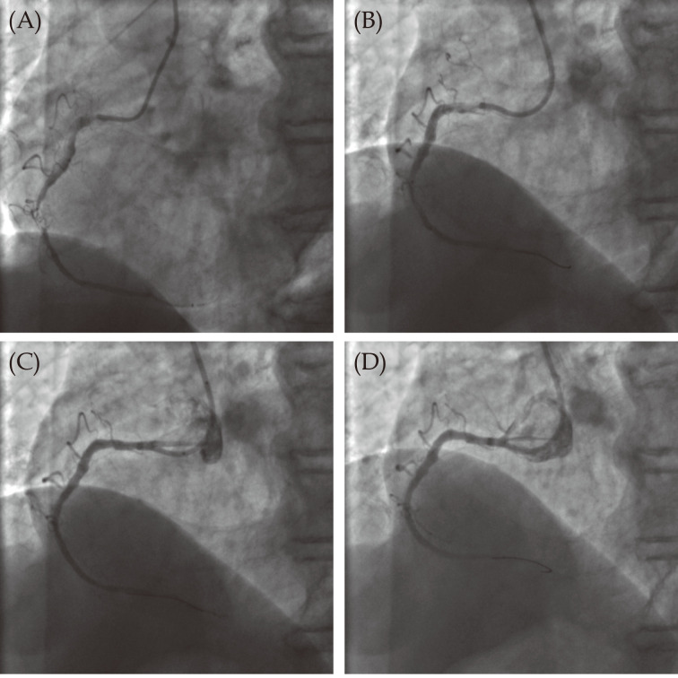
The clinical success rate was 100%. No PCI-related complications, such as vascular dissection and perforation, were observed on coronary angiography immediately after and 28 days after the surgery (Figure 4).
Figure 4.
Images of percutaneous coronary intervention performed in experimental pigs using the robot-assisted system.
(A): The image of the robot-assisted system performing pre-percutaneous coronary intervention angiography; (B): the image after the robot-assisted system performs balloon pre-dilation; (C): the image after the robot-assisted system completing the stent implantation; and (D): the angiographic image of the follow-up 28 day after stent implantation by the robot-assisted system. LAD: left anterior descending artery.
There was no significant difference in the contrast volume between the R-PCI group and M-PCI group (24.80 ± 5.88 mL vs. 31.20 ± 6.43 mL, P = 0.172). The PCI procedure time in the R-PCI group was longer than that in the M-PCI group (6.60 ± 1.74 min vs. 3.00 ± 0.00 min, P = 0.005). The cumulative exposure time (6.60 ± 2.58 min vs. 45.00 ± 10.56 min, P = 0.009) and air kerma dose (4.40 ± 1.36 mGy vs. 7.20 ± 1.72 mGy, P = 0.034) in the R-PCI group were both better than those in the M-PCI group (Table 1).
Table 1. Summary of the preclinical trial.
| Variables | Robot-assisted group | Traditional manual group | P-value |
| Data are presented as means ± SD. | |||
| Contrast volume, mL | 24.80 ± 5.88 | 31.20 ± 6.43 | 0.172 |
| Cumulative exposure time, min | 6.60 ± 2.58 | 45.00 ± 10.56 | 0.009 |
| Air kerma dose, mGy | 4.40 ± 1.36 | 7.20 ± 1.72 | 0.034 |
| Percutaneous coronary intervention procedure time, min | 6.60 ± 1.74 | 3.00 ± 0.00 | 0.005 |
Clinical Trial
Baseline characteristics of the patients are presented in Table 2. Eleven patients who underwent elective PCI were included in this trial. The mean age of the patients was 63 years (IQR: 52–72 years), and ten patients (91%) were men. Seven patients (64%) had hypertension, six patients (55%) had diabetes mellitus, three patients (27%) had hyperlipidemia, and one patient (9%) had previously undergone PCI. None of the patients had a history of MI or coronary artery bypass grafting. The liver and kidney functions of the enrolled patients were roughly normal, and they had no decreased left ventricular ejection fraction.
Table 2. Baseline characteristics of patients.
| Variables | Patients (n = 11) |
| Data are presented as means ± SD or n (%). *Presented as median (interquartile range). NYHA: New York Heart Association. | |
| Age, yrs | 63 (52–72)* |
| Male | 10 (91%) |
| Body mass index, kg/m2 | 25.7 ± 3.4 |
| Hypertension | 7 (64%) |
| Diabetes mellitus | 6 (55%) |
| Hyperlipidemia | 3 (27%) |
| Prior myocardial infarction | 0 |
| Prior percutaneous coronary intervention | 1 (9%) |
| Prior coronary artery bypass grafting | 0 |
| Medical treatment | |
| Aspirin | 11 (100%) |
| Clopidogrel | 5 (45%) |
| Ticagrelor | 6 (55%) |
| Statins | 11 (100%) |
| Nitrate | 5 (45%) |
| Beta-blockers | 4 (36%) |
| NYHA functional class | |
| Class I | 7 (64%) |
| Class II | 4 (36%) |
| Class III | 0 |
| Class IV | 0 |
| Left ventricular ejection fraction, % | 65 (65–68)* |
| Heart rate, beat/min | 73.9 ± 7.9 |
| Systolic blood pressure, mmHg | 137.2 ± 19.3 |
| C-reative protein, mg/dL | 1.75 (0.93–2.97)* |
| Leukocyte, × 109/L | 6.23 (5.76–7.38)* |
| Red blood cells, × 1012/L | 4.74 (4.47–5.01)* |
| Hemoglobin, g/L | 150 (139–152)* |
| Platelets, × 109/L | 234 (164–250)* |
| Neutrophil percentage, % | 68.9 (66.9–75.5)* |
| B-type natriuretic peptide, ng/L | 37 (9–140)* |
| Albumin, U/L | 42 (41.2–46)* |
| Aspartate aminotransferase, U/L | 18 (16–21)* |
| Alanine aminotransferase, U/L | 21 (15–24)* |
| Total bilirubin, μmol/L | 11.66 (8.24–12.39)* |
| Urea, μmol/L | 6.03 (5.78–7.48)* |
| Creatinine, μmol/L | 79 (74.6–83.9)* |
| Estimated glomerular filtration rate, mL/min per 1.73 m2 | 91.8 (85.6–94.7)* |
Characteristics of coronary lesions are presented in Table 3. There were seventeen lesions in eleven patients, including eleven target lesions (65%) and six non-target lesions (35%). The target lesions included six left anterior descending artery lesions (55%), one left circumflex artery disease (9%), and four right coronary artery lesions (36%). Based on the American College of Cardiology/American Heart Association (ACC/AHA) classification of coronary lesions in 1988, among the eleven target lesions, there was one type B1 lesion (9%), two type B2 lesions (18%), and eight type C lesions (73%). The reference vessel diameter of the target lesion was 3 mm (IQR: 2.75–3.5 mm), the minimum lumen diameter was 1 mm (IQR: 0.5–1.5 mm), the lesion length was 20 mm (IQR: 15–25 mm), and the preoperative visual estimate of lumen stenosis was 90% (IQR: 85%–95%).
Table 3. Baseline angiographic characteristics of the patients.
| Variables | Patients (n = 11) |
| Data are presented as n (%). *Presented as median (interquartile range). | |
| Target lesions | 11 (65%) |
| Non-target lesions | 6 (35%) |
| Target vessel | |
| Left anterior descending artery | 6 (55%) |
| Left circumflex artery | 1 (9%) |
| Right coronary artery | 4 (36%) |
| Lesion type | |
| A | 0 |
| B1 | 1 (9%) |
| B2 | 2 (18%) |
| C | 8 (73%) |
| Reference vessel diameter, mm | 3 (2.75–3.5)* |
| Minimum lumen diameter, mm | 1 (0.5–1.5)* |
| Lesion stenosis, mm | 20 (15–25)* |
| Lesion stenosis, % | 90 (85–95)* |
Primary outcomes and result analysis are presented in Figure 5. The R-PCI was used to operate the devices to reach the target lesions and successfully complete balloon pre-dilation, stent deployment, and balloon post-dilation. Dissection, thrombosis, reflow, or other complications were not observed.
Figure 5.
Images of the robot-assisted system used for percutaneous coronary intervention on patients.
(A): The image of the robot-assisted system for pre-percutaneous coronary intervention angiography; (B): the image after pre-dilation; (C): the image after stent deployment; and (D): the image after post-dilation.
The residual stenosis in all eleven patients was 0. One day after the procedure, creatine kinase, creatine kinase-myocardial band and myoglobin showed no significant changes, and high-sensitivity troponin I was elevated compared with the preoperative level (P = 0.0033) (Table 4); however, there were no in-hospital MACEs. All patients were asymptomatic and had no MACEs during the 30-day follow-up period. The clinical success rate was 100%.
Table 4. Myocardial enzyme levels one day before and after the procedure.
| Variables | Pre-procedure | Post-procedure | P-value |
| Data are presented as median (interquartile range). | |||
| Creatine kinase, U/L | 103.3 (64–179) | 123 (60–165) | 0.8589 |
| Creatine kinase-myocardial band, ng/mL | 1.2 (0.8–2.6) | 2.3 (0.9–2.6) | 0.2296 |
| High-sensitivity troponin I, pg/mL | 3.9 (1.9–7.5) | 40.1 (10.7–310.6) | 0.0033 |
| Myoglobin, ng/mL | 26.8 (16.6–31.8) | 25.1 (18.6–48.6) | 0.8589 |
The guidewire was pushed smoothly during the surgery and successfully reached a predetermined position. PCI procedures were completed by the robot-assisted system independently, and there was no need to switch to M-PCI. The technical success rate was 100% (Table 5).
Table 5. The results of primary outcomes.
| Variables | Patients (n = 11) |
| Data are presented as n (%). | |
| Clinical success | 11 (100%) |
| Residual stenosis | 0 |
| Major adverse cardiovascular events | 0 |
| Nonfatal stroke | 0 |
| Nonfatal myocardial infarction | 0 |
| Cardiovascular death | 0 |
| Technical success | 11 (100%) |
Secondary outcomes are presented in Table 6. The total procedure time was 53 min (IQR: 39–67 min), the PCI procedure time was 33 min (IQR: 24–46 min), and the fluoroscopy time was 18 min (IQR: 12–20 min). The contrast volume used in the procedure was 88 mL (IQR: 55–160 mL). The air kerma dose was 1674 mGy (IQR: 1056–4360 mGy) and the dose-area product was 7210 cGy × cm2 (IQR: 6272–12,545 cGy × cm2). All eleven patients had a drug-eluting stent with a diameter of 3 mm (IQR: 2.75–3.5 mm), and the stent length was 26 mm (IQR: 22–28 mm).
Table 6. The results of secondary outcomes.
| Variables | Patients (n = 11) |
| Data are presented as median (interquartile range). | |
| Total procedure time, min | 53 (39–67) |
| Percutaneous coronary intervention procedure time, min | 33 (24–46) |
| Fluoroscopy time, min | 18 (12–20) |
| Contrast volume, mL | 80 (55–160) |
| Radiation | |
| Air kerma dose, mGy | 1674 (1056–4360) |
| Dose-area product, cGy × cm2 | 7210 (6272–12,545) |
| Stent | |
| Diameter, mm | 3 (2.75–3.5) |
| Length, mm | 26 (22–28) |
DISCUSSION
This is the first clinical trial of an R-PCI system in China. In this trial, we evaluated the safety and feasibility of an independently developed robot-assisted system from China for PCI. The clinical and the technical success rates were 100%.
M-PCI is one of the major methods for treating CHD. Over the years, PCI technology and devices have developed rapidly; however, interventionists are still required to wear lead aprons and perform complete procedures under X-ray exposure.[8]
Beyar, et al.[9] developed the first-generation robot-assisted remote navigation system 16 years ago and performed the first clinical trial. During this period, the ability to perform PCI using a robot-assisted system continued to be demonstrated. The PRECISE (Percutaneous Robotically-Enhanced Coronary Intervention) study[5] demonstrated the safety and feasibility of the robot-assisted system, CorPath200, for the treatment of simple lesions (based on the 1988 ACC/AHA classification of coronary lesions, mainly type A lesions). The CORA-PCI (Complex Robotically Assisted Percutaneous Coronary Intervention) study in 2017[6] proved that using a robot-assisted system in some complex lesions (according to the 1988 ACC/AHA classification of coronary lesions, mainly type B2/C lesions) could achieve the same effect as M-PCI.
The use of robot-assisted systems can reduce the radiation exposure of interventionists, thereby reducing the occurrence of radiation-induced lens opacities, nervous system tumors, and other diseases.[3] Similarly, the incidence of orthopedic diseases, such as lumbar spine lesions caused by heavy lead aprons is reduced.[2] Longitudinal geographic miss can lead to poor prognosis,[10] and robot-assisted systems can accurately measure the length of lesions,[7] thereby reducing the incidence of longitudinal geographic miss. Moreover, recent studies have shown that R-PCI can reduce the radiation dose of patients to a certain extent.[11] However, the existing robot-assisted systems possess some limitations, such as lack of haptic feedback. The guidewire is subject to multiple resistances during interventional procedures were as follows: (1) the contact force between the tip of the guidewire and the vessel wall; (2) friction between the guidewire and the vessel wall; and (3) viscous resistance of the blood to the guidewire.[12] In the traditional procedure, the interventionalist can feel these resistances, which can aid in the understanding of the position and status of the guidewire and therefore use it to guide their next step. Haptic feedback also limits the force with which the interventionalist can manipulate the guidewire and protects the patient from injuries. Without haptic feedback warnings, complications such as dissection and perforation may occur. In addition, the existing robot-assisted systems can only use the rapid exchange system. Hence, technologies such as rotational atherectomy and orbital rotational atherectomy cannot be used. Meanwhile, existing robot-assisted systems do not support the use of microcatheters and could not manipulate multiple guidewires and or balloons simultaneously. Therefore, their application in complex lesions is limited.
The application of robot-assisted systems in China started relatively late. The earliest report was published on March 15, 2017. Professor Dou from Fuwai Hospital, Chinese Academy of Medical Sciences and Peking Union Medical College, Beijing, China successfully completed the first case of R-PCI in Asia using the CorPath GRX robot-assisted system.[13] Recently, there has been rapid progress in China’s independent research and development of robot-assisted systems. The Institute of Modern Medical Engineering Systems team of the Beijing Institute of Technology[14] proposed a master-slave medical robot scheme that can achieve a catheter positioning accuracy of 8 mm. At the same time, a catheter with three-dimensional force feedback was developed. Feng, et al.[15] proposed a master-slave control method for motion scaling, which solves the problem of adjusting the speed of the interventional devices. However, the above technologies are all in the early stages of research and development and have not been commercialized, and clinical reports are unavailable.
The ETcath200 robot-assisted system is the first robot system for PCI developed by a Chinese team and has completely gained independent intellectual property rights. The interventionist sits in the control room and remotely controls the guidewire, balloon, stent, and other devices by manipulating the control unit of the robot-assisted system. The interventionist does not need to enter the operating room and wear heavy lead aprons; therefore, the use of a robot-assisted system for PCI can significantly reduce the radiation exposure of the interventionist and the occurrence of orthopedic diseases. Additionally, the robot-assisted system can accurately measure the length of the lesion and deploy the stent, thereby reducing longitudinal geographic miss occurrence. The previous robot-assisted system lacked haptic feedback, whereas this robot-assisted system could record the resistance of the guidewire or the device through the pressure sensor, convert it into a pressure curve, and form visual feedback through the pressure curve. This reduces the impact of haptic feedback loss to a large extent. The robot-assisted system can also be used to simulate the manner in which the interventionist manipulates the guidewire (such as dotting and rotation), so that precision medical treatments can be performed.
Eleven patients were enrolled in this trial, and PCI was completed using a robot-assisted system. The patients enrolled in this trial were aged 63 years (IQR: 52–72 years), and the lesions were mainly complex lesions [two patients (18%) with type B2 lesions and eight patients (73%) with type C lesions].
The results of this trial showed that one day after the procedure, creatine kinase, creatine kinase-myocardial band, and myoglobin of the patients showed no significant changes compared with those before the procedure (P > 0.05), and high-sensitivity troponin I increased compared with that before the procedure (P = 0.0033). PCI-related MI (type 4a MI)[16] is defined as a normal baseline cardiac troponin level, postoperative cardiac troponin levels > 5 times the upper reference limit 99th percentile, and should provide new evidence of myocardial ischemia, including electrocardiogram changes or imaging evidence, or procedure-related complications resulting in decreased coronary blood flow.
None of the patients experienced chest tightness, chest pain, and other symptoms after the procedure, significant changes in electrocardiogram, or evidence of procedure-related complications, such as dissection, as determined by self-report or postoperative angiography. None of the patients exhibited in-hospital MACEs or PCI-related MI after the procedure. All patients were asymptomatic during the 30-day follow-up period and had no out-of-hospital 30-day MACEs. Postoperative residual stenosis was less than 30% in all the patients. The clinical success rate was 100%. All eleven patients were treated completely with the independently developed PCI robot-assisted system, and there was no need to convert to M-PCI due to the inability of a guidewire or balloon/stent catheter to reach the target position or a poorly supported catheter. The technical success rate was 100%.
This trial was a preliminary demonstration of the safety and feasibility of using the independently developed robot-assisted system in China for PCI in partially complex lesions.
The total procedure time in this trial was 53 min (IQR: 39–67 min), and the PCI procedure time was 33 min (IQR: 24–46 min), most of which was spent on installing the bedside equipment and placing or fixing guidewires, balloons, stents, or other devices into a sterile single-use cassette. This led to an increase in the procedure time, consistent with observations from previous studies.[4,5] However, this part of the procedure is overshadowed by the longer procedure time when using robot-assisted systems in complex lesions.[6] As mentioned above, the current robot-assisted system for PCI does not support haptic feedback. This disadvantage limits the safety of R-PCI. The ETcath200 robot-assisted system for PCI used in this trial has the first flexible guidewire resistance detection technology, which can detect resistance when the guidewire encounters an obstacle or deformation during the procedure and identify resistance as low as 0.01 N and convert the resistance into a pressure curve in real time. Thus, the trend of resistance changes is visible to the interventionalist, and the impact of the loss of haptic feedback can be reduced to an extent, thereby improving the safety of R-PCI and patients’ benefits.
LIMITATIONS
This clinical trial has some limitations. Firstly, it is a single-arm, single-center, prospective trial with a small sample size and has not compared the new robot-assisted system with M-PCI or other existing robot-assisted systems. It’s still unclear whether ETcath200 robot-assisted system for PCI could bring more benefits for patients than M-PCI or other existing robot-assisted systems. Secondly, the long-term safety and efficacy outcomes of the ETcath200 robot-assisted system for PCI are unknown due to the lack of long-term follow-up. However, it provides strong evidence that the clinical use of China’s independently developed robot-assisted system for PCI is safe and feasible. Last but not least, a larger, multicenter, prospective clinical trial is in preparation to evaluate the safety and feasibility of applying the ETcath200 robot-assisted system in a larger population and multiple centers and its effectiveness compared with that of M-PCI.
CONCLUSIONS
In this study, we reported the results of the preclinical and clinical trials of an independently developed robot-assisted system in China for PCI, which proved safe and feasible. A larger, prospective, multicenter clinical trial is currently being prepared to further verify its safety, feasibility, and effectiveness compared with those of existing robot-assisted systems for PCI or M-PCI.
ACKNOWLEDGMENTS
This study was supported by the National Natural Science Foundation of China (No.7212027 & No.7214223), the National Key Research and Development Program of China (2017YFC0908800), and the Beijing Municipal Health Commission (PXM2020_026272_000002 & PXM2020_026272_000014). All authors had no conflicts of interest to disclose.
References
- 1.Klein LW, Tra Y, Garratt KN, et al Occupational health hazards of interventional cardiologists in the current decade: results of the 2014 SCAI membership survey. Catheter Cardiovasc Interv. 2015;86:913–924. doi: 10.1002/ccd.25927. [DOI] [PubMed] [Google Scholar]
- 2.Goldstein JA, Balter S, Cowley M, et al Occupational hazards of interventional cardiologists: prevalence of orthopedic health problems in contemporary practice. Catheter Cardiovasc Interv. 2004;63:407–411. doi: 10.1002/ccd.20201. [DOI] [PubMed] [Google Scholar]
- 3.Roguin A Brain tumours among interventional cardiologists: a call for alarm? Eur Heart J. 2012;33:1850–1851. doi: 10.1093/eurheartj/ehs159. [DOI] [PubMed] [Google Scholar]
- 4.Granada JF, Delgado JA, Uribe MP, et al First-in-human evaluation of a novel robotic-assisted coronary angioplasty system. JACC Cardiovasc Interv. 2011;4:460–465. doi: 10.1016/j.jcin.2010.12.007. [DOI] [PubMed] [Google Scholar]
- 5.Weisz G, Metzger DC, Caputo RP, et al Safety and feasibility of robotic percutaneous coronary intervention: PRECISE (Percutaneous Robotically-Enhanced Coronary Intervention) study. J Am Coll Cardiol. 2013;61:1596–1600. doi: 10.1016/j.jacc.2012.12.045. [DOI] [PubMed] [Google Scholar]
- 6.Mahmud E, Naghi J, Ang L, et al Demonstration of the safety and feasibility of robotically assisted percutaneous coronary intervention in complex coronary lesions: results of the CORA-PCI study (Complex Robotically Assisted Percutaneous Coronary Intervention) JACC Cardiovasc Interv. 2017;10:1320–1327. doi: 10.1016/j.jcin.2017.03.050. [DOI] [PubMed] [Google Scholar]
- 7.Bezerra HG, Mehanna E, W Vetrovec G, et al Longitudinal geographic miss (LGM) in robotic assisted versus manual percutaneous coronary interventions. J Interv Cardiol. 2015;28:449–455. doi: 10.1111/joic.12231. [DOI] [PubMed] [Google Scholar]
- 8.Grüntzig AR, Senning A, Siegenthaler WE Nonoperative dilatation of coronary-artery stenosis: percutaneous transluminal coronary angioplasty. N Engl J Med. 1979;301:61–68. doi: 10.1056/NEJM197907123010201. [DOI] [PubMed] [Google Scholar]
- 9.Beyar R, Gruberg L, Deleanu D, et al Remote-control percutaneous coronary interventions: concept, validation, and first-in-humans pilot clinical trial. J Am Coll Cardiol. 2006;47:296–300. doi: 10.1016/j.jacc.2005.09.024. [DOI] [PubMed] [Google Scholar]
- 10.Costa MA, Angiolillo DJ, Tannenbaum M, et al Impact of stent deployment procedural factors on long-term effectiveness and safety of sirolimus-eluting stents (final results of the multicenter prospective STLLR trial) Am J Cardiol. 2008;101:1704–1711. doi: 10.1016/j.amjcard.2008.02.053. [DOI] [PubMed] [Google Scholar]
- 11.Patel TM, Shah SC, Soni YY, et al Comparison of robotic percutaneous coronary intervention with traditional percutaneous coronary intervention: a propensity score-matched analysis of a large cohort. Circ Cardiovasc Interv. 2020;13:e008888. doi: 10.1161/CIRCINTERVENTIONS.119.008888. [DOI] [PubMed] [Google Scholar]
- 12.Yang X, Wang H, Sun L, et al Operation and force analysis of the guide wire in a minimally invasive vascular interventional surgery robot system. Chin J Mech Eng. 2015;28:249–257. doi: 10.3901/CJME.2014.1229.181. [DOI] [Google Scholar]
- 13.Dou KF, Song CX, Mu CW, et al Feasibility and safety of robotic PCI in China: first in man experience in Asia. J Geriatr Cardiol. 2019;16:401–405. doi: 10.11909/j.issn.1671-5411.2019.05.004. [DOI] [PMC free article] [PubMed] [Google Scholar]
- 14.Guo SX [Current systems and research directions for robot-based endovascular intervention] Life Sci Instru. 2013;11:3–12. doi: 10.3969/j.issn.1671-7929.2013.05.001. [DOI] [Google Scholar]
- 15.Feng ZQ, Hou ZG, Bian GB, et al [Master-slave interactive control and implementation for minimally invasive vascular interventional robots] Acta Autom Sin. 2016;42:696–705. doi: 10.16383/j.aas.2016.c150577. [DOI] [Google Scholar]
- 16.Thygesen K, Alpert JS, Jaffe AS, et al Fourth universal definition of myocardial infarction (2018) J Am Coll Cardiol. 2018;72:2231–2264. doi: 10.1016/j.jacc.2018.08.1038. [DOI] [PubMed] [Google Scholar]


