. 2022 Sep 9;42(7):1925–1939. doi: 10.1148/rg.220073

Figure 14.

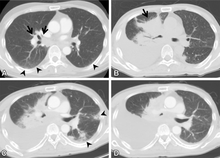
ICI-related pneumonitis in a 62-year-old man with squamous cell cancer. The patient was treated with nivolumab as the sixth line of therapy. (A) Axial pretreatment CT image shows subtle ground-glass abnormality (arrowheads) in the subpleural area, indicating ILA. Right hilar lymphadenopathy (arrows) also is apparent. (B) Axial CT image 10 months later shows a mass (arrow) in the right lung and pleural effusion, suggesting tumor exacerbation. (C) Axial CT image 20 days after the patient started nivolumab therapy shows a newly emergent patchy ground-glass abnormality and consolidation (arrowheads), representing ICI-related pneumonitis with an organizing pneumonia pattern; nivolumab was discontinued. (D) Axial CT image 3 months after the discontinuation of nivolumab shows that the patchy opacities have disappeared.

ICI-related pneumonitis in a 62-year-old man with squamous cell cancer. The patient was treated with nivolumab as the sixth line of therapy. (A) Axial pretreatment CT image shows subtle ground-glass abnormality (arrowheads) in the subpleural area, indicating ILA. Right hilar lymphadenopathy (arrows) also is apparent. (B) Axial CT image 10 months later shows a mass (arrow) in the right lung and pleural effusion, suggesting tumor exacerbation. (C) Axial CT image 20 days after the patient started nivolumab therapy shows a newly emergent patchy ground-glass abnormality and consolidation (arrowheads), representing ICI-related pneumonitis with an organizing pneumonia pattern; nivolumab was discontinued. (D) Axial CT image 3 months after the discontinuation of nivolumab shows that the patchy opacities have disappeared.