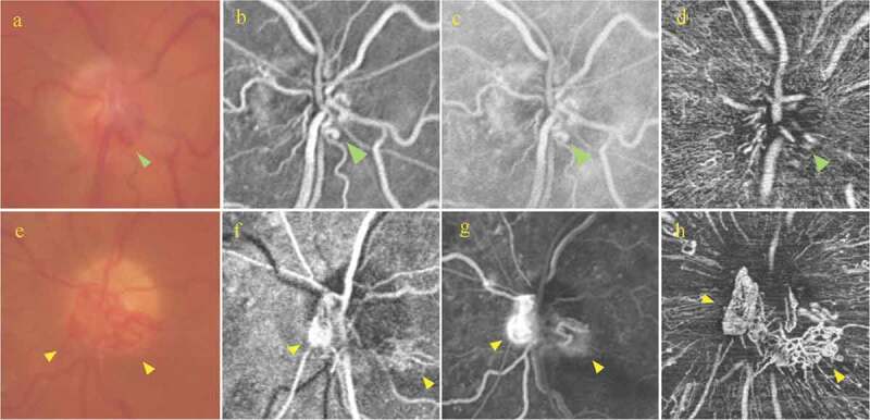
Figure 2.

Right eye: (a) Magnified colour optic disc photograph demonstrating irregular vascular loops at the optic disc (green arrow); (b, c) Magnified early and late fluorescein angiography, respectively showing that the vessels at the optic disc show no leakage (green arrow); (d) Radial peripapillary capillary (RPC) slab of optical coherence tomography angiography (OCTA) showing an irregular looking vascular network (green arrow).
Left eye: (e) Magnified colour optic disc photograph demonstrating vascular loops located at the nasal and inferior optic disc quadrants (yellow arrows); (f, g) Early and late phases of fluorescein angiography showing the presence of progressive leakage from the optic disc (yellow arrows); (h) RPC slab of OCTA showing the presence of multiple irregular and looped vessels in a mesh-like structure (yellow arrows).
