. 2022 Nov 4;11:e68745. doi: 10.7554/eLife.68745

Figure 5. Condensin dosage compensation (DC) is recruited to and spreads in either direction from the ectopically inserted recruitment elements on the X-chromosome (rex) sites on chromosome-II in L3.

(A) Snapshot of a region on chromosome-II where rex sites are inserted. Shown are four different conditions in L3 stage: wild-type, ERC61 (single rex-8 insertion) (Albritton et al., 2017), ERC63 (rex-8, rex-1, rex-8) [33], and ERC90 super rex, an array of six truncated, midsection (150 bp) of three strong rex’s: rex-40, rex-8, rex-35, repeated. Binding surrounding the insertion sites in both directions increases with increased number and strength of inserted rexes. The levels of condensin DC ectopically recruited to chromosome-II remain weaker than the endogenous binding at X-chromosome (lower panel). (B) The enrichment of condensin DC on each autosome plotted as chromosome-wide mean enrichment and total number of binding peaks calculated from DPY-27 ChIP-seq data. The ChIP-seq values or the total number of peaks are rescaled for each data, such that the mean of autosomes excluding chromosome-II is fixed to 0 and that of X-chromosome is at 1. The plot highlights weak but reproducible recruitment of more condensin DC to chromosome-II by rex insertion. (C) The level of condensin DC binding at the inserted strong rex-8 (indicated by arrow in each strain) is comparable to the endogenous, as indicated by the ChIP-seq signal at inserted rex site (400 bp) normalized to the mean of all endogenous strong rex sites on the X.

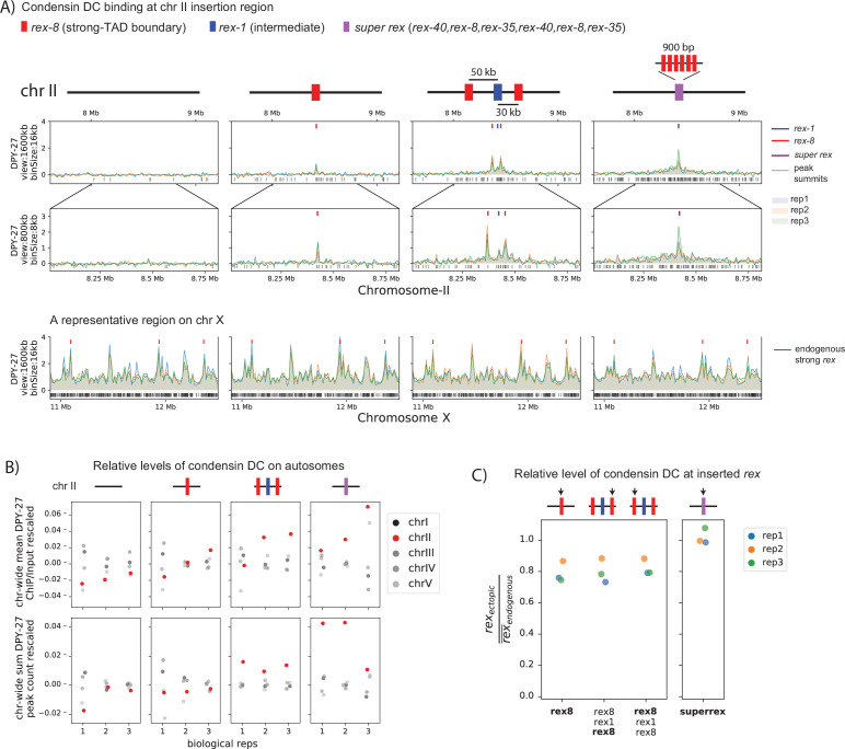
Figure 5.

Figure 5—figure supplement 1. Condensin dosage compensation (DC) is recruited to and spreads in either direction from the ectopically inserted recruitment elements on the X-chromosome (rex) sites on chromosome-II in embryo.

Figure 5—figure supplement 1.

(A) Snapshot of a region on chromosome II where rex sites are inserted. Shown are four different conditions in embryo stage: wild-type, ERC06 (single rex-1 insertion) (Albritton et al., 2017), ERC61 (single rex-8 insertion) [33], ERC62 (rex-1, rex-8) [33], and ERC63 (rex-8, rex-1, rex-8) (Albritton et al., 2017). Binding surrounding the insertion sites in both directions increases with increased number and strength of inserted rexes. The levels of condensin DC ectopically recruited to chromosome-II remain weaker than the endogenous binding at X-chromosome (lower panel). (B) The enrichment of condensin DC on each autosome plotted as chromosome-wide mean enrichment and total number of binding peaks calculated from DPY-27 ChIP-seq data. The ChIP-seq values or the total number of peaks are rescaled for each data, such that the mean of autosomes excluding chromosome-II is fixed to 0 and that of X-chromosome is at 1. The plot highlights weak but reproducible recruitment of more condensin DC to chromosome-II by rex insertion. (C) The level of condensin DC binding at the inserted rex-1 or strong rex-8 (indicated by arrow in each strain) is comparable to the endogenous, as indicated by the ChIP-seq signal at inserted rex site (400 bp) normalized to the mean of all endogenous strong rex sites on the X.
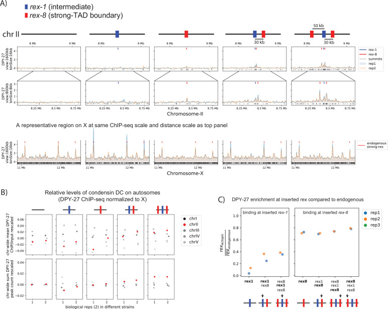
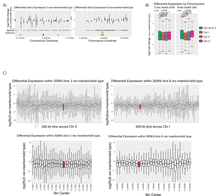
Figure 5—figure supplement 2. mRNA-seq data comparing recruitment elements on the X-chromosome (rex) insertion strains to wildtype.

Figure 5—figure supplement 2.

(A) Log2 fold change between the insertion and wildtype for each gene in the 100 kb region surrounding the insertion sites. Two insertion conditions used are ERC63 (rex-8, rex-1, rex-8 on chromosome-II) [33] (left panel) and ERC08 (eight rex-1 on chromosome-I) (right panel). The gray shading underneath the points indicates gene bodies. (B) Boxplots of the log2 fold change in the two strains. Autosomes III, IV, and V are grouped as autosomal control. Two-tailed student’s t-test p-values are given. (C) Boxplots log2 fold change values are binned into 200 kb (top panels) or 500 kb (bottom panels) domains tiled across the chromosome where the rex sites are inserted. Bins containing a significant level of repression are marked with asterisks (Fisher exact test; * indicates p-value<0.05, ** indicates p-value<0.01, *** indicates p-value<0.001). The red bins indicate bins that contain an inserted rex site.